Spanish Fly for two - how they affect libido in women and men
Contents Biologically active additive based on an extract obtained from a beetle with a fly (or fly...
From this article you will learn:
Everyone knows that a person is about 80% water. After all, water is the basis of blood (91%), gastric juice (98%), mucous membranes and other fluids in the human body. Our muscles also have water (74%), in the skeleton it is about 25%, and, of course, it is present in the brain (82%). Therefore, water definitely affects the ability to remember, thinking and physical capabilities of a person. How to keep the body's water balance at a normal level so that there are no health problems? You will learn about this from our article.
Water and electrolyte balance of the body- this is a set of processes of assimilation and distribution of water throughout the human body and its subsequent withdrawal.
When the water balance is normal, then the amount of fluid secreted by the body is adequate to the incoming volume, that is, these processes are balanced. If there is not enough water drunk, the balance will turn out to be negative, which means that the metabolism will be significantly slowed down, the blood will become too thick and will not be able to distribute oxygen throughout the body in the right volume, the body temperature will rise and the pulse will increase. It follows from this that the total load on the body will be higher, but the performance will drop.
But if you drink more water than you need, that too can be harmful. The blood will become too thin, and the cardiovascular system will get a big load. The concentration of gastric juice will also decrease, and this will lead to disruption of the digestive processes. Excess water causes a violation of the water balance in the human body, and makes the excretory system work with an increased load - excess fluid is excreted with sweat and urine. This not only leads to additional work of the kidneys, but also contributes to the excessive loss of nutrients. All these processes eventually disrupt the water-salt balance and significantly weaken the body.
Also, you can not drink a lot during physical exertion. Your muscles will tire quickly and you may even get cramps. You have probably noticed that athletes do not drink a lot of water during training and performances, but only rinse their mouths so as not to overload the heart. You can also use this technique during jogging and training.

The causes of imbalance are the incorrect distribution of fluid throughout the body or its large losses. As a result, there is a deficiency of trace elements that are actively involved in metabolic processes.
One of the main elements is calcium, its concentration in the blood may decrease, in particular, for the following reasons:
The concentration of another equally important trace element - sodium- may decrease for the following reasons:

deficit potassium occurs with alcohol abuse, taking corticosteroids, as well as with a number of other pathologies, for example:
However, potassium levels can also rise, which also upsets the balance.
If during the day the body has spent more fluid than it has received, then this is called negative water balance or dehydration. At the same time, tissue nutrition is disturbed, brain activity decreases, immunity decreases, and you may feel unwell.
Symptoms of negative water balance:
Minerals (dissolved in water, they are called electrolytes) also affect the water-salt balance.
The most important are calcium (Ca), sodium (Na), potassium (K), magnesium (Mg), compounds with chlorine, phosphorus, bicarbonates. They are responsible for the most important processes in the body.
Negative consequences for the body will be both with an insufficient amount of water and trace elements, and with an excess. You may not have enough water in your body if you have had vomiting, diarrhea, or heavy bleeding. Most of all, the lack of water in the diet is felt by children, especially newborns. They have an increased metabolism, as a result of which the concentration of electrolytes and metabolic products can increase very quickly in the tissues. If the excess of these substances is not removed in time, it can pose a serious threat to health.
Many pathological processes in the kidneys and liver lead to fluid retention in the tissues, causing a violation of the water balance in the body. If a person drinks too much, then water will also accumulate. As a result, the water-salt balance is disturbed, and this, in turn, causes not only malfunctions in the functioning of various organs and systems, but can also lead to more serious consequences, such as pulmonary and cerebral edema, and collapse. In this case, there is already a threat to human life.

In the case of hospitalization of the patient, the analysis of the water and electrolyte balance of his body is not carried out. Usually, drugs with electrolytes are prescribed immediately (of course, depending on the underlying diagnosis and severity of the condition), and further therapy and research are based on the body's response to these drugs.
When a person is admitted to the hospital, the following information is collected and entered into his card:
The sooner you establish the cause of the disease, the sooner you can eliminate problems with your water-salt balance and quickly organize the necessary treatment.

The average person needs about two liters of water per day. You can accurately calculate the required volume of liquid using the formula below. About one and a half liters a person receives from drinks, almost a liter comes from food. Also, part of the water is formed due to the oxidation process in the body.
To calculate the amount of water you need per day, you can use the following formula: multiply 35-40 ml of water by body weight in kilograms. That is, it is enough to know your own weight to instantly calculate the individual need for water.
For example, if your weight is 75 kg, then using the formula we calculate the volume you need: multiply 75 by 40 ml (0.04 l) and get 3 liters of water. This is your daily volume of fluid intake to maintain the normal water-salt balance of the body.
Every day the human body loses a certain amount of water: it is excreted in the urine (about 1.5 l), with sweat and breath (about 1 l), through the intestines (about 0.1 l). On average, this amount is 2.5 liters. But the water balance in the human body is very dependent on external conditions: ambient temperature and the amount of physical activity. Increased activity and heat cause thirst, the body itself tells you when you need to make up for the loss of fluid.

At high air temperatures, our body heats up. And overheating can be very dangerous. Therefore, the mechanism of thermoregulation immediately turns on, based on the evaporation of liquid by the skin, due to which the body cools. Approximately the same thing happens during an illness with an elevated temperature. In all cases, a person needs to replenish the loss of fluid, take care of restoring the water-salt balance in the body by increasing water intake.
Under comfortable conditions, at an air temperature of about 25 ° C, the human body releases about 0.5 liters of sweat. But as soon as the temperature begins to rise, the secretion of sweat increases, and each additional degree causes our glands to part with another hundred grams of fluid. As a result, for example, in a 35-degree heat, the amount of sweat excreted by the skin reaches 1.5 liters. The body in this case, thirst reminds of the need to replenish the supply of fluid.
So, we have already found out how much water a person needs to consume during the day. However, it is important in what mode the fluid enters the body. It is necessary to evenly distribute the intake of water during wakefulness. Thanks to this, you will not provoke swelling, do not make the body suffer from a lack of water, which will bring it the maximum benefit.
How to normalize the water balance in the body? Many people only drink water when they are thirsty. This is a big mistake. Thirst indicates that you are already dehydrated. Even when it is very small, it still has a strong effect on the body. Remember that you should not drink a lot at breakfast, lunch and dinner, as well as immediately after meals. This will significantly reduce the concentration of gastric juice and worsen the digestive process.
How to restore the water balance in the body?
It is best to draw up a water intake schedule for yourself, for example:
In addition, one glass can be drunk during a meal. As a result, we get the right amount of water in twenty-four hours. The proposed drinking schedule ensures a uniform flow of water into the body, which means that there will be no need to worry about swelling or dehydration.
To maintain a normal water-salt balance, one should not forget about the following factors:
With a uniform water supply, your metabolism will improve, energy will be generated constantly during the period of activity and you will not get so tired from work. Also, maintaining the water balance in the body will not accumulate toxins, which means that the liver and kidneys will not be overloaded. Your skin will become more elastic and firm.

Excessive loss of fluid or insufficient intake for a person is fraught with failures of various systems. How to restore the water-salt balance in the body? It must be understood that at one time the water deficit cannot be replenished, therefore it is not required to drink large portions. The fluid in the body should flow evenly.
The state of dehydration is also accompanied by sodium deficiency, so you need to drink not just water, but various solutions with electrolytes. They can be bought at a pharmacy and simply dissolved in water. But if dehydration is severe enough, you should immediately seek medical help. This is especially important in relation to children, with any signs of dehydration in a small child, it is necessary to call an ambulance. The same applies to older people.
In case of oversaturation of tissues and organs with water, it is not necessary to independently restore the water-salt balance in the body. Consult a doctor and find out the cause of the failure that caused this condition. Often it is a symptom of a disease and requires treatment.
What to do to stay hydrated:
A person who, without due responsibility, approaches the use of liquids, is threatened with dehydration or swelling. In no case do not disturb the water balance in the body. Keep a close eye on the amount of fluid in your body.
Restoring the water-salt balance in the body is very important for the well-being and functioning of organs. Below is a general scheme by which the health status of patients with these problems is normalized in medical institutions.
If a person is prescribed intravenous saline solutions, a preliminary calculation is made of the degree of disturbance of the water and electrolyte balance and, taking into account these data, a plan of therapeutic measures is drawn up. There are simple formulas based on the normative and actual indicators of sodium concentration in the blood. This technique allows you to determine the violations of the water balance in the human body, the calculation of fluid deficiency is carried out by a doctor.

The Ecocenter company supplies coolers, pumps and related equipment to Russia for bottling water from bottles of various sizes. All equipment is supplied under the trademark "ECOCENTER".
We provide the best ratio of price and quality of equipment, as well as offer our partners excellent service and flexible terms of cooperation.
You can be convinced of the attractiveness of collaboration by comparing our prices with the cost of similar equipment from other suppliers.
All our equipment complies with the standards established in Russia and has quality certificates. We deliver dispensers to customers, as well as all the spare parts and accessories they need in the shortest possible time.
Metabolic diseases. Effective methods of treatment and prevention Tatyana Vasilievna Gitun
Violations of water and electrolyte balance
Hypokalemia is a low concentration of potassium in the blood serum. It develops with a decrease in the amount of this mineral substance in the blood serum below 3.5 mmol / l and in cells (hypocalhystia), in particular in erythrocytes and muscles, below 40 mmol / l.
The cause of the disease is the loss of potassium in:
repeated vomiting;
Intoxication with acetylsalicylic acid (aspirin);
Polyuria (excessive urination) accompanying certain diseases or associated with prolonged use of diuretics.
With hypokalemia, disturbances in the metabolism of carbohydrates and proteins, acid-base, and also water balance are noted.
Treatment of the disease is aimed at eliminating its cause and restoring potassium deficiency.
The patient is recommended a vegetable diet and potassium preparations (potassium chloride, panangin, potassium orotate) orally or parenterally. The same drugs, along with potassium-sparing drugs (veroshpiron, triampur), are used for prophylaxis in patients receiving diuretics for a long time.
Dehydration of the body (exicosis) is a pathological condition that is caused by a decrease in the water content in the patient's body. The loss of water, which leads to a decrease in body weight by 10-20%, is life-threatening. A common cause of dehydration is diarrhea, persistent vomiting, polyuria (in diabetes, certain kidney diseases, hypervitaminosis D, hyperparathyroidism, Addison's disease, improper use of diuretics). It occurs with profuse sweating and evaporation of water with exhaled air, as well as acute blood loss and plasma loss (with extensive burns).
Dehydration can be provoked by water starvation as a result of a violation of the drinking regime, associated with disorders of consciousness of helpless patients and children with improper care for them, patients with loss of a sense of thirst of a psychogenic nature and people deprived of access to water (for example, during natural disasters).
The loss of water is accompanied by the removal of sodium and other active substances from it. With the predominance of its loss over the loss of salts and water starvation, a hyperosmotic, or water-deficient, type of dehydration develops, which is characterized by a pronounced decrease in the water content in the cells of organs and tissues (hypohydration, or dehydration, of cells). If the primary loss of sodium (for example, with adrenal insufficiency, some forms of nephritis), hypoosmotic, or salt-deficient, type of dehydration is noted, in which water from the intercellular space is redistributed in the cells, accumulating in them in large quantities.
For all types of dehydration, the common features are:
Decrease in body weight by more than 5%;
Dryness and flabbiness of the skin;
The appearance of wrinkles on the skin of the face;
The sharpness of his features;
Decreased blood pressure.
With any of the exicoses, urgent hospitalization is necessary. With the iso-osmotic type of dehydration, isotonic solutions of sodium chloride and glucose are injected intravenously, with plasma loss - plasma, as well as its substitutes. Mineral water is used for drinking, food should be liquid (for example, juices, broths, kefir), which includes products that are not contraindicated due to the patient's underlying illness.
A patient with a hyperosmotic type of dehydration should be given water without sugar and salt, or intravenously administered 1 liter of a 5% glucose solution (with the addition of 8 IU of insulin for injection), with the first 200 ml by jet, the rest by drip.
In the future, it is recommended to give the patient berry fruit drinks (for example, lingonberries or cranberries) without sugar or slightly sweetened. In the hypoosmotic type of dehydration, adults are first intravenously injected with a hypertonic solution of sodium chloride (up to 20 ml of a 10% solution) and glucose (40 ml of a 20% solution), after which treatment is continued by drip administration of isotonic solutions of these substances with a total volume of 1.5 -2 l. Use deoxycorticosterone acetate (DOXA) and other drugs that have the properties of adrenal hormones. Provide a diet high in salt. Children are prescribed oralit and pedialitis tablet solutions (1 tablet per 1 liter of water), which contain sodium and potassium salts in a proportion close to their ratio in blood plasma, subcutaneous or intravenous infusions of isotonic glucose-salt solutions under the control of central venous pressure and urine specific gravity. Indicators of the effectiveness of measures against dehydration of the hypoosmotic type are considered to be an increase in pulse pressure and normalization of blood pressure, as well as an improvement in patient tolerance to orthostatic load.
Prevention of dehydration consists in the prevention and timely treatment of diseases accompanied by loss of water, in the correct use of diuretics.
This text is an introductory piece. From the book Propaedeutics of childhood diseases author O. V. Osipova author From the book Propaedeutics of childhood diseases: lecture notes author O. V. Osipova From the book Pathological Physiology author Tatyana Dmitrievna Selezneva From the book Heat Disorders in Newborns author Dmitry Olegovich Ivanov From the book Colorpuncture. 40 effective treatment regimens by Ki Sheng Yu From the book Improvement of the spine and joints: methods of S. M. Bubnovsky, the experience of readers of the Bulletin of the Healthy Lifestyle author Sergei Mikhailovich Bubnovsky From the book We remove salt from the body: effective ways to cleanse with diets and folk remedies author Irina Ilyinichna Ulyanova From the book Life Support for Aircraft Crews after a Forced Landing or Splashing author Vitaly Georgievich Volovich From the book Features of the National Hangover author A. Borovsky From the book Heart Treatment with Herbs author Ilya Melnikov From the book Metabolic Diseases. Effective methods of treatment and prevention author Tatyana Vasilievna Gitun From the book Juice Treatment author Ilya Melnikov From the book Real recipes against cellulite. 5 min a day author Kristina Alexandrovna Kulagina From the book Learning to understand your analyzes author Elena V. Poghosyan From the book Clinical Nutrition for Chronic Diseases author Boris Samuilovich KaganovViolation of water-electrolyte metabolism is an extremely common pathology in seriously ill patients. The resulting water content disorders in various body media and the associated changes in the content of electrolytes and CBS create the prerequisites for the occurrence of dangerous disorders of vital functions and metabolism. This determines the importance of an objective assessment of the exchange of water and electrolytes both in the preoperative period and during intensive care.
Water with substances dissolved in it is a functional unity both in biological and physico-chemical terms and performs diverse functions. Metabolic processes in the cell proceed in the aquatic environment. Water serves as a dispersion agent for organic colloids and an indifferent basis for the transport of building and energy substances to the cell and the evacuation of metabolic products to the excretory organs.
In newborns, water accounts for 80% of body weight. With age, the water content in tissues decreases. In a healthy man, water is on average 60%, and in women 50% of body weight.
The total volume of water in the body can be divided into two main functional spaces: intracellular, whose water makes up 40% of body weight (28 liters in men with a weight of 70 kg), and extracellular - about 20% of body weight.
The extracellular space is a fluid that surrounds cells, the volume and composition of which is maintained by regulatory mechanisms. The main cation of the extracellular fluid is sodium, the main anion is chlorine. Sodium and chloride play a major role in maintaining the osmotic pressure and fluid volume of this space. The extracellular fluid volume consists of a rapidly moving volume (functional extracellular fluid volume) and a slowly moving volume. The first of these includes plasma and interstitial fluid. The slowly moving volume of extracellular fluid includes fluid found in bones, cartilage, connective tissue, subarachnoid space, and synovial cavities.
The concept of "third water space" is used only in pathology: it includes fluid accumulating in the serous cavities with ascites and pleurisy, in the subperitoneal tissue layer with peritonitis, in the closed space of intestinal loops with obstruction, especially with volvulus, in the deep layers of the skin in the first 12 hours after the burn.
The extracellular space includes the following water sectors.
Intravascular water sector - plasma serves as a medium for erythrocytes, leukocytes and platelets. The protein content in it is about 70 g/l, which is much higher than in the interstitial fluid (20 g/l).
The interstitial sector is the environment in which cells are located and actively function, it is a fluid of the extracellular and extravascular spaces (together with lymph). The interstitial sector is not filled with a freely moving liquid, but with a gel that holds the water in a fixed state. The basis of the gel is glycosaminoglycans, mainly hyaluronic acid. Interstitial fluid is a transport medium that does not allow substrates to spread throughout the body, concentrating them in the right place. Through the interstitial sector, the transit of ions, oxygen, nutrients into the cell and the reverse movement of toxins into the vessels, through which they are delivered to the excretory organs, take place.
Lymph, which is an integral part of the interstitial fluid, is mainly intended for the transport of chemical large-molecular substrates (proteins), as well as fatty conglomerates and carbohydrates from the interstitium into the blood. The lymphatic system also has a concentration function, since it reabsorbs water in the area of the venous end of the capillary.
The interstitial sector is a significant "capacity" containing? all body fluid (15% of body weight). Due to the fluid of the interstitial sector, the plasma volume is compensated for in acute blood and plasma loss.
Intercellular water also includes transcellular fluid (0.5-1% of body weight): fluid of serous cavities, synovial fluid, fluid of the anterior chamber of the eye, primary urine in the tubules of the kidneys, secretions of the lacrimal glands, secretions of the glands of the gastrointestinal tract.
The general directions of water movement between body media are shown in Fig. 3.20.
The stability of the volumes of liquid spaces is ensured by the balance of inputs and losses. Usually, the vascular bed is replenished directly from the gastrointestinal tract and lymphatics, emptied through the kidneys and sweat glands, and exchanged with the interstitial space and the gastrointestinal tract. In turn, the interstitial sector exchanges water with the cellular, as well as with the circulatory and lymphatic channels. Free (osmotically bound) water - with the interstitial sector and intracellular space.
The main causes of water and electrolyte balance disorders are external fluid losses and their non-physiological redistribution between the main fluid sectors of the body. They can occur due to pathological activation of natural processes in the body, in particular with polyuria, diarrhea, excessive sweating, with profuse vomiting, due to losses through various drains and fistulas, or from the surface of wounds and burns. Internal movement of fluids is possible with the development of edema in injured and infected areas, but is mainly due to changes in the osmolality of fluid media. Specific examples of internal movements are the accumulation of fluids in the pleural and abdominal cavities in pleurisy and peritonitis, blood loss in tissues with extensive fractures, the movement of plasma into injured tissues in crush syndrome, etc. A special type of internal fluid movement is the formation of so-called transcellular pools in the gastrointestinal tract (with intestinal obstruction, volvulus, intestinal infarction, severe postoperative paresis).
Fig.3.20. General directions of water movement between body media
An imbalance of water in the body is called dyshydria. Dyshydria is divided into two groups: dehydration and hyperhydration. In each of them, three forms are distinguished: normosmolal, hypoosmolal and hyperosmolal. The classification is based on the osmolality of the extracellular fluid, because it is the main factor determining the distribution of water between cells and the interstitial space.
Differential diagnosis of various forms of dyshydria is carried out on the basis of anamnestic, clinical and laboratory data.
Finding out the circumstances that led the patient to a particular dyshydria is of paramount importance. Indications of frequent vomiting, diarrhea, taking diuretic and laxative drugs suggest that the patient has a water-electrolytic imbalance.
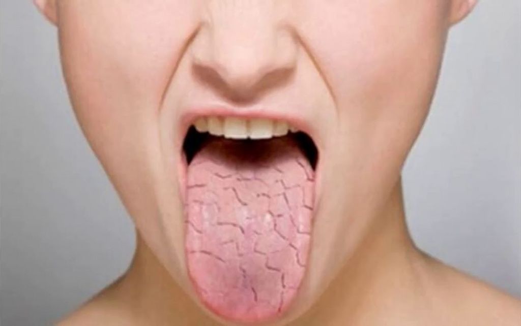
Thirst is one of the early signs of water deficiency. The presence of thirst indicates an increase in the osmolality of the extracellular fluid, followed by cellular dehydration.
Dryness of the tongue, mucous membranes and skin, especially in the axillary and inguinal regions, where the sweat glands are constantly functioning, indicate significant dehydration. At the same time, the turgor of the skin and tissues decreases. Dryness in the axillary and inguinal areas indicates a pronounced water deficit (up to 1500 ml).
The tone of the eyeballs may indicate, on the one hand, dehydration (decreased tone), on the other hand, hyperhydration (eyeball tension).
Edema is more often caused by an excess of interstitial fluid and sodium retention in the body. No less informative in interstitial hyperhydria are signs such as puffiness of the face, smoothness of the reliefs of the hands and feet, the predominance of transverse striation on the back surface of the fingers, and the complete disappearance of longitudinal striation on their palmar surfaces. It should be taken into account that edema is not a highly sensitive indicator of the balance of sodium and water in the body, since the redistribution of water between the vascular and interstitial sectors is due to a high protein gradient between them.
Changes in soft tissue turgor in relief zones: face, hands and feet are reliable signs of interstitial dyshydria. Interstitial dehydration is characterized by: retraction of the periocular tissue with the appearance of shadow circles around the eyes, sharpening of facial features, contrasting reliefs of the hands and feet, especially noticeable on the back surfaces, accompanied by a predominance of longitudinal striation and wrinkling of the skin, highlighting the articular areas, which gives them the appearance of a bean pod, flattening of the fingertips.
The appearance of “hard breathing” during auscultation is due to increased sound conduction on exhalation. Its appearance is due to the fact that excess water is quickly deposited in the interstitial tissue of the lungs and leaves it when the chest is elevated. Therefore, it should be sought in those areas that occupied the lowest position for 2-3 hours before listening.
Changes in turgor and volume of parenchymal organs are a direct sign of cellular hydration. The most accessible for research are the tongue, skeletal muscles, liver (sizes). The dimensions of the tongue, in particular, must correspond to its place, limited by the alveolar process of the lower jaw. With dehydration, the tongue noticeably decreases, often does not reach the front teeth, the skeletal muscles are flabby, foam rubber or gutta-percha consistency, the liver is reduced in size. With hyperhydration, teeth marks appear on the lateral surfaces of the tongue, skeletal muscles are tense, painful, and the liver is also enlarged and painful.
Body weight is a significant indicator of fluid loss or gain. In young children, severe fluid deficiency is indicated by a rapid decrease in body weight of more than 10%, in adults - more than 15%.
Laboratory studies confirm the diagnosis and complement the clinical picture. Of particular importance are the following data: osmolality and concentration of electrolytes (sodium, potassium, chloride, bicarbonate, sometimes calcium, phosphorus, magnesium) in plasma; hematocrit and hemoglobin, blood urea, total protein and albumin to globulin ratio; results of a clinical and biochemical analysis of urine (amount, specific gravity, pH values, sugar level, osmolality, protein, potassium, sodium, acetone bodies, sediment examination; concentration of potassium, sodium, urea and creatinine).
Dehydration. Isotonic (normoosmolal) dehydration develops due to loss of extracellular fluid, similar in electrolyte composition to blood plasma: with acute blood loss, extensive burns, abundant discharge from various parts of the gastrointestinal tract, with leakage of exudate from the surface of extensive superficial wounds, with polyuria, with excessively energetic therapy with diuretics, especially against the background of a salt-free diet.
This form is extracellular, since, with its inherent normal osmolality of the extracellular fluid, the cells are not dehydrated.
A decrease in the total content of Na in the body is accompanied by a decrease in the volume of the extracellular space, including its intravascular sector. Hypovolemia occurs, hemodynamics are disturbed early, and with severe isotonic losses, dehydration shock develops (example: cholera algid). Loss of 30% or more of plasma volume is directly life threatening.
There are three degrees of isotonic dehydration: I degree - loss of up to 2 liters of isotonic fluid; II degree - loss up to 4 liters; III degree - loss of 5 to 6 liters.
The characteristic signs of this dyshydria are a decrease in blood pressure when the patient is kept in bed, compensatory tachycardia, and orthostatic collapse is possible. With an increase in isotonic fluid loss, both arterial and venous pressure decrease, peripheral veins collapse, slight thirst arises, deep longitudinal folds appear on the tongue, the color of the mucous membranes is not changed, diuresis is reduced, urinary excretion of Na and Cl is reduced due to increased intake into the blood vasopressin and aldosterone in response to a decrease in plasma volume. At the same time, the osmolality of blood plasma remains almost unchanged.
Microcirculation disorders arising on the basis of hypovolemia are accompanied by metabolic acidosis. With the progression of isotonic dehydration, hemodynamic disturbances are aggravated: CVP decreases, blood thickening and viscosity increase, which increases the resistance to blood flow. Pronounced disturbances of microcirculation are noted: "marble", cold skin of the extremities, oliguria turns into anuria, arterial hypotension increases.
Correction of the considered form of dehydration is achieved mainly by infusion of normosmolal fluid (Ringer's solution, lactasol, etc.). In case of hypovolemic shock, in order to stabilize hemodynamics, a 5% glucose solution (10 ml / kg), normosmolal electrolyte solutions are first administered, and only then a colloidal plasma substitute is transfused (at the rate of 5-8 ml / kg). The rate of transfusion of solutions in the first hour of rehydration can reach 100-200 ml/min, then it is reduced to 20-30 ml/min. Completion of the stage of urgent rehydration is accompanied by an improvement in microcirculation: marbling of the skin disappears, limbs become warmer, mucous membranes turn pink, peripheral veins fill up, diuresis is restored, tachycardia decreases, and blood pressure normalizes. From this point on, the rate is reduced to 5 ml/min or less.
Hypertonic (hyperosmolal) dehydration differs from the previous variety in that against the background of a general fluid deficiency in the body, a lack of water predominates.
This type of dehydration develops when there is a loss of electrolyte-free water (perspiration loss), or when water loss exceeds electrolyte loss. The molal concentration of the extracellular fluid increases, and then the cells also dehydrate. The reasons for this condition can be an absolute lack of water in the diet, insufficient intake of water in the patient's body with defects in care, especially in patients with impaired consciousness, with loss of thirst, impaired swallowing. It can lead to increased water loss during hyperventilation, fever, burns, polyuric stage of acute renal failure, chronic pyelonephritis, diabetes and diabetes insipidus.
Together with water from the tissues, potassium enters, which, with preserved diuresis, is lost in the urine. With moderate dehydration, hemodynamics is disturbed little. With severe dehydration, BCC decreases, resistance to blood flow increases due to increased blood viscosity, increased release of catecholamines, and increased afterload on the heart. Blood pressure and diuresis decrease, while urine with a high relative density and an increased concentration of urea is released. Plasma Na concentration rises above 147 mmol/l, which accurately reflects the lack of free water.
The clinic of hypertensive dehydration is caused by dehydration of cells, especially brain cells: patients complain of weakness, thirst, apathy, drowsiness, with deepening dehydration, consciousness is disturbed, hallucinations, convulsions, hyperthermia appear.
Water deficit is calculated by the formula:
C (Napl.) - 142
X 0.6 (3.36),
Where: s (Napl.) - the concentration of Na in the patient's blood plasma,
0.6 (60%) - the content of all water in the body in relation to body weight, l.
The therapy is aimed not only at eliminating the cause of hypertensive dehydration, but also at replenishing the cellular fluid deficiency by infusion of 5% glucose solution with the addition of up to 1/3 of the volume of isotonic NaCl solution. If the patient's condition allows, rehydration is carried out at a moderate pace. Firstly, it is necessary to be wary of increased diuresis and additional fluid loss, and secondly, the rapid and abundant administration of glucose can reduce the molar concentration of the extracellular fluid and create conditions for the movement of water into the brain cells.
In severe dehydration with symptoms of dehydration hypovolemic shock, impaired microcirculation and centralization of blood circulation, urgent restoration of hemodynamics is necessary, which is achieved by replenishing the volume of the intravascular bed not only with glucose solution, which quickly leaves it, but also with colloidal solutions that retain water in the vessels, reducing the rate of fluid inflow into brain. In these cases, infusion therapy begins with an infusion of 5% glucose solution, adding to it up to 1/3 of the volume of reopoliglyukin, 5% albumin solution.
The ionogram of the blood serum is initially uninformative. Along with an increase in the concentration of Na +, the concentration of other electrolytes is also increased, and normal indicators of the concentration of K + always make one think about the presence of true hypocaligistia, which manifests itself after rehydration.
As diuresis is restored, it is necessary to prescribe an intravenous infusion of K + solutions. As the rehydration proceeds, a 5% glucose solution is poured in, periodically adding electrolyte solutions. The effectiveness of the rehydration process is controlled according to the following criteria: restoration of diuresis, improvement of the general condition of the patient, moistening of the mucous membranes, and a decrease in the concentration of Na + in the blood plasma. An important indicator of the adequacy of hemodynamics, especially venous flow to the heart, can be the measurement of CVP, which is normally equal to 5-10 cm of water. Art.
Hypotonic (hypoosmolal) dehydration is characterized by a predominance of a lack of electrolytes in the body, which leads to a decrease in the osmolality of the extracellular fluid. True Na+ deficiency can be accompanied by a relative excess of "free" water while maintaining dehydration of the extracellular space. The molar concentration of the extracellular fluid is reduced, conditions are created for the fluid to enter the intracellular space, including the brain cells with the development of its edema.
The volume of circulating plasma is reduced, blood pressure, CVP, pulse pressure are reduced. The patient is lethargic, drowsy, apathetic, he has no feeling of thirst, a characteristic metallic taste is felt.
There are three degrees of Na deficiency: I degree - deficiency up to 9 mmol/kg; II degree - deficiency 10-12 mmol / kg; III degree - deficiency up to 13-20 mmol/kg of body weight. With III degree of deficiency, the general condition of the patient is extremely severe: coma, blood pressure is reduced to 90/40 mm Hg. Art.
With moderately severe violations, it is enough to limit the infusion of 5% glucose solution with isotonic sodium chloride solution. With a significant deficiency of Na +, half of the deficiency is compensated with a hypertonic (molar or 5%) solution of sodium chloride, and in the presence of acidosis, the correction of Na deficiency is carried out with a 4.2% solution of sodium bicarbonate.
The calculation of the required amount of Na is carried out according to the formula:
Na + deficiency (mmol / l) \u003d x 0.2 x m (kg) (3.37),
Where: s(Na)pl. - Na concentration in the patient's blood plasma, mmol/l;
142 - the concentration of Na in the blood plasma is normal, mmol / l,
M - body weight (kg).
Infusions of solutions containing sodium are carried out at a decreasing rate. During the first 24 hours, 600-800 mmol Na + is injected, in the first 6-12 hours - approximately 50% of the solution. In the future, isotonic electrolyte solutions are prescribed: Ringer's solution, lactasol.
The identified deficiency of Na is replenished with solutions of NaCl or NaHCO3. In the first case, it is assumed that 1 ml of a 5.8% NaCl solution contains 1 mmol of Na, and in the second (used in the presence of acidosis), it is assumed that an 8.4% solution of bicarbonate in 1 ml contains 1 mmol. The calculated amount of one or another of these solutions is administered to the patient along with the transfused normosmolal saline solution.
Hyperhydration. It can also be normo-, hypo- and hyperosmolal. Anesthesiologists-resuscitators have to meet with her much less frequently.
Isotonic hyperhydration often develops as a result of excessive administration of isotonic saline solutions in the postoperative period, especially in case of impaired renal function. The causes of this hyperhydration can also be heart disease with edema, cirrhosis of the liver with ascites, kidney disease (glomerulonephritis, nephrotic syndrome). The development of isotonic hyperhydration is based on an increase in the volume of extracellular fluid due to a proportional retention of sodium and water in the body. The clinic of this form of hyperhydration is characterized by generalized edema (edematous syndrome), anasarca, a rapid increase in body weight, reduced blood concentrations; tendency to hypertension. The therapy of this dyshydria is reduced to the exclusion of the causes of their occurrence, as well as to the correction of protein deficiency by infusions of native proteins with the simultaneous removal of salts and water with the help of diuretics. With insufficient effect of dehydration therapy, hemodialysis with blood ultrafiltration can be performed.
Hypotonic hyperhydration is caused by the same factors that cause the isotonic form, but the situation is aggravated by the redistribution of water from the intercellular to the intracellular space, transmineralization and increased cell destruction. With hypotonic overhydration, the water content in the body increases significantly, which is also facilitated by infusion therapy with electrolyte-free solutions.
With an excess of "free" water, the molal concentration of body fluids decreases. "Free" water is evenly distributed in the fluid spaces of the body, primarily in the extracellular fluid, causing a decrease in the Na+ concentration in it. Hypotonic hyperhydration with hyponatriplasmia is observed with excessive intake of "free" water in quantities exceeding the possibility of excretion, if a) the bladder and prostate bed are washed with water (without salts) after its transurethral resection, b) drowning in fresh water occurs, c) an excessive infusion of glucose solutions is carried out in the oligoanuric stage of the SNP. This dyshydria may also be due to a decrease in glomerular filtration in the kidneys in acute and chronic kidney failure, congestive heart failure, liver cirrhosis, ascites, glucocorticoid deficiency, myxedema, Barter's syndrome (congenital insufficiency of the kidney tubules, a violation of their ability to retain Na + and K + with increased production of renin and aldosterone, hypertrophy of the juxtaglomerular apparatus). It occurs with ectopic production of vasopressin by tumors: thymoma, oat-round cell lung cancer, adenocarcinoma of the 12th duodenum and pancreas, with tuberculosis, increased production of vasopressin in lesions of the hypothalamic region, meningoencephalitis, hematoma, congenital anomalies and brain abscess, prescribing drugs drugs that increase the production of vasopressin (morphine, oxytocin, barbiturates, etc.).
Hyponatremia is the most common violation of water and electrolyte metabolism, accounting for 30-60% of all electrolyte imbalances. Often this violation is iatrogenic in nature - when an excess amount of a 5% glucose solution is infused (glucose is metabolized and "free" water remains).
The clinical picture of hyponatremia is diverse: disorientation and stupor in elderly patients, convulsions and coma in the acute development of this condition.
Acute development of hyponatremia is always clinically manifested. In 50% of cases, the prognosis is unfavorable. With hyponatremia up to 110 mmol / l and hypoosmolality up to 240-250 mosmol / kg, conditions are created for hyperhydration of brain cells and its edema.
The diagnosis is based on an assessment of the symptoms of damage to the central nervous system (fatigue, delirium, confusion, coma, convulsions) that occur against the background of intensive infusion therapy. It clarifies the fact of the elimination of neurological or mental disorders as a result of the preventive administration of solutions containing sodium. Patients with acute development of the syndrome, with severe clinical manifestations of the nervous system, primarily with the threat of developing cerebral edema, need emergency treatment. In these cases, intravenous administration of 500 ml of a 3% sodium chloride solution is recommended in the first 6-12 hours, followed by repeated administration of the same dose of this solution during the day. When sodium reaches 120 mmol/l, the administration of hypertonic sodium chloride solution is stopped. With possible decompensation of cardiac activity, it is necessary to prescribe furosemide with simultaneous administration of hypertonic solutions - 3% potassium chloride solution and 3% sodium chloride solution to correct Na + and K + losses.
The treatment of choice for hypertensive hyperhydration is ultrafiltration.
In hyperthyroidism with glucocorticoid deficiency, the administration of thyroidin and glucocorticoids is useful.
Hypertonic hyperhydration occurs as a result of excessive administration of hypertonic solutions into the body by the enteral and parenteral route, as well as infusions of isotonic solutions to patients with impaired renal excretory function. Both major water sectors are involved in the process. However, an increase in osmolality in the extracellular space causes dehydration of the cells and the release of potassium from them. The clinical picture of this form of hyperhydration is characterized by signs of edematous syndrome, hypervolemia and lesions of the central nervous system, as well as thirst, skin hyperemia, agitation, and a decrease in blood concentration parameters. Treatment consists in adjusting infusion therapy with the replacement of electrolyte solutions with native proteins and glucose solutions, in the use of osmodiuretics or saluretics, in severe cases - hemodialysis.
There is a close relationship between the severity of deviations in the water-electrolyte status and nervous activity. The peculiarity of the psyche and the state of consciousness can help to navigate in the direction of the tonic shift. With hyperosmia, there is a compensatory mobilization of cellular water and replenishment of water reserves from the outside. This is manifested by the corresponding reactions: suspiciousness, irritability and aggressiveness up to hallucinosis, severe thirst, hyperthermia, hyperkinesis, arterial hypertension.
On the contrary, with a decrease in osmolality, the neurohumoral system is brought into an inactive state, which provides the cell mass with rest and the opportunity to assimilate part of the water unbalanced by sodium. More often there are: lethargy and hypodynamia; aversion to water with its profuse losses in the form of vomiting and diarrhea, hypothermia, arterial and muscular hypotension.
Imbalance of K+ ions. In addition to disorders related to water and sodium, a seriously ill patient often has an imbalance of K + ions, which plays a very important role in ensuring the vital activity of the body. Violation of the content of K + in cells and in the extracellular fluid can lead to serious functional disorders and adverse metabolic changes.
The total supply of potassium in the body of an adult is from 150 to 180 g, that is, approximately 1.2 g / kg. Its main part (98%) is located in the cells, and only 2% - in the extracellular space. The largest amounts of potassium are concentrated in intensively metabolizing tissues - kidney, muscle, brain. In a muscle cell, some of the potassium is in a state of chemical bonding with protoplasmic polymers. Significant amounts of potassium are found in protein deposits. It is present in phospholipids, lipoproteins and nucleoproteins. Potassium forms a covalent type of bond with phosphoric acid residues, carboxyl groups. The significance of these bonds lies in the fact that complex formation is accompanied by a change in the physicochemical properties of the compound, including solubility, ionic charge, and redox properties. Potassium activates several dozen enzymes that provide metabolic cellular processes.
The complexing abilities of metals and competition between them for a place in the complex itself are fully manifested in the cell membrane. Competing with calcium and magnesium, potassium facilitates the depolarizing action of acetylcholine and the transfer of the cell to an excited state. With hypokalemia, this translation is difficult, and with hyperkalemia, on the contrary, it is facilitated. In the cytoplasm, free potassium determines the mobility of the energy cell substrate - glycogen. High concentrations of potassium facilitate the synthesis of this substance and at the same time hinder its mobilization for energy supply of cellular functions, low concentrations, on the contrary, inhibit glycogen renewal, but contribute to its breakdown.
Regarding the effect of potassium shifts on cardiac activity, it is customary to dwell on its interaction with cardiac glycosides. The result of the action of cardiac glycosides on Na + / K + - ATPase is an increase in the concentration of calcium, sodium in the cell and the tone of the heart muscle. A decrease in the concentration of potassium, a natural activator of this enzyme, is accompanied by an increase in the action of cardiac glycosides. Therefore, dosing should be individual - until the desired inotropism is achieved or until the first signs of glycoside intoxication.
Potassium is a companion of plastic processes. Thus, the renewal of 5 g of protein or glycogen needs to be provided by 1 unit of insulin, with the introduction of about 0.1 g of dibasic potassium phosphate and 15 ml of water from the extracellular space.
Potassium deficiency refers to the lack of its total content in the body. Like any deficit, it is the result of losses not offset by revenues. Its severity sometimes reaches 1/3 of the total content. The reasons may be different. Decreased dietary intake may be due to forced or conscious fasting, loss of appetite, damage to the masticatory apparatus, stenosis of the esophagus or pylorus, consumption of potassium-poor food, or infusion of potassium-depleted solutions during parenteral nutrition.
Excessive losses may be associated with hypercatabolism, increased excretory functions. Any massive and uncompensated loss of body fluids leads to massive potassium deficiencies. This can be vomiting with gastric stenosis or intestinal obstruction of any localization, loss of digestive juices in intestinal, biliary, pancreatic fistulas or diarrhea, polyuria (polyuric stage of acute renal failure, diabetes insipidus, abuse of saluretics). Polyuria can be stimulated by osmotically active substances (high glucose concentration in diabetes or steroid mellitus, use of osmotic diuretics).
Potassium practically does not undergo active resorption in the kidneys. Accordingly, its loss in the urine is proportional to the amount of diuresis.
A deficiency of K+ in the body may be indicated by a decrease in its content in the blood plasma (normally about 4.5 mmol / l), but provided that catabolism is not increased, there is no acidosis or alkalosis and a pronounced stress reaction. Under such conditions, the level of K + in plasma 3.5-3.0 mmol / l indicates its deficiency in the amount of 100-200 mmol, within 3.0-2.0 - from 200 to 400 mmol and at a content of less than 2, 0 mmol / l - 500 mmol or more. To some extent, the lack of K + in the body can be judged by its excretion in the urine. The daily urine of a healthy person contains 70-100 mmol of potassium (equal to the daily release of potassium from tissues and consumption from food). A decrease in potassium excretion to 25 mmol per day or less indicates a profound potassium deficiency. With potassium deficiency resulting from its large losses through the kidneys, the potassium content in daily urine is above 50 mmol, with potassium deficiency as a result of insufficient intake into the body - below 50 mmol.
Potassium deficiency becomes noticeable if it exceeds 10% of the normal content of this cation, and threatening - when the deficiency reaches 30% or more.
The severity of clinical manifestations of hypokalemia and potassium deficiency depends on the rate of their development and the depth of disorders.
Disorders of neuromuscular activity are leading in the clinical symptoms of hypokalemia and potassium deficiency and are manifested by changes in the functional state, the central and peripheral nervous system, the tone of striated skeletal muscles, smooth muscles of the gastrointestinal tract and bladder muscles. When examining patients, hypotension or atony of the stomach, paralytic ileus, stagnation in the stomach, nausea, vomiting, flatulence, bloating, hypotension or atony of the bladder are revealed. On the part of the cardiovascular system, a systolic murmur is recorded at the apex and an expansion of the heart, a decrease in blood pressure, mainly diastolic, bradycardia or tachycardia. With acutely developing deep hypokalemia (up to 2 mmol / l and below), atrial and ventricular extrasystoles often occur, myocardial fibrillation and circulatory arrest are possible. The immediate danger of hypokalemia lies in the disinhibition of the effects of antagonistic cations - sodium and calcium, with the possibility of cardiac arrest in systole. ECG signs of hypokalemia: low biphasic or negative T, the appearance of a V wave, QT expansion, PQ shortening. Typically, the weakening of tendon reflexes up to their complete disappearance and the development of flaccid paralysis, a decrease in muscle tone.
With the rapid development of deep hypokalemia (up to 2 mmol / l and below), generalized weakness of the skeletal muscles comes to the fore and can result in paralysis of the respiratory muscles and respiratory arrest.
When correcting potassium deficiency, it is necessary to ensure that potassium enters the body in the amount of physiological need, to compensate for the existing deficiency of intracellular and extracellular potassium.
K + deficiency (mmol) \u003d (4.5 - K + square), mmol / l * body weight, kg * 0.4 (3.38).
The elimination of potassium deficiency requires the exclusion of any stress factors (strong emotions, pain, hypoxia of any origin).
The amount of nutrients, electrolytes and vitamins prescribed under these conditions should exceed the usual daily requirements so as to cover both losses to the environment (during pregnancy - to the needs of the fetus) and a certain proportion of the deficiency.
To ensure the desired rate of restoration of the level of potassium in the composition of glycogen or protein, every 2.2 - 3.0 g of chloride or disubstituted potassium phosphate should be administered along with 100 g of glucose or pure amino acids, 20 - 30 units of insulin, 0.6 g of calcium chloride, 30 g of sodium chloride and 0.6 g of magnesium sulfate.
To correct hypocaligistia, it is best to use dipotassium phosphate, since glycogen synthesis is impossible in the absence of phosphates.
The complete elimination of cellular potassium deficiency is tantamount to a complete restoration of proper muscle mass, which is rarely achievable in a short time. It can be considered that a deficit of 10 kg of muscle mass corresponds to a deficiency of potassium of 1600 mEq, that is, 62.56 g of K+ or 119 g of KCI.
When K+ deficiency is eliminated intravenously, its estimated dose in the form of a KCl solution is infused together with a glucose solution, based on the fact that 1 ml of a 7.45% solution contains 1 mmol K., 1 meq of potassium = 39 mg, 1 gram of potassium = 25 meq. , 1 gram of KCl contains 13.4 meq of potassium, 1 ml of a 5% solution of KCl contains 25 mg of potassium or 0.64 meq of potassium.
It must be remembered that the entry of potassium into the cell requires some time, so the concentration of infused K + solutions should not exceed 0.5 mmol / l, and the infusion rate should not exceed 30-40 mmol / h. 1 g of KCl, from which a solution for intravenous administration is prepared, contains 13.6 mmol of K+.
If the deficiency of K+ is large, its replenishment is carried out within 2-3 days, given that the maximum daily dose of intravenously administered K+ is 3 mmol/kg.
The following formula can be used to determine the safe infusion rate:
Where: 0.33 - the maximum allowable safe infusion rate, mmol / min;
20 is the number of drops in 1 ml of crystalloid solution.
The maximum rate of potassium administration is 20 meq/h or 0.8 g/h. For children, the maximum rate of potassium administration is 1.1 meq / h or 43 mg / h. The adequacy of correction, in addition to determining the content of K + in plasma, can be determined by the ratio of its intake and excretion into the body. The amount of K + excreted in the urine in the absence of aldesteronism remains reduced relative to the administered dose until the deficiency is eliminated.
Both K+ deficiency and excess K+ content in plasma pose a serious danger to the body in case of renal insufficiency and its very intensive intravenous administration, especially against the background of acidosis, increased catabolism and cellular dehydration.
Hyperkalemia may be the result of acute and chronic renal failure in the stage of oliguria and anuria; massive release of potassium from tissues against the background of insufficient diuresis (deep or extensive burns, injuries); prolonged positional or tourniquet compression of the arteries, late restoration of blood flow in the arteries during their thrombosis; massive hemolysis; decompensated metabolic acidosis; the rapid introduction of large doses of relaxants of the depolarizing type of action, diencephalic syndrome in traumatic brain injury and stroke with convulsions and fever; excessive intake of potassium in the body against the background of insufficient diuresis and metabolic acidosis; the use of excess potassium in heart failure; hypoaldosteronism of any origin (interstitial nephritis; diabetes; chronic adrenal insufficiency - Addison's disease, etc.). Hyperkalemia can occur with rapid (within 2-4 hours or less) transfusion of massive doses (2-2.5 liters or more) of donor erythrocyte-containing media with long periods of preservation (more than 7 days).
Clinical manifestations of potassium intoxication are determined by the level and rate of increase in plasma potassium concentration. Hyperkalemia does not have well-defined, characteristic clinical symptoms. The most common complaints are weakness, confusion, various kinds of parasthesia, constant fatigue with a feeling of heaviness in the limbs, muscle twitching. In contrast to hypokalemia, hyperreflexias are recorded. Intestinal spasms, nausea, vomiting, diarrhea are possible. From the side of the cardiovascular system, bradycardia or tachycardia, a decrease in blood pressure, extrasystoles can be detected. The most typical ECG changes. Unlike hypokalemia, in hyperkalemia there is a certain parallelism of ECG changes and the level of hyperkalemia. The appearance of a high, narrow, pointed positive T wave, the onset of the ST interval below the isoelectric line, and shortening of the QT interval (ventricular electrical systole) are the first and most characteristic ECG changes in hyperkalemia. These signs are especially pronounced with hyperkalemia close to the critical level (6.5-7 mmol / l). With a further increase in hyperkalemia above the critical level, the QRS complex expands (especially the S wave), then the P wave disappears, an independent ventricular rhythm occurs, ventricular fibrillation and circulatory arrest occurs. With hyperkalemia, there is often a slowdown in atrioventricular conduction (an increase in the PQ interval) and the development of sinus bradycardia. Cardiac arrest with high hyperglycemia, as already indicated, can occur suddenly, without any clinical symptoms of a threatening condition.
If hyperkalemia occurs, it is necessary to intensify the excretion of potassium from the body by natural means (stimulation of diuresis, overcoming oligo- and anuria), and if this path is impossible, to carry out artificial excretion of potassium from the body (hemodialysis, etc.).
If hyperkalemia is detected, any oral and parenteral administration of potassium is immediately stopped, drugs that contribute to the retention of potassium in the body (capoten, indomethacin, veroshpiron, etc.) are canceled.
When high hyperkalemia (more than 6 mmol / l) is detected, the first therapeutic measure is the appointment of calcium preparations. Calcium is a functional potassium antagonist and blocks the extremely dangerous effect of high hyperkalemia on the myocardium, which eliminates the risk of sudden cardiac arrest. Calcium is prescribed in the form of a 10% solution of calcium chloride or calcium gluconate, 10-20 ml intravenously.
In addition, it is necessary to carry out therapy that reduces hyperkalemia by increasing the movement of potassium from the extracellular space into the cells: intravenous administration of a 5% solution of sodium bicarbonate at a dose of 100-200 ml; the appointment of concentrated (10-20-30-40%) glucose solutions at a dose of 200-300 ml with simple insulin (1 unit per 4 g of glucose administered).
Alkalinization of the blood promotes the movement of potassium into the cells. Concentrated glucose solutions with insulin reduce protein catabolism and thereby the release of potassium, help reduce hyperkalemia by increasing the current of potassium into cells.
In case of hyperkalemia not corrected by therapeutic measures (6.0-6.5 mmol/l and above in acute renal failure and 7.0 mmol/l and above in chronic renal failure) with simultaneously detectable ECG changes, hemodialysis is indicated. Timely hemodialysis is the only effective method of direct excretion of potassium and toxic products of nitrogen metabolism from the body, which ensures the preservation of the patient's life.
Electrolytes are substances that allow the transmission of electrical impulses. They also perform many other functions, so they play a special role in the human body. There are several essential electrolytes for humans. If there is a shortage of them, there will be serious problems. Along with the loss of fluid, a person also loses useful salts, so it is important to maintain their amount in the norm, making up for the deficiency through or special medicines.
Not all people understand what it is. Human electrolytes are salts that are capable of conducting electrical impulses. These substances perform several important functions, among which is the transmission of nerve impulses. In addition, they perform the following functions:
Each electrolyte performs its function. There are the following types:
There are norms for the content of electrolytes in the blood. If there is a lack or excess of substances, problems arise with the body. The salts influence each other, thereby creating a balance.
In addition to the fact that they affect the transmission of nerve impulses, each electrolyte has an individual function. For example, it helps in the work of the heart muscle and brain. Sodium helps the body's muscles respond to nerve impulses and do their job. The normal amount of chlorine in the body helps the digestive system to function properly. Calcium affects the strength of bones and teeth.
Based on this, it becomes clear that electrolytes perform many functions, so it is important to maintain their optimal content in the body. Lack or excess of one of the substances leads to serious pathologies that lead to health problems in the future.
Electrolytes are strongly lost along with the liquid. If a person, he must keep in mind that it will be necessary to replenish not only water, but also salt. There are special drinks that restore the water and electrolyte balance in the human body. They are used to avoid dangerous pathologies due to the loss of a large amount of salts and fluids.
If there is a deficiency or excess of electrolytes, then this will necessarily affect human health. There are various symptoms that you need to pay attention to. Deficiency occurs due to a large loss of fluids, disease and malnutrition. An excess of substances occurs due to the use of foods that contain salts in large quantities, as well as when certain organs are affected by diseases.
If an electrolyte deficiency occurs, the following symptoms occur:

If these symptoms occur, you should consult a doctor. A blood test for electrolytes will help determine the exact cause of their appearance. With its help, the amount of salts that affect the water and electrolyte balance in the body at the time of blood donation is determined.
1 Water and electrolyte disorders in surgical patients, infusion therapy.
2 Plan: Introduction Violation of water and electrolyte metabolism: dehydration, hyperhydration. Dehydration: Hypertonic, isotonic, hypotonic. infusion therapy. Conclusion.
3 Introduction. A clinician of any profile often has to treat patients with severe disorders of water and electrolyte balance - the most important system of the body's internal environment, the constancy of which, according to Claude Bernard, "is the condition for a free life." Mild degrees of water and electrolyte imbalance can be compensated by the body's reserve capabilities and not manifest clinically. More severe changes in water-electrolyte metabolism cannot be compensated even by excessive stress of all body systems and lead to pronounced disorders of the body's vital functions. This is due to the fact that a change in the amount of water and electrolytes disrupts the course of physicochemical processes, since water acts as a universal solvent and serves as the main “transport system” of the body and mediates its connection with the external environment and body cells. Being a severe pathological syndrome, the imbalance of water and electrolytes affects the subtle processes of metabolism, diffusion, osmosis, filtration and active movement of ions. The resulting changes in the concentration of electrolytes in the intracellular fluid leads to disruption of the activity of excitable tissues (nerve and muscle). In addition, the change in osmolarity leads to the movement of water between the extracellular and intracellular sectors, which compromises the viability of the cell. In the conditions of a whole organism, all organs and systems are involved in the process. Being unrecognized and not eliminated, violations of the hydroelectrolyte balance largely determine the results of the treatment of the underlying disease. Particularly severe disorders of water and electrolyte balance are found in patients with surgical and therapeutic clinics.
4 For competent diagnosis and treatment of water-electrolyte disorders, you need to have an idea about the fluid spaces of the body, electrolyte metabolism and acid-base balance. Water-electrolyte composition and body fluid spaces Water makes up 45–80% of body weight, depending on body fat content (see Table 13.1). Water is distributed in intracellular and extracellular spaces. The extracellular fluid bathes the outside of the cells and contains most of the body's sodium. Extracellular fluid is divided into interstitial and intravascular (plasma). For life support, the water-electrolyte balance of the intravascular fluid is most important, so treatment should be aimed primarily at restoring it. The composition of the intracellular and extracellular fluid: o Sodium is the main cation and osmotically active component of the extracellular fluid. o Potassium is the main cation and osmotically active component of the intracellular fluid. o Water passes freely through cell membranes, equalizing the osmotic pressure of intracellular and extracellular fluids. By measuring the osmolarity of one space (for example, plasma), we estimate the osmolarity of all fluid spaces in the body. Osmolarity is usually determined from plasma sodium concentration using the formula: plasma osmolarity (mosmol/kg) = 2 + glucose (mg%)/18 + BUN (mg%)/2.8. o An increase in plasma sodium concentration (osmolarity) indicates a relative lack of water. o A decrease in plasma sodium concentration (osmolarity) indicates a relative excess of water. The osmotic constancy of the body is provided by the consumption and excretion of water, which are regulated by ADH and thirst mechanisms. Many surgical patients are unable to drink (nothing by mouth prescription, nasogastric tube, etc.) and lose control of fluid intake. Osmotic disorders are not uncommon and often iatrogenic.
5 Sodium, as the main osmotically active component of extracellular fluid, plays an important role in maintaining BCC. o The volume of extracellular fluid is maintained at a constant level due to the retention of sodium and water by the kidneys. o Diagnosis of sodium deficiency must be clinical, i.e. based on physical examination findings and assessment of central hemodynamics (CVP and PAWP). A decrease in the total sodium content in the body is accompanied by symptoms of hypovolemia (tachycardia, orthostatic hypotension, shock). The severity of symptoms depends on the degree of hypovolemia and should be taken into account when planning treatment. o Plasma sodium concentration does not provide an indication of total body sodium. o With an excess of sodium, edema, arterial hypertension, weight gain, ascites, and in some cases heart failure are observed. Edema of the legs, leaving a hole when pressed, appears with an excess of 2-4 liters of 0.9% NaCl. Anasarca occurs when the volume of extracellular fluid increases by 80-100% (that is, by about 15 liters with a weight of 70 kg). To prevent the accumulation of sodium in the body, it is necessary to take into account all the details of infusion therapy, the function of the cardiovascular system and the kidneys of the patient.
6 Potassium is the main cation of the intracellular fluid. In a healthy adult, only about 2% (60–80 meq) of total body potassium (3000–4000 meq; 35–55 meq/kg body weight) is found in the extracellular fluid. The total content of potassium in the body depends mainly on muscle mass: in women it is less than in men, and is reduced with muscle atrophy (for example, in severely emaciated and long-term bedridden patients). Assessment of total potassium plays an important role in the treatment of hypokalemia and hyperkalemia. Both of these conditions adversely affect the function of the heart. With hypokalemia, hyperpolarization of the membranes of nerve and muscle cells occurs and their excitability decreases. In patients receiving cardiac glycosides, hypokalemia increases the risk of supraventricular tachyarrhythmias and is considered a life-threatening condition. With hypokalemia, the sensitivity of the kidneys to ADH decreases and their concentration function is impaired. This explains the polyuria often observed in patients with chronic potassium deficiency. With hyperkalemia, depolarization of the membranes of nerve and muscle cells occurs and their excitability increases. Hyperkalemia is a critical condition in which circulatory arrest is possible. The distribution of potassium changes with violations of the acid-base balance. Acidosis causes the release of potassium from cells and an increase in its concentration in plasma. Alkalosis causes the movement of potassium into the cells and a decrease in its concentration in plasma. On average, every 0.1 unit change in arterial blood pH causes an oppositely directed change in plasma potassium concentration of 0.5 mEq/L. For example, in a patient with a potassium concentration of 4.4 meq/l and a pH of 7.00, an increase in pH to 7.40 should be expected to decrease the potassium concentration to 2.4 meq/l. Thus, a normal plasma potassium concentration in acidosis indicates a potassium deficiency, and a normal potassium concentration in alkalosis indicates an excess of potassium.
8 2. Violation of water and electrolyte metabolism. Disorders of water metabolism (dysgridia) can be manifested by dehydration (lack of water in the body, dehydration), hyperhydration (syndrome of excess water in the body). Hypovolemia is characteristic of dehydration, hypervolemia is characteristic of hyperhydration
9 Dehydration Fluid deficiency can occur either as a result of insufficient intake of fluids or as a result of increased loss of body fluids, or as a result of abnormal movement of fluids in the body. Insufficient intake of fluids may be due to the inability of oral nutrition, when the patient cannot or should not take food by mouth, with deficient oral intake, tube or parenteral administration of fluids. These situations can arise after surgery or injuries, with various diseases of the gastrointestinal tract, psycho-neurological diseases, etc.
10 Third body of water The third body of water is the area of the body where, as a result of injury, operations or diseases are temporarily moved and excluded from active exchange of body fluids. The third body of water does not normally occur!
11 Formation of the third body of water. The third body of water is formed in 2 ways. The first way: This is the movement of body fluids into natural body cavities with the exclusion of fluids from active circulation. For example, the movement of fluids in the gastrointestinal tract with intestinal obstruction, into the abdominal cavity with peritonitis, into the pleural cavity with pleurisy. The second way: Loss of fluids from the active circulation when they move into the cavity of its functional loss - in edema, the essence of which is the sequestration of interstitial fluid in the foci and zones of the disease, injury, surgery. The third water space can also be formed due to edema alone. For example, in diseases with local or generalized edema, in case of injury or inflammation of tissues.
12 Variants of dehydration syndromes 1. Hypertonic dehydration (water depletion) is characterized by a predominant loss of extracellular water, which increases the osmolar pressure of the interstitial and intravascular fluid. Cause: loss of electrolyte-free (pure) water or electrolyte-poor water.
13 2. Isotonic dehydration a syndrome of dehydration and desalting, which develops with significant losses of both water and salts. The osmolarity and tonicity of the interstitial fluid do not change!
14 Hypotonic dehydration. Dehydration syndrome with a predominant deficiency of salts and, above all, sodium chloride. Characteristic: Decrease in osmolarity, decrease in the volume of extracellular, interstitial, extracellular fluids, increase in the volume of intracellular fluid (swelling of cells).
15 Infusion therapy The basis of post-traumatic and postoperative medication prescriptions is the use of painkillers, antibacterial and infusion agents. Infusion therapy (from lat. infusio injection, injection; and other Greek. ???????? treatment) is a treatment method based on the introduction into the bloodstream of various solutions of a certain volume and concentration, in order to correct pathological losses of the body or their prevention. In other words, this is the restoration of the volume and composition of the extracellular and intracellular water space of the body by introducing fluid from the outside, often parenterally (from other Greek ???? - near, about, with and ?????? gut) funds into the body, bypassing the gastrointestinal tract). lat.dr.-Greek.
16 Infusion therapy plays an important role in modern medicine, since no serious disease can be treated without infusion therapy. Infusions of various solutions solve a wide range of problems: from the local administration of medicinal substances to maintaining the vital activity of the whole organism. Resuscitation, surgery, obstetrics, gynecology, infectious diseases, therapy, include infusion of various solutions and substances in a number of their therapeutic measures. It is difficult to find a field of medicine where infusion therapy would not be used. Resuscitation surgeon and obstetrics gynecology infectious diseases therapy
17 Conclusion Violation of the concentration of electrolytes largely determines the development of shifts in the water balance. Severe disorders in the function of respiration, the cardiovascular system, and even the death of patients can occur due to impaired water metabolism. These disorders are expressed by hyperhydration or dehydration. There are the following four types of possible violations of water and electrolyte balance: extracellular and cellular overhydration, extracellular and cellular dehydration.
Oliguria and polyuria, hypernatremia and hyponatremia - these disorders are recorded in more than 30% of patients with severe cerebral lesions. They have different origins.
A significant part of these disorders is associated with the usual causes of water and electrolyte disorders (VAN) - inadequate fluid intake by a person, excessive or insufficient infusion therapy, the use of diuretics, the composition of the drugs used for enteral and parenteral nutrition, and so on.
Doctors should try to eliminate the violations that have arisen by correcting infusion therapy, medication prescriptions, and the patient's diet. If the actions taken have not brought the expected result, and violations of the water and electrolyte balance are still noted, physicians may assume that they are based on central neurogenic disorders.
Water-electrolyte disorders, as a manifestation of CNS dysfunction, can occur with brain lesions of various etiologies: trauma, stroke, hypoxic and toxic brain damage, inflammatory diseases of the central nervous system, etc. In this article, we will focus on the three most significant clinical and outcome disorders: central diabetes insipidus (CDI), increased secretion of antidiuretic hormone (SIADH), and cerebral salt wasting syndrome (CSWS).
Central diabetes insipidus (CDI, cranial diabetes insipidus) is a syndrome that occurs as a result of a decrease in plasma levels of antidiuretic hormone (ADH). The appearance of this syndrome is associated with an unfavorable overall outcome and brain death. Its occurrence suggests that the deep structures of the brain are involved in the pathological process - the hypothalamus, the legs of the pituitary gland or the neurohypophysis.
In terms of symptoms, polyuria over 200 ml/h and hypernatremia over 145 mmol/l appear, signs of hypovolemia. Urine has a low specific gravity (<1010), низкую осмолярность (< 200 мосм/л) и низкое содержание натрия (< 50 ммоль/л).
Treatment of diabetes insipidus
It is necessary to control hourly diuresis and compensate for fluid losses with 0.45% sodium chloride solution, 5% glucose, enteral water administration. Enter desmopressin ( Minirin ):
In the absence of desmopressin or its insufficient effect, doctors prescribe hypothiazide. It paradoxically reduces diuresis (mechanism of action is unclear). Take 25-50 mg 3 times a day. Carbamazepine reduces diuresis and reduces the feeling of thirst in the patient. The average dose of carbamazepine for adults is 200 mg 2-3 times a day. It is also necessary to monitor and correct plasma electrolytes.
Syndrome of increased secretion of antidiuretic hormone (SIADH-syndrome of inappropriate secretion of antidiuretic hormone). This disease is based on excessive secretion of antidiuretic hormone (ADH).
In this condition, the kidneys are able to excrete significantly less water. The osmolarity of urine, as a rule, exceeds the osmolarity of plasma. The severity of these manifestations may be different. In the absence of restrictions on fluid intake, in some cases, hyponatremia and overhydration can progress rapidly. The result may be an increase in cerebral edema, a deepening of neurological symptoms. With severe hyponatremia (110-120 mmol / l), the patient may develop convulsive syndrome.
Blockers of V2-vasopressin receptors conivaptan, tolvaptan effectively eliminate fluid retention and lead to a rapid restoration of sodium levels in the blood. Conivaptan: loading dose of 20 mg over 30 minutes, followed by a continuous infusion at a rate of 20 mg/day for 4 days. Tolvaptan is given to the patient inside 1 time per day in the morning, 15-30 mg. Patients receiving these drugs should stop any previous fluid restriction. If necessary, treatment with vaptans can be carried out indefinitely.
It should be noted that the cost of these drugs is high, which makes them inaccessible for widespread use. If vaptans are not available, spend "traditional" treatment:
Sodium levels in the blood must be monitored frequently to avoid neurological complications. Rapid correction of hyponatremia can lead to the development of focal brain demyelination. During treatment, it is necessary to ensure that the daily increase in the level of sodium in the blood does not exceed 10-12 mmol.
When using hypertonic solutions of sodium chloride, as a result of the redistribution of fluid into the vascular bed, there is a possibility of developing pulmonary edema. Intravenous administration of furosemide 1 mg/kg immediately after the start of sodium chloride infusion serves to prevent this complication. The effect of the introduction of hypertonic sodium chloride solution does not last too long, the infusion has to be repeated periodically. The introduction of less concentrated solutions of sodium chloride does not reliably eliminate hyponatremia and increases fluid retention.
Cerebral salt wasting syndrome (CSWS). The pathophysiology of this syndrome is associated with impaired secretion of atrial natriuretic peptide and cerebral natriuretic factor.
A person shows high diuresis and signs of BCC deficiency. Also typical is a high specific gravity of the urine, an increase in urinary sodium greater than 50-80 mmol/l, hyponatremia, and an elevated or normal serum uric acid level. This syndrome often occurs in patients with subarachnoid hemorrhage. Develops during the first week after cerebral damage. Lasts up to 4 weeks (on average - 2 weeks). Expression can be from minimal to very strong.
Treatment consists of adequate replacement of water and sodium losses. Restriction in the introduction of liquid does not apply. To make up for losses, in most cases, a 0.9% sodium chloride solution is used. Sometimes very large infusion volumes are required, reaching 30 or more liters per day. If hyponatremia is not eliminated by the introduction of 0.9% sodium chloride, which indicates a large sodium deficiency, physicians use an infusion of 1.5% sodium chloride solution.
Reduce the volume of infusion therapy and accelerate the stabilization of the bcc, allows the appointment of mineralocorticoids - the patient is given fludrocortisone(Kortineff), 0.1-0.2 mg orally 2 times a day. Hydrocortisone effective in doses of 800-1200 mg / day. Large volumes of infusion, the use of mineralocorticoid drugs, polyuria can lead to hypokalemia, which also requires timely correction.
Disorders of water and electrolyte metabolism in TBI are multidirectional changes. They arise due to reasons that can be divided into three groups:
1. Violations typical for any resuscitation situation (the same for TBI, peritonitis, pancreatitis, sepsis, gastrointestinal bleeding).
2. Violations specific to brain lesions.
3. Iatrogenic disorders caused by forced or erroneous use of pharmacological and non-pharmacological treatments.
It is difficult to find another pathological condition in which such a variety of water and electrolyte disturbances would be observed, as in TBI, and the threat to life was so great if they were not diagnosed and corrected in time. To understand the pathogenesis of these disorders, let us dwell in more detail on the mechanisms that regulate water-electrolyte metabolism.
Three "pillars" on which the regulation of water-electrolyte metabolism is based are antidiuretic hormone (ADH), renin-angiotensin-aldosterone system (RAAS) and atrial natriuretic factor (PNF) (Fig. 3.1).
ADH affects the reabsorption (i.e. reabsorption) of water in the renal tubules. When the triggers (hypovolemia, arterial hypotension and hypoosmolality) are turned on, ADH is released into the blood from the posterior pituitary gland, which leads to water retention and vasoconstriction. The secretion of ADH is stimulated by nausea and angiotensin II, and inhibited by PNP. With excessive production of ADH, the syndrome of excessive production of antidiuretic hormone (SIVADH) develops. To realize the effects of ADH, in addition to adequate functioning of the posterior pituitary gland, normal sensitivity of specific ADH receptors located in the kidneys is required. With a decrease in the production of ADH in the pituitary gland, the so-called central diabetes insipidus develops, with a violation of the sensitivity of receptors - nephrogenic diabetes insipidus.
RAAS affects the excretion of sodium by the kidneys. When the trigger mechanism (hypovolemia) is turned on, there is a decrease in blood flow in the juxtamedullary glomeruli, which leads to the release of renin into the blood. An increase in renin levels causes the conversion of inactive angiotensin I to active angiotensin II. Angiotensin II induces vasoconstriction and stimulates the adrenal glands to release the mineralocorticoid aldosterone. Aldosterone causes water and sodium retention, in exchange for sodium it ensures the excretion of potassium and calcium due to the reversible blockade of their tubular reabsorption.
To a certain extent, PNP can be considered as a hormone-antagonist for ADH and RAAS. With an increase in the volume of circulating blood (hypervolemia), the pressure in the atria rises, which leads to the release of PNP into the blood and promotes the excretion of sodium by the kidneys. According to modern data, ouabain, a low molecular weight compound formed in the hypothalamus, acts similarly to PNP. Most likely, excess ouabain is responsible for the development of cerebral salt wasting syndrome.
3.1.1. Mechanisms of dysregulation of water-electrolyte metabolism in TBI
Volemic disorders are observed in any resuscitation situation. TBI is no exception to this rule. Activation of all links in the regulation of water-electrolyte metabolism in brain damage occurs due to the development of hypovolemia. In TBI, mechanisms of dysregulation specific to brain lesions are also activated. They are triggered by damage to the diencephalic areas of the brain and disruption of the connections between the hypothalamus and the pituitary gland due to direct trauma, an increase in brain dislocation, or vascular disorders. The result of the activity of these specific mechanisms are changes in the production of ADH, ouabain, and tropic hormones of the anterior pituitary gland (for example, adrenocorticotropic hormone, which indirectly affects the level of aldosterone), characteristic of cerebral pathology.
Hypertonic solutions, optimized hyperventilation, hypothermia used to relieve intracranial hypertension are forced iatrogenic measures that deepen water and electrolyte disorders. The use of saluretics in TBI most often (but not always!) Is an example of the use of drugs for erroneous indications, which causes gross violations of the water and electrolyte balance.
Dysfunction of hormones that regulate water and electrolyte balance leads to disturbances in volemic status (hypo- and hypervolemia), sodium content (hypo- and hypernatremia), osmolality (hypo- and hyperosmolality). Violations of the content of potassium, magnesium, calcium, acid-base state are noted. All of these disorders are interrelated. However, we will begin with a description of disturbances in the concentration of sodium, which is the central ion that regulates the osmotic pressure of the blood and determines the balance of water between the intravascular bed and the interstitial space of the brain.
Sodium disorders
Table 5. Types of sodium disorders
Hypernatremia, depending on the presence of volemic disorders, is divided into hypovolemic, euvolemic and hypervolemic. Hypernatremia is always accompanied by an increase in the effective osmolality of the blood, that is, it is hypertonic.
Hypovolemic hypernatremia is most often observed in the initial stages of TBI. The causes of hypovolemic hypernatremia at this stage are renal and extrarenal fluid losses that are not compensated by its sufficient intake into the body. Often there is blood loss, as well as associated injuries. Since the victim is in an altered consciousness, he loses the ability to adequately respond to water losses through the kidneys and skin. Vomiting is a common symptom of intracranial hypertension. Therefore, fluid loss through the gastrointestinal tract can also play a significant role in the development of hypovolemia. It is also possible for fluid to move into the so-called third space due to sequestration in the paretic intestine.
The result of the activation of the described mechanisms is hypovolemia. The body tries to compensate for the loss of intravascular volume by attracting fluid from the interstitial space. This space is dehydrated, but the attracted fluid is not enough to "fill" the intravascular space. The result is extracellular dehydration. Since it is mainly water that is lost, the sodium level in the extracellular sector (interstitial and intravascular space) increases.
Hypovolemia triggers another mechanism of hypernatremia: hyperaldosteronism develops, which leads to sodium retention in the body (J.J. Marini, A.P. Wheeler, 1997). This reaction is also adaptive, since the osmotically active properties of sodium make it possible to retain water in the body and compensate for hypovolemia. At the same time, sodium retention leads to compensatory potassium excretion, which is accompanied by a number of negative consequences.
The inclusion of the described pathological mechanism is also possible in later periods of TBI, however, such a pronounced hypovolemia as in the early stages is not observed, since the patient is already receiving treatment by this time.
This type of hypernatremia occurs when water loss predominates over sodium loss. It is observed with a deficiency or inefficiency of ADH, the use of diuretics, osmostat reset syndrome.
Deficiency of ADH is called tasteless, salt-free diabetes, diabetes insipidus (because urine contains little salt) and otherwise central diabetes insipidus. Central diabetes insipidus occurs due to direct damage to the pituitary gland or a violation of its blood supply. The syndrome is characterized by impaired ADH production and is accompanied by hypernatremia due to excessive excretion of hypotonic, low-sodium urine. The treatment of the syndrome is reduced to the use of synthetic substitutes for antidiuretic hormone and the correction of water losses.
The inefficiency of ADH, otherwise called nephrogenic diabetes insipidus, can develop with concomitant kidney diseases, hypercalcemia, hypokalemia. Chronic use of certain drugs (for example, lithium for depressive disorders) may reduce the sensitivity of renal receptors to the action of ADH.
Loop diuretics, such as furosemide, have an unpredictable effect on sodium and water excretion. In some situations, more water than sodium may be lost, resulting in hypernatremia. It is assumed that the mechanism of this phenomenon is associated with the effect of a loop diuretic on the sensitivity of renal ADH receptors, that is, in fact, it is a variant of nephrogenic diabetes insipidus. In other cases, more sodium is lost than water, and hyponatremia develops.
Osmostat reset syndrome is a peculiar condition characterized by the establishment of a new normal blood sodium level and a corresponding change in its osmolality. According to our data, in TBI, the osmostat reset syndrome often leads to a lower rather than a higher sodium norm, so we will consider it in more detail in the section on hyponatremia.
This form of hypernatremia in TBI is rare. It always occurs iatrogenically. The main reason is the introduction of an excess of sodium-containing solutions - hypertonic (3-10%) solutions of sodium chloride, as well as a 4% solution of sodium bicarbonate. The second reason is the exogenous administration of corticosteroids, which to some extent have mineralocorticoid properties. Due to an excess of aldosterone, sodium and water are retained by the kidneys, and potassium is lost in exchange for sodium. As a result, hypervolemic hypernatremia and hypokalemia develop.
To clarify the mechanisms of hypernatremia, it is very important to study the osmolality of urine and the sodium content in it.
The osmolality of urine, like the total osmolality of blood, depends on the concentration of sodium, glucose and urea. In contrast to the value of blood osmolality, it varies widely: it can increase (more than 400 mOsm / kg of water), be normal (300 - 400 mOsm / kg of water) and reduced (less than 300 mOsm / kg of water). If urine osmolality cannot be measured, the specific gravity of urine can be used as a rough estimate.
The combination of high urinary osmolality and hypernatremia suggests three possible conditions:
Dehydration and reduced water intake (hypodipsia),
Significant exogenous administration of sodium.
For the differential diagnosis of these conditions, it is useful to study the sodium content in the urine. The sodium concentration in the urine is low with dehydration and other extrarenal causes of hypernatremia, high with an excess of mineralocorticoids and exogenous sodium administration.
Normal urine osmolality and hypernatremia are noted with the use of diuretics, with mild diabetes insipidus. Low urine osmolality and hypernatremia are indicative of severe central or nephrogenic diabetes insipidus. The content of sodium in the urine in all these cases is variable.
Hyponatremia is not an early symptom in TBI. Its development, as a rule, is noted already in the conditions of treatment, therefore, with hyponatremia, the volume of circulating blood is almost normal or slightly increased. Unlike hypernatremia, which is always accompanied by a hyperosmolal state of the blood, hyponatremia can be combined with both hyperosmolality and normo- and hypoosmolality.
Hypertensive hyponatremia is the rarest and least logical form of sodium depletion in the blood. The level of sodium, the main agent that provides the osmotic properties of the blood, is reduced, and the osmolality is increased! This type of hyponatremia can develop only with the accumulation in the blood of a significant amount of other osmotically active substances - glucose, urea, starch, dextrans, alcohol, mannitol. These agents may be introduced externally or produced endogenously. An example of an endogenous mechanism for the development of hypertensive hyponatremia is hyperglycemia due to decompensation of diabetes mellitus. This situation is often found in elderly patients with TBI. With an increase in blood osmolality, the level of sodium in it decreases compensatory. If the osmolality exceeds 295 mOsm/kg of water, the mechanisms that remove sodium from the body are activated. As a result, not only the concentration of sodium in the blood decreases, but also its absolute amount.
Hypo- and normotonic hyponatremia
Hypo- and normotonic hyponatremia reflect different degrees of activity of the same pathological processes. In milder cases, normoosmolality is observed. More often, a decrease in the level of sodium in the blood is accompanied by its hypoosmolality. Five mechanisms can lead to hypotonic hyponatremia in TBI:
2. Syndrome of excessive production of ADH.
3. Renal and cerebral salt-wasting syndromes.
5. Osmostat's reset syndrome.
The first two mechanisms cause an excess of water, the second two cause a lack of sodium. The latter mechanism most likely reflects the so-called "stress norm".
Water intoxication develops more often iatrogenically, as a result of inadequate correction of hypovolemia, accompanied by loss of water and sodium. Adequate replenishment of water losses and insufficient correction of sodium losses lead to water intoxication. One of the arguments of supporters of limiting the use of glucose solutions in TBI is the development of water intoxication when using these agents. The explanation is as follows: glucose is metabolized to carbon dioxide and water. As a result, when transfusing glucose solutions, only water is actually introduced. How important this mechanism is for the development of cerebral edema and increased ICP remains unclear.
ADH overproduction syndrome
Syndrome of excessive production of ADH, also called the syndrome of inappropriate secretion of ADH, leads to water retention in the body due to its increased reabsorption in the renal tubules. As a result, the volume of urine and the sodium content in the blood decrease. Despite hyponatremia, urinary sodium concentrations exceed 30 mEq/L due to compensatory stimulation of atrial natriuretic factor and suppression of aldosterone secretion.
Salt-wasting syndromes and mineralocorticoid deficiency
With renal and cerebral salt-wasting syndromes, as well as with mineralocorticoid insufficiency, excessive sodium losses in the urine are noted. Their immediate culprit in cerebral salt wasting syndrome is ouabain, which enhances sodium excretion by the kidneys.
The causes of renal salt-wasting syndrome often remain unclear. Pre-existing kidney disease or genetic defects with impaired sensitivity to PNP and ouabain may be relevant. Excess sodium loss compared to water loss can be observed with the use of saluretics. In mineralocorticoid deficiency, low levels of aldosterone cause a violation of sodium reabsorption in the renal tubules with the development of natriuresis and hyponatremia.
Osmostat's reset syndrome
In this syndrome, for unclear reasons, a new normal sodium level is established, so the kidneys do not respond to this level with compensatory changes in the excretion of sodium and water.
Diagnosis of hypotonic hyponatremia
For the differential diagnosis of the causes of hypotonic hyponatremia in our clinic, the following algorithm is used (Fig. 3.2). According to this algorithm, in addition to studying the osmolality of blood and the level of sodium in it, it is mandatory to determine the osmolality of urine and the concentration of sodium in it. Sometimes pharmacological tests are necessary to clarify the diagnosis. In all cases, treatment begins with the introduction of hypertonic (3%) solutions of sodium chloride.
High urine osmolality (more than 400 mOsm/kg of water) in combination with hyponatremia indicates syndrome of excessive production of ADH. At the same time, there is an increase in the concentration of sodium in the urine - more than 30 meq / l. The osmolality of urine remains almost constant with changes in the amount of fluid and the rate of its introduction. This is a very important symptom, since in other cases of hyponatremia, fluid loading and fluid restriction cause corresponding changes in urinary osmolality. The introduction of a 3% solution of sodium chloride allows you to temporarily increase the level of sodium in the blood without a significant effect on the sodium content in the urine.
Hyponatremia and low urinary osmolality can be associated with both low and high urinary sodium levels. A low sodium level (less than 15 mEq/L) is indicative of water intoxication or osmostat reset syndrome. To diagnose water intoxication, it is necessary to conduct a thorough analysis of the clinical picture, the composition of the administered drugs, the study of kidney function and biochemical blood tests. The diagnosis of water intoxication is based on the exclusion of all possible causes of sodium loss, except sodium restriction in the diet and as part of fluid therapy. For the differential diagnosis between these syndromes, the administration of a hypertonic solution of sodium chloride is necessary. With water intoxication, this pharmacological test leads to the restoration of sodium concentration in the blood with a gradual increase in the level of sodium in the urine.
The osmolality of urine gradually normalizes. The administration of hypertonic sodium chloride solution in osmostat reset syndrome has a temporary effect on the level of sodium in the blood. In the urine after this test, transient hypernatremia and hyperosmolality are noted.
Low or normal urine osmolality with high urinary sodium (more than 30 mEq/L) indicates either salt-wasting syndromes (including due to the use of saluretics) or mineralocorticoid deficiency. The introduction of a 3% solution of sodium chloride causes a temporary increase in the level of sodium in the blood. At the same time, sodium loss in the urine increases. For the differential diagnosis of mineralocorticoid insufficiency and salt-wasting syndromes, the administration of drugs with mineralocorticoid effects (for example, fludrocortisone) is used.
After the use of exogenous mineralocorticoids in mineralocorticoid insufficiency, the concentration of sodium in the urine decreases and its content in the blood increases, with salt-losing syndromes, these indicators remain unchanged.
For a correct assessment of the causes of hypokalemia, it is necessary to use the Gamble rule and the concept of an anion gap.
According to Gamble's rule, the body always maintains the electrical neutrality of the blood plasma (Fig. 3.3). In other words, the blood plasma should contain the same number of oppositely charged particles - anions and cations.
The main plasma cations are sodium and potassium. The main anions are chlorine, bicarbonate and proteins (mainly albumin). In addition to them, there are many other cations and anions, the concentration of which is difficult to control in clinical practice. Normal plasma concentration of sodium is 140 meq/l, potassium is 4.5 meq/l, calcium is 5 meq/l, magnesium is 1.5 meq/l, chloride is 100 meq/l and bicarbonate is 24 meq/l. Approximately 15 meq / l is provided by the negative charge of albumin (at its normal level). The difference between the content of cations and anions is:
(140 + 4.5 + 5 + 1.5) - (100 + 24 + 15) \u003d 12 (meq / l).
The remaining 12 meq/L is provided by undetectable anions and is called the "anion gap". Undetectable anions are ions of mineral acids excreted by the kidneys (sulfate ion, phosphate ion, etc.). When calculating the anion gap, the level of albumin must be taken into account. With a decrease in the level of this protein for every 10 g / l, the charge created by it decreases by 2-2.5 meq / l. Accordingly, the anion gap increases.
The most common cause of hypokalemia is hypovolemia. A decrease in circulating blood volume causes activation of aldosterone secretion, which provides a compensatory sodium retention. In order to maintain electrical neutrality of the blood plasma during sodium retention in the body, the kidneys remove another cation - potassium (Fig. 3.4).
Another cause of hypokalemia is an iatrogenic excess of the mineralocorticoid hormone aldosterone. In TBI, this cause can lead to hypokalemia with exogenous administration of hydrocortisone, prednisolone, dexamethasone, and other corticosteroid drugs with mineralocorticoid properties (Fig. 3.5).
Similar mechanisms lead to hypokalemia with saluretics. Furosemide and other saluretics cause sodium and water loss by blocking the reabsorption of these substances in the renal tubules. Water loss leads to secondary hyperaldosteronism, sodium retention, and potassium excretion (Fig. 3.6).
Another cause of hypokalemia in TBI can be vomiting and constant active aspiration of gastric contents through a probe (Fig. 3.7). In these cases, hydrochloric acid is lost, that is, hydrogen and chlorine ions, as well as water. A decrease in the plasma content of each of them can cause hypokalemia by activating various mechanisms.
Water loss induces secondary aldosteronism, and the kidneys compensatory retain sodium and excrete potassium.
A decrease in the concentration of hydrogen and chlorine ions in the blood plasma causes hypochloremic alkalosis.
Alkalosis is an excess of bicarbonate ions. To compensate for this excess and maintain normal plasma pH, hydrogen ions are attracted, which come from the intracellular space. In exchange for the lost hydrogen ions, cells take up potassium from the plasma, and it passes into the cells. As a result, hypokalemia develops. Metabolic alkalosis and hypokalemia are a very common combination, regardless of which of them is the cause and which is the effect.
The frequent use of ?-agonists in TBI also leads to hypokalemia as a result of the activation of the mechanisms for the redistribution of potassium from plasma to the cell (Fig. 3.8).
To clarify the etiology of hypokalemia, the study of chlorides in the urine is informative. Their high content (more than 10 meq/l) is characteristic of an excess of mineralocorticoids (hyperaldosteronism, hypovolemia). Low chloride content (less than 10 meq/l) is characteristic of other mechanisms of hypokalemia.
The main extracellular cation is sodium. The main intracellular cation is potassium. Normal concentrations of ions in blood plasma: sodium - 135-145 meq / l, potassium - 3.5-5.5 meq / l. Normal concentrations of ions inside cells: sodium - 13-22 meq / l, potassium - 78-112 meq / l. Maintaining a gradient of sodium and potassium on both sides of the cell membrane ensures the vital activity of the cell.
This gradient is maintained by the sodium-potassium pump. During cell membrane depolarization, sodium enters the cell and potassium leaves it according to a concentration gradient. Inside the cell, the concentration of potassium decreases, the level of sodium rises. Then the level of ions is restored. The potassium-sodium pump “pumps” potassium against the concentration gradient into the cell, and sodium “pumps” out of it (Fig. 3.9). Due to the fact that the level of potassium in the blood plasma is low, insignificant changes in the concentration of this cation significantly affect its absolute value. An increase in plasma potassium from 3.5 to 5.5 meq/l, i.e. 2 meq/l, means an increase of more than 50%. An increase in the concentration of potassium inside the cell from 85 to 87 meq / l, that is, by the same 2 meq / l, is an increase of only 2.5%! It would not be worth doing these arithmetic operations if it were not for the constant confusion with hypokalemia and hypocaligistia in textbooks, journal publications and during professional discussions. You can often find "scientific" reasoning of this kind: "You never know what level of potassium in the plasma, it is important - what it is in the cells!". Apart from the fact that in clinical practice it can be difficult to assess the level of potassium inside cells, it is fundamentally important to understand that most of the known physiological effects of potassium are related to its content in blood plasma and do not depend on the concentration of this cation in cells.
Hypokalemia leads to the following negative consequences.
Weakness of striated and smooth muscles develops. The muscles of the legs are the first to suffer, then the arms, up to the development of tetraplegia. At the same time, violations of the functions of the respiratory muscles are noted. Even with moderate hypokalemia, intestinal paresis appears due to impaired smooth muscle function.
The sensitivity of vascular muscles to catecholamines and angiotensin worsens, resulting in instability of blood pressure.
The sensitivity of the renal epithelium to ADH is impaired, resulting in the development of polyuria and polydipsia.
A very important negative consequence of hypokalemia is a decrease in the threshold for the occurrence of ventricular fibrillation and an acceleration of the mechanisms of circulation of the excitatory impulse through the conduction system of the heart - re-entry. This leads to an increase in the frequency of cardiac arrhythmias triggered by this mechanism. The ECG shows depression of the ST segment, the appearance of U waves, smoothing and inversion of the T waves (Fig. 3.10). Contrary to popular belief, changes in potassium levels do not significantly affect the rate of normal (sinus) rhythm.
Long-term maintenance of hypovolemia leads to the depletion of not only potassium reserves in the blood, but also in cells, that is, hypokalemia may be accompanied by hypocaligistia. Hypocaligistia has less obvious negative consequences than hypokalemia. These consequences do not develop for a long time due to the large reserves of potassium in the cells, but, in the end, they disrupt the metabolic processes in the cell due to disruption of the potassium-sodium pump.
These pathophysiological mechanisms explain the feeling of a "black hole" known to many resuscitators, when the daily administration of large doses of exogenous potassium allows maintaining the level of potassium in the blood plasma only at the lower limit of normal. Exogenously administered potassium is directed to the relief of hypocaligistia and it takes a lot of time to make up for the potassium deficiency in the body. An increase in the rate of administration of exogenous potassium does not allow solving this problem, since this raises the threat of hyperkalemia with persistent hypocaligistia.
Hyperkalemia in isolated TBI is rare. Two mechanisms can lead to its development. The first is iatrogenic. Ineffective attempts to control hypokalemia may prompt the physician to excessively increase the rate of administration of potassium-containing solutions. The intracellular sector can hold a lot of potassium. But it takes a certain time for potassium to enter the intracellular space, so the clinical effects develop not due to changes in the level of potassium in the cells, but due to a temporary increase in the content of this ion in the blood plasma.
The second cause of hyperkalemia in TBI is kidney damage due to trauma, circulatory disorders, or the use of nephrotoxic drugs. In this case, hyperkalemia is necessarily combined with oliguria and is one of the signs of the true form of acute renal failure.
Clinical manifestations of hyperkalemia are mainly associated with cardiac arrhythmias and conduction disturbances. The ECG shows an expansion of the QRS complex, narrowing and growth of the T wave. PQ and QT intervals increase (Fig. 3.11). Muscle weakness is noted, as well as arterial hypotension due to peripheral vasodilation and a decrease in the pumping function of the heart.
Other electrolyte disorders
Violations of the content of calcium, magnesium, phosphate should be assumed in the event of unexplained neuromuscular disorders. Hypomagnesemia is more common. In this regard, in case of malnutrition, alcoholism, inflammatory bowel disease and diarrhea, diabetes, the use of a number of drugs (saluretics, digitalis, aminoglycosides), it is necessary to remember to compensate for a possible magnesium deficiency.
www.reancenter.ru
Violation of the exchange of water and electrolytes is expressed in 1) excess or 2) deficiency of intracellular and extracellular water, always associated with a change in the content of electrolytes.
An increase in the total amount of water in the body, when its intake and formation is greater than excretion, is called a positive water balance (hyperhydration). A decrease in total water reserves, when its losses exceed intake and formation, is called negative water balance (hypohydration) or dehydration of the body. Similarly, positive and negative salt balance are distinguished.
In addition to changes in the total amount of water and salts in the body, a violation of the WSM can also be manifested by a pathological redistribution of water and basic electrolytes between the blood plasma, interstitial and intracellular spaces.
In violation of the WSM, the volume and osmotic concentration of extracellular water, especially its interstitial sector, changes first of all.
Classification of disorders of water-salt metabolism.
1. Deficiency of water and electrolytes.
Deficiency is one of the most common types of violation of the GUS. Occurs when the body loses fluids containing electrolytes: 1) urine (diabetes and non-diabetes mellitus, kidney disease accompanied by polyuria, prolonged use of natriuretic diuretics, adrenal insufficiency); 2) increased secretion of intestinal and gastric juice (diarrhea, intestinal and gastric fistulas, indomitable vomiting); 3) transudate, exudate (high fistulas). (burns, inflammation of the serous membranes, etc.).
Negative VSO is also established during complete water starvation.
Calcium- the most important structural component of bones.
Clinically pronounced hypocalcemia develops only in acute alkalosis (with psychogenic hyperventilation) and hypoparathyroidism.
In hypohydria, extracellular water and sodium are primarily lost.
Sodium- the main osmotically active component of the extracellular fluid - plays an important role in maintaining BCC.
The volume of extracellular fluid is maintained at a constant level due to the retention of sodium and water by the kidneys.
Diagnosis sodium deficiency - must be clinical, i.e. based on data from a physical examination and assessment of central hemodynamics (CVD, pulmonary artery pressure). The causes of deficiency are loss through the gastrointestinal tract (vomiting, diarrhea, loss of extracellular fluid - burns, increased sweating, sequestration of extracellular fluid into the third space (peritonitis, ascites, intestinal obstruction).
Excessive urinary loss (diuretics, nephritis, adrenal insufficiency).
Blood loss, salt-free diet.
Treatment- restore the volume of extracellular fluid with solutions containing sodium.
Potassium - in a healthy person, the total amount of potassium in the body is 3-4000 meq. The total potassium content depends mainly on muscle mass, in women it is less than in men, and is reduced with muscle atrophy. Assessment of total potassium plays an important role in the treatment of hypokalemia and hyperkalemia. Both of these conditions are incidentally reflected in the function of the heart.
With hypokalemia, hyperpolarization of the membranes of nerve and muscle cells occurs and their excitability decreases. In patients receiving cardiac glucosides, hypokalemia increases the risk of supraventricular tachyarrhythmias and is considered a life-threatening condition.
With hypokalemia, the sensitivity of the kidneys to antidiuretic hormones decreases and their concentration function is impaired. This explains the polyuria often observed in patients with chronic potassium deficiency.
Acidosis causes the release of potassium from the cell and an increase in its concentration in plasma.
Early symptoms of potassium deficiency are general malaise, weakness, paralytic ileus, and bloating. Muscle paresis is observed only with a very deep potassium deficiency. Potassium deficiency predisposes to the development of hepatic coma (in liver disease) and polyuria. The degree of deficiency can be assessed by the general state of potassium in the plasma, or rather in the cell.
Treatment - prescribe intravenous administration of potassium salts with a low infusion rate. Co-administration with glucose and insulin is an effective treatment for hypokalemia.
Significant deficiency of electrolytes - demineralization of the body - occurs when the loss of biological fluids containing electrolytes is replaced by fresh water or glucose solution. In this case, the osmotic concentration of the extracellular fluid falls, water partially moves into the cells and their excessive hydration occurs.
Dehydration of the body. Clinically, dehydration is manifested by a decrease in body weight, severe thirst, loss of appetite, and nausea. The mucous membranes of the oral cavity, the conjunctiva dry up, hoarseness appears. The skin becomes flabby, wrinkled, loses elasticity, the fold of the skin of the abdomen does not smooth out for a long time. Blood pressure decreases, the pulse quickens and weakens. Decreased diuresis. Weakness increases, headache, dizziness, unsteady gait occur, coordination of movements is disturbed. Weakening muscle strength, attention. There are complaints of tingling in the muscles, paresthesia. With the aggravation of the clinical picture, a further decrease in body weight occurs, the eyeballs sink, facial features become sharper, vision and hearing weaken.
Signs of severe dehydration organisms occur in adults after the loss of approximately 1/3, and in children 1/5 of the volume of extracellular water.
The greatest danger is the collapse due to hypovolemia and dehydration of the blood with an increase in its viscosity. With improper treatment (for example, with a salt-free liquid), the development of collapse is also facilitated by a decrease in the concentration of sodium in the blood - hyponatremia. Significant arterial hypotension can impair filtration in the renal glomeruli, causing oliguria, hyperazotemia, and acidosis. When water loss predominates, extracellular hyperosmia and cell dehydration occur.
To judge the presence and severity of dehydration, it is necessary to monitor body weight daily. It is also important to accurately determine the amount of urine output and fluid intake.
The degree of dehydration of the body and ways to correct it depend not only on the volume of water consumed, but also on the volume of water lost, as well as on the state of the water and electrolyte balance. The characteristic clinical signs of this condition are excruciating thirst, dry mucous membranes, loss of skin elasticity (skin fold does not smooth out for a long time); sharpening of facial features.
Dehydration of brain cells is manifested by an increase in body temperature, a violation of the rhythm of breathing, clouding of consciousness, hallucinations. Decreased body weight. The hematocrit is increased, the concentration of sodium in the blood plasma increases. Severe dehydration causes hyperkalemia.
In cases of abuse of salt-free liquid and excessive hydration of cells, the sensation of thirst does not occur, despite the negative balance of water; mucous membranes are moist; drinking fresh water causes nausea. Hydration of brain cells causes severe headache, muscle cramps. Deficiency of water and salts in these cases is compensated by long-term administration of a liquid containing basic electrolytes, taking into account the magnitude of their loss and under the control of VSO indicators.
Water deficiency with a relatively small loss of electrolytes occurs when the body overheats or during heavy physical work due to increased sweating.
A relative excess of electrolytes is observed during the period of water starvation - with insufficient water supply for weakened patients who are in an unconscious state and receive forced nutrition, with swallowing disorders.
Excess water and electrolytes- a frequent form of violation of the VSM, manifested mainly in the form of edema and dropsy of various origins.
An excess of sodium leads to sodium retention by the kidneys (renal, heart, liver failure). With a large salt load - increased reabsorption (aldosterone hypersecretion).
The only reliable symptom of an increase in total body sodium is edema, which impairs wound healing and increases the risk of heart failure and pulmonary edema.
Treatment - limit the intake of sodium salts, prescribe diuretics. If edema is accompanied by severe hyperproteinemia, protein deficiency must be eliminated.
Life-threatening hyperkalemia occurs only in renal failure.
With hyperkalemia depolarization of the membranes of nerve and muscle cells occurs and their excitability increases. Hyperkalemia is a critical condition in which circulatory (heart) arrest is possible.
Increasing the concentration of potassium in plasma, up to 5 meq / l stimulates the secretion of aldosterone, which enhances the secretion of potassium. When the concentration of potassium in plasma exceeds 7 mEq / l, intracardiac conduction slows down, arrhythmias occur, blood pressure and heart rate decrease, and cardiac arrest is possible. For diagnosis - ECG.
Treatment is intravenous calcium gluconate, sodium bicarbonate (alkalinization stimulates the return of potassium to cells) and glucose with insulin (potassium is deposited in the liver along with glycogen). If the concentration of potassium does not decrease, then urgent hemodialysis is necessary.
Hypercalcemia - with hyperparathyroidism, sarcoidosis, hypervitaminosis D, malignant neoplasms. Chronic hypercalcemia leads to the formation of urinary stones and calcification of soft tissues.
Treatment - use saline diuresis in / in infusion of 0.9% NaCl in the amount of 2.5 - 4 l / day, furasemide, calciotonin, indomethacin, glucocorticoids.
The main causes of a positive water-electrolyte balance are violations of the excretory functions of the kidneys (glomerulonephritis), secondary hyperaldosteronism (with heart failure, nephrotic syndrome, liver cirrhosis, starvation).
Excess water with a relative deficiency of electrolytes occurs when a large amount of fresh water or glucose solutions is introduced into the body with insufficient fluid excretion (oliguria in kidney pathology or when using vasopressin or its hypersecretion after injury or surgery).
Hypoosmolarity of blood and interstitial fluid is accompanied by cell hydration, body weight increases. There is nausea and vomiting. Mucous membranes are moist. Hydration of brain cells is evidenced by apathy, drowsiness, headache, convulsions. Oliguria develops. In severe cases, pulmonary edema, ascites, hydrothorax develop.
Acute manifestations of water intoxication are eliminated by increasing the osmotic concentration of the extracellular fluid by intravenous administration of hypertonic saline. Water consumption is severely limited. One of the regulators of water filtration with substances dissolved in it (except for proteins) into the interstitial space from reabsorption capillaries (in the venous area) is the colloid-osmotic (oncotic) blood pressure created by plasma proteins.
Filtration and reabsorption of fluid at the capillary level is carried out by the interaction of the following biophysical forces: intracapillary blood pressure and oncotic pressure of the interstitial fluid (30 mm Hg and 10 mm Hg)
The difference between the forces of filtration and reabsorption in the arterial section of the capillary reaches 7 mm Hg.
A decrease in blood oncotic pressure in hypoproteinemia significantly disrupts transcapillary metabolism.
For example, at the level of total protein 6Og/l, the oncotic blood pressure is approximately 20 mm Hg, while the filtration force increases from 10 to 12 mm Hg, and the resorption force decreases from 7 to 3 mm Hg, i.e. conditions are created for water retention in the tissues.
Infusion therapy is a type of treatment based on the intravenous infusion of a large amount of fluid for a long time (several hours or even days).
The objectives of infusion therapy, and, consequently, the indications are:
maintaining the normal volume and composition of extracellular fluid, including BCC
Normalization of the electrolyte balance of the body, taking into account the natural daily need for electrolytes and their pathological losses.
· Correction of shifts of KShchR.
Normalization of homeostatic and rheological properties of blood.
Maintenance of normal macro- and microcirculation.
Prevention and treatment of dysfunctions of the heart, lungs, kidneys, gastrointestinal tract, endocrine glands.
· Ensuring adequate metabolism, i.e., reimbursement of the body's energy costs, correction of protein, fat and carbohydrate metabolism.
For infusion therapy, fluids are used that provide the patient's need for water and electrolytes (0.9% saline solution, 0.6% potassium chloride solution, 0.9% ammonium chloride solution, 4.5 - 8.4% sodium bicarbonate solution) , combined solutions (Ringer-Locke solution, Hartmann, Butler solution, etc.)
Other solutions deliver plastic materials - essential and non-essential amino acids (protein hydrolysates, amino blood, codeine protein hydrolyzate, aminosol, synthetic amino acid mixtures).
The following are a source of replenishment of energy costs: fat emulsions (intralipid, lipofundim), glucose in a 5-40% solution, ethyl alcohol. Drugs administered for the purpose of detoxification are also used. The introduction of infusion solutions is carried out by catheterization of the main veins (subclavian, jugular, femoral, umbilical).
The amount of fluid and electrolytes administered must be strictly controlled, based on the need of a healthy person for water, electrolytes, and taking into account losses to patients with urine, sweat, vomiting, drainage, and through external fistulas.
Complications of infusion therapy are general and local. General complications are associated with 1) individual intolerance of the patient to administered drugs, allergic and pyrogenic reactions; 2) excessive transfusion of fluid or electrolytes.
Local complication - damage to the walls of blood vessels, phlebitis, thrombophlebitis, as well as infectious complications (prolonged standing of the catheter).
Infusion therapy can be effective and safe only under strict clinical and biochemical control. Of the laboratory tests, the hematocrit, specific gravity of urine, plasma proteins, sugar, urea, concentration of potassium, Na, C1, blood serum and K in erythrocytes are of the greatest importance.
Fluid overload - an increase in body weight, the appearance of edema, an increase in CVP, an increase in the size of the liver.
Fluid deficiency is judged: by a decrease in CVP, by a decrease in blood pressure, orthostat, by a decrease in diuresis, by a decrease in skin turgor.
The activity of enzyme systems involved in all metabolic processes is optimal with normal blood acid-base balance (7.36 - 7.4). If Ph is reduced, then the activity of enzymes is disturbed and severe disorders of metabolic processes occur. It is possible to normalize Ph effectively and quickly by infusing buffer solutions - soda, sodium lactate or sorbamine for alkalosis. It should be borne in mind that 56% of the total buffer capacity of the blood is due to erythrocytes and 44% to plasma systems. Therefore, the buffering capacity of the blood decreases with anemia, which predisposes to a violation of blood acid-base balance.
Fluid deficiency is indicated by a decrease in CVP, a decrease in blood pressure, orthostatic collapse, a decrease in diuresis, a decrease in skin turgor
Fluid overload - an increase in body weight, the appearance of edema, an increase in CVP, an increase in the size of the liver.
For each of these indications, standard programs have been developed, i.e. a certain set of infusion agents with specific doses, sequential administration. As a rule, a patient may have several indications for infusion therapy, so an individual program is drawn up for him. This should take into account, first of all, the total volume of water that the patient should receive per day, the need for other ingredients (electrolytes) and their content in infusion media. Infusion therapy can be effective and safe only under strict clinical and biochemical control. Of the laboratory tests, the hematocrit, specific gravity of urine, plasma proteins, sugar, urea, concentration of potassium, Na, CI, blood serum and K in erythrocytes are of the greatest importance.
Fluid deficiency - a decrease in CVP, a decrease in blood pressure, a decrease in diuresis, a decrease in skin turgor.
Trauma, injury. Classification. General principles of diagnostics. stages of assistance.
trauma, or damage, is the effect on the body of agents that causes disturbances in the anatomical structure and physiological functions of organs and tissues and is accompanied by local and general reactions of the body.
Types of agents: mechanical, chemical, thermal, electrical, radiation, mental, etc.).
traumatism- a set of injuries in a certain territory or among a certain contingent of people (in industry, agriculture, etc.) for a certain period of time.
Non-productive injuries:
transport (air, rail, road, etc.);
Industrial injuries:
According to the nature of the injury, there are: open and closed injuries.
open – injuries in which there is damage to the outer integument (skin, mucous membrane).
Types of closed injuries: bruise, sprain, rupture, concussion, prolonged compression syndrome, dislocations, fractures.
According to the ratio of the localization of damage and the point of action of the agent: direct and indirect.
Superficial (skin) - bruise, wound; subcutaneous (rupture of ligaments, muscles, dislocations, fractures) and abdominal (concussions and ruptures of internal organs)
Penetrating into the cavity and non-penetrating.
Isolated, combined, combined.
Combined injuries (polytrauma) - damage to 2 or more anatomical regions.
Combined damage - the impact of two or more damaging factors.
The mechanism of injury depends on:
- the magnitude of the external force;
- points of application of force;
- the direction of the force;
- the nature of the changes
Separate types of traumatism.
Industrial injuries (5-6%). The nature of injuries at work is different and largely depends on the characteristics of production.
In the engineering industry, injuries and bruises prevail, most often of the distal extremities.
In the chemical and metallurgical industry - burns.
In the mining industry - soft tissue injuries, fractures of long tubular bones, pelvic bones and spine.
Agricultural injuries - ranges from 23 to 36%.
The peculiarity is seasonality: the greatest number of injuries is observed during the period of mass field work during the sowing and harvesting campaigns.
The most typical injuries:
- injuries to the head, spine, pelvic bones, limbs, resulting from a fall from a height, when hit under the wheels of agricultural machines.
- lacerated and bruised wounds inflicted by animals, etc.
Also, in most cases, it occurs as a result of safety violations.
Street injuries are one of the most severe types of injuries, while its share is constantly growing.
Injuries resulting from street injuries are usually divided into two groups:
1) injuries caused by transport (40-60%); Feature - the maximum severity of damage and high mortality.
2) injuries caused by the unimprovement of sidewalks, streets, yards.
Household injuries (40-50%) - associated with the performance of various household chores. A special group is made up of injuries associated with alcohol intoxication (fights, domestic excesses).
Sports injuries (5-6%). Causes:
- insufficient material and technical equipment of sports halls and playgrounds;
- admission of persons to sports without standard clothing and shoes;
- insufficient physical training and technical illiteracy of athletes;
- Violations of the rules of conducting training sessions.
The most frequent injuries: bruises and abrasions; damage to the ligamentous apparatus; fractures and fractures of bones.
Traumatic disease is the totality of all pathological and adaptive changes that occur in the body after an injury.
In the system of body reactions to aggression, two phases are distinguished - catabolic and anabolic.
In the catabolic phase, due to the activation of the sympathetic-adrenal and pituitary-corticoadrenal systems, the catabolism of proteins, fats and carbohydrates is significantly enhanced. The duration of the phase is up to 3 days.
In the anabolic phase, the neurohumoral response of the body subsides and the processes of assimilation and proliferation begin to predominate. The duration of the phase is 1-2 weeks.
Local tissue changes in the damaged area undergo the following phases:
Melting and removal of necrotic tissues (up to 3-4 days).
Proliferation of connective tissue elements with the formation of granulation tissue (from 2-3 days to 2 weeks).
Classification of traumatic disease (periods).
1. Acute reaction to trauma, shock period (up to 2 days).
2. Period of relative adaptation, early manifestations (up to 14 days).
3. Late manifestations (more than 14 days).
4 Recovery period.
According to the severity of the course - 3 forms:
Clinical variants of traumatic disease:
1) head injuries; 2) spine; 3) chest; 4) abdomen; 5) pelvis;
Features of examination of the patient with trauma.
- Depend on the severity of the patient's condition, the nature of the injuries received.
- In most cases, the victims arrive in the acute period, immediately after the injury, against the background of pain, stress.
- In some cases, the victims need emergency medical care.
- The severity of the condition of the victim in some cases does not allow for the collection of anamnesis.
- Inadequate assessment by the patient of his condition (alcohol or drug intoxication, mental status disorders, etc.).
1. Before establishing the final diagnosis, exclusion of life-threatening conditions: bleeding, damage to internal organs, traumatic shock (consciousness, pulse, blood pressure, the nature of respiratory movements, the presence of paralysis, etc.);
2. Assessment of the state of the functions of vital organs (brain, heart, respiratory organs);
3. Study of the damaged area.
During local inspection pay attention to the following points:
- the presence of a forced position of the patient;
- identification of zones of deformation, edema, the presence of hematomas, damage to integumentary tissues;
- identification of areas of tissue soreness during palpation;
- determination of range of motion (active and passive) and sensitivity;
- assessment of peripheral circulation (color of the limb, the presence of pulsation of the main arteries, skin temperature);
In the process of examining a trauma patient, all known methods of laboratory and instrumental diagnostics can be used. Among the instrumental methods, the most commonly used are: X-ray examination, ultrasound diagnostics, computed tomography, video endoscopy.
The main objectives of treatment:
Saving the life of the patient (in the presence of life-threatening conditions: stopping bleeding, anti-shock measures, etc.;
preservation and restoration of the anatomical structure, function of the damaged organ and the patient's ability to work;
prevention of wound infection.
Timely provision of first aid for any injury is crucial in its outcome, as well as in the timing and quality of treatment. The most effective four-stage treatment:
The first stage is the medical post, where self-help and mutual assistance is provided, i.e. first aid to the victim (bandage with antiseptic, temporary stop of bleeding).
The second stage - a health center, ambulance teams - transport immobilization, the introduction of tetanus toxoid, antibiotics, painkillers.
The third stage is a trauma center, a polyclinic, where qualified medical assistance is provided.
The fourth stage is the inpatient department of the traumatology department, where specialized neurosurgical, general surgical, and thoracic medical care is provided.
Separate types of damage.
Compression (compressio) occurs if the force that caused the injury acts for a long time. Clinical manifestations of light compression are manifested by pain and hemorrhages.
With prolonged compression, accompanied by a violation of the blood circulation of tissues, necrosis of the skin, subcutaneous tissue and muscles (bedsores) is formed.
Small compressions cause only local damage and do not pose an immediate threat to the life of the victim.
Dangerous tissue compression, accompanied by a kink of large vessels (brachial, popliteal, femoral arteries) in an uncomfortable position of the body with an arm or lower limb turned back, sharply bent at the knee and hip joints, in persons who are unconscious, intoxicated or intoxicated (syndrome positional pressure). As a result of this compression, limb edema, paresis and paralysis of the corresponding nerves, kidney damage, etc.
Closed soft tissue injuries. Bruises, sprains, tears. Clinic, diagnosis, treatment.
Closed soft tissue injuries include:
Syndrome of prolonged compression
A bruise (contusio) is a closed mechanical damage to soft tissues and organs without a visible violation of their anatomical integrity.
Bruises are the most common injuries. They can occur both independently and accompany other more severe injuries (dislocations, fractures, damage to internal organs), be one of the components of polytrauma. A bruise is usually the result of a fall from a small height or a blow inflicted by a blunt object with low kinetic energy.
The severity of the bruise is determined both by the nature of the traumatic object (its mass, speed, point of application and direction of action of the force), and the type of tissue affected (skin, subcutaneous tissue, muscles), as well as their condition (blood filling, contraction, tone) .
Most often, superficially located soft tissues - skin and subcutaneous tissue - are subjected to bruising. However, bruising of internal organs (bruising of the brain, heart, lungs) is also possible. Such injuries are referred to as damage to internal organs.
The main clinical manifestations of a bruise are pain, swelling, hematoma, and impaired function of the damaged organ.
Pain occurs immediately at the time of injury and can be very significant, which is associated with damage to a large number of pain receptors. Bruises are especially painful when the periosteum is damaged. Within a few hours, the pain subsides, and its further appearance is usually associated with an increase in hematoma.
Swelling becomes noticeable almost immediately after injury. , painful on palpation, without clear boundaries, gradually turning into unchanged tissues.
The swelling increases within a few hours (until the end of the first day), which is associated with the development of traumatic edema and inflammatory changes.
The time of manifestation of a hematoma (hemorrhage) depends on its depth. With a bruise of the skin and subcutaneous tissue, the hematoma becomes visible almost immediately (imbibition, impregnation of the skin - intradermal hematoma). With a deeper location, the hematoma can appear outside in the form of a bruise only on the 2nd-3rd day.
The color of the bruise changes due to the breakdown of hemoglobin. A fresh bruise is red in color, then its color turns purple, and after 3-4 days it turns blue. After 5-6 days, the bruises turn green and then yellow, after which they gradually disappear. Thus, by the color of the bruise, it is possible to determine the age of damage and the simultaneity of their receipt, which is especially important for forensic medical examination.
Violation of the function during bruising usually does not occur immediately, but as the hematoma and edema increase. In this case, there are restrictions in active movements, which is associated with a pronounced pain syndrome. Passive movements can be saved, although they are also very painful. This distinguishes bruises from fractures and dislocations, in which the loss of range of motion occurs immediately after the injury and affects both active and passive movements.
Before starting treatment for a bruise, you need to make sure that there are no other more severe injuries.
Treatment for bruises is fairly simple. To reduce the development of hematoma and traumatic edema, cold and rest should be applied as early as possible. To do this, an ice pack is applied to the injury site, which is desirable to keep intermittently during the first day. For sports injuries, for the same purpose, spraying the skin in the area of \u200b\u200bdamage with chloroethyl is used. If the limb is damaged, it can be placed under cold running water and bandaged with a wet bandage.
To reduce movements in case of bruises in the area of \u200b\u200bthe joints, a pressure bandage is applied (as early as possible from the moment of injury). To reduce edema, an elevated position of the limb is used.
Starting from 2-3 days, thermal procedures (heating pad, ultraviolet irradiation, UHF therapy) are used to accelerate the resorption of the hematoma and stop the edema.
In some cases, with the formation of large hematomas, especially deep ones, they are punctured, after which a pressure bandage is applied. Punctures in some cases have to be repeated. Evacuation of such hematomas is necessary because of the risk of infection (festering hematoma) or its organization (organized hematoma).
With bruises, significant detachment of the subcutaneous tissue is also possible, which usually leads to the accumulation of serous fluid and requires repeated punctures and the application of pressure bandages, and sometimes the introduction of sclerosing agents.
Stretching (distorsio) is tissue damage with partial tears while maintaining anatomical continuity.
A sprain usually occurs with a sharp, sudden movement. The mechanism of injury consists in the action of forces with opposite directions or is created by the action of a force with a fixed organ, limb. The ligaments of the joints are most often damaged, especially the ankle (when the foot is twisted).
The clinical picture during stretching resembles a bruise with localization in the area of the joints. Pain, swelling and hematoma are also observed here, and the dysfunction of the joint is even more pronounced than with a bruise.
Treatment consists of cooling the injured area and applying a pressure bandage to reduce range of motion and increase hematoma. From the 3rd day, thermal procedures begin and gradually restore the load.
A rupture (ruptura) is a closed injury to tissues or an organ with a violation of their anatomical integrity.
The mechanisms for the occurrence of ruptures and sprains are similar. But when ruptured, a sudden strong movement or muscle contraction leads to tissue stretching that exceeds the elasticity barrier, which causes a violation of the integrity of the organ.
Allocate ruptures of ligaments, muscles and tendons.
Ligament rupture can be either an independent injury or accompany more serious injuries (dislocation or fracture). In the latter cases, diagnosis and treatment determine the most severe damage.
Ligament rupture most often occurs in the ankle and knee joint. In this case, there is severe pain, swelling and hematoma, as well as a significant dysfunction of the joint. Rupture of the ligaments of the knee joint is often accompanied by the development of hemarthrosis (especially with damage to the intra-articular cruciate ligaments). The presence of blood in the joint is determined using the symptom of balloting the patella (they cover the joint with brushes, while the first fingers of both hands press on the patella and feel its floating-spring displacement by palpation), as well as by radiography (expansion of the joint space).
Treatment of ligament rupture is to cool during the first day and provide rest. For this, tight bandaging is used, and in some cases, the imposition of a plaster splint.
Careful movements are started 2-3 weeks after the injury, gradually restoring the load.
With hemarthrosis, a joint puncture is performed with the evacuation of the spilled blood. With the accumulation of blood in the future, punctures may be repeated, but this is required quite rarely. After the puncture, a plaster splint is applied for 2-3 weeks, and then rehabilitation begins.
Some types of ligament injuries require emergency or elective surgery (for example, torn cruciate ligaments of the knee).
Muscle ruptures are usually observed with excessive stress on them (impact of gravity, rapid strong contraction, strong blow to the contracted muscle).
When damaged, the victim feels severe pain, after which swelling and hematoma appear in the rupture zone, the function of the muscle is completely lost. The most common rupture of the quadriceps femoris, gastrocnemius, biceps brachii.
There are incomplete and complete muscle tears.
With an incomplete rupture, a hematoma and severe pain in the damaged area are observed. Treatment usually consists of cooling (1st day), creating rest in the position of muscle relaxation for 2 weeks. (plaster cast).
From the 3rd day it is possible to carry out physiotherapy procedures. With repeated injuries (sports injury), treatment may be longer.
A distinctive feature of a complete rupture is the palpatory definition of a defect ("failure", "retraction") in the muscle in the area of damage, which is associated with a contraction of the torn ends of the muscle. A hematoma is determined in the defect zone.
Treatment of complete ruptures is surgical: the muscles are sutured, after which immobilization is necessary in the relaxed position of the sutured muscle for 2-3 weeks (gypsum bandage). Restoration of function and loads is carried out under the supervision of a physical therapy methodologist.
The mechanism of tendon ruptures is the same as for muscle ruptures. Rupture (tearing) of tendons usually occurs either at the point of attachment to the bone, or at the point of transition of the muscle into the tendon. The most common ruptures are the extensor tendons of the fingers, the Achilles tendon, and the long head of the biceps brachii.
When a tendon is ruptured, patients complain of pain, local pain and swelling in the tendon area is noted, the function of the corresponding muscle (flexion or extension) completely drops out while maintaining passive movements.
Treatment of tendon ruptures is operative: the tendons are sutured with special sutures, after which they are immobilized for 2-3 weeks with a plaster cast in the position of relaxation of the corresponding muscle, and then gradually begin rehabilitation.
Only in some cases, when the extensor tendon of the finger is torn off, conservative treatment is possible (immobilization in the extension position).
Traumatic toxicosis. Pathogenesis, clinical picture. Modern methods of treatment.
Synonym - long-term compression syndrome (SDS), crash syndrome.
SDS is a pathological condition caused by prolonged (more than 2 hours) tissue compression.
Characterized by the fact that after the elimination of mechanical compression occurs traumatic toxicosis, due to the entry into the systemic circulation of the decay products of damaged tissues.
For the first time in the world, the SDS clinic was described by N.I. Pirogov in the "Beginnings" of general military field surgery.
Mortality in SDS and already developed acute renal failure reaches 85-90%.
According to the localization of damage in SDS, the limbs predominate (81%), more often the lower ones (59%).
In 39%, SDS is combined with fractures of the spine and bones of the skull.
By severity The clinical course of SDS is divided into mild, moderate and severe degrees:
TO mild degree include cases of damage to limited areas of the limb, trunk without the development of shock. In this form, intoxication manifests itself in the form of minor myoglobinuria with the development of reversible renal dysfunction.
At medium degree the scale of soft tissue damage is greater, but still limited within the lower leg or forearm, which is clinically manifested by more pronounced intoxication and the development of impaired renal function of II-III degree.
Severe degree- usually occurs when the entire upper or lower limb is damaged and proceeds with severe endogenous intoxication and impaired renal function.
Classification by periods of clinical course:
1. Compression period.
2. Post-compression period:
A) early (1-3 days) - an increase in edema and vascular insufficiency;
B) intermediate (4-18 days) - acute renal failure;
C) late (over 18 days) - convalescence.
Local symptoms of severe compression appear after the release of the limb.
In the first hours after decompression, the patient's condition may seem satisfactory. This can lead to serious errors in diagnosis and treatment, fraught with death.
The patient notes pain in the area of damage, difficulty in movement, weakness, nausea. The pulse is accelerated, blood pressure is lowered, excitement, euphoria are often observed.
Already in the first hours, the following local tissue changes are noted:
- discoloration of the limb - at first pallor, then the skin becomes purple-bluish;
- a rapid increase in edema, blisters appear, filled with serous and hemorrhagic contents.
- there is no pulse on the main arteries, movements in the limbs are minimal or impossible.
As tissue edema develops, the general condition worsens. The patient becomes lethargic, blood pressure decreases markedly, tachycardia increases. The clinical picture corresponds to traumatic shock. A feature of shock in SDS is elevated hematocrit, red blood cell count, and hemoglobin.
The following factors contribute to the development of shock:
- plasma loss in crushed tissues;
- a sharp increase in hematocrit, hemoglobin, the number of red blood cells.
The amount of urine progressively decreases, it becomes dark due to myo- and hemoglobinuria, contains protein, erythrocytes. Within a few days, acute renal failure and uremia may develop.
Uremia is a pathological condition caused by the retention of nitrogenous wastes in the blood, acidosis, electrolyte, water and osmotic imbalance in renal failure. Most often, patients with SDS die from acute renal failure on the 8-12th day after the injury.
At the same time, there is an increase in liver failure.
If the functions of the kidneys and liver are restored, a late stage occurs, characterized by tissue necrosis.
When providing first aid, even before the release of the victim from compression, it is necessary to introduce painkillers (narcotic and non-narcotic analgesics).
After a gentle release from compression, first of all, if necessary, ensure the patency of the respiratory tract, stop external bleeding, apply an aseptic bandage to the wound and immobilize the limb.
The imposition of a tourniquet on a limb is indicated in two situations: in order to stop arterial bleeding and with obvious signs of non-viability of the limb.
The following degrees of limb ischemia are distinguished (V.A. Kornilov, 1989):
1. Compensated ischemia, in which there is no complete cessation of blood supply, active movements, pain and tactile sensitivity are preserved. If the tourniquet was applied at the site of injury, it must be removed.
2. Uncompensated ischemia. Pain and tactile sensitivity are absent, passive movements are preserved, active ones are absent. The tourniquet is not applied.
3. Irreversible ischemia. There is no tactile and pain sensitivity, as well as active and passive movements. The picture corresponds to the "rigor mortis" of the muscles. In this case, a tourniquet is needed. Amputation of a limb is indicated.
4. Explicit dry or wet gangrene. The tourniquet is left or, in its absence, applied. Amputation shown.
Immediately after the release of the limb, it is mandatory to bandage it all over with elastic or conventional bandages while maintaining arterial blood flow.
The bandaging of the limb along with the imposition of the splint is carried out during transportation.
It is permissible to cool the affected part of the body, conduct circular novocaine blockades.
Starting from the moment the victim is released from compression, transfusion therapy should be carried out through a catheter installed in the central vein (hemodez, polyglucin, reopoliglyukin).
Antihistamines are prescribed.
In hemodynamic disorders, norepinephrine, mezaton, dopamine are administered, blood products are transfused.
Apply hyperbaric oxygenation, extracorporeal methods of detoxification.
- if the edema continues to grow and the symptoms of ischemia do not disappear, strip incisions are made to unload the tissues with dissection of the fascia.
- with necrosis of the limb - necrectomy, amputation.
With the development of acute renal failure, a decrease in diuresis below 600 ml per day, regardless of the level of urea and creatinine, hemodialysis is indicated. Emergency indications for hemodialysis are: anuria, hyperkalemia more than 6 mmol/l, pulmonary edema, cerebral edema.
Critical disorders of vital activity in surgical patients. Fainting. Collapse. Shock.
The terminal state poses a direct threat to the life of the patient and is the initial stage of thanatogenesis. In the terminal state, a complex of severe changes develops in the patient's body: there is a violation of the regulation of vital functions, characteristic general syndromes and organ disorders develop.
Fainting, or syncope (from the Latin "to weaken, exhaust") - attacks of short-term loss of consciousness, caused by a temporary violation of cerebral blood flow. Fainting is a symptom of some primary disease. There are a large number of pathological conditions accompanied by the formation of syncope: firstly, these are diseases accompanied by a decrease in cardiac output - cardiac arrhythmias, stenosis of the aorta or pulmonary arteries, myocardial infarction, angina attacks; secondly, these are conditions accompanied by a violation of the nervous regulation of blood vessels - for example, fainting when swallowing, when quickly rising from a horizontal position; thirdly, these are states of low oxygen content in the blood - anemia and other blood diseases, hypoxia at altitude in rarefied air or in stuffy rooms.
The clinical manifestations of syncope can be described as follows. Loss of consciousness with it, as a rule, is preceded by a state of nausea, nausea. blurred vision or flickering "flies" before the eyes, ringing in the ears. There is weakness, sometimes yawning, sometimes legs give way and a feeling of impending loss of consciousness approaches. patients turn pale, covered with sweat. After that, the patient loses consciousness. The skin is ash-gray, the pressure drops sharply, heart sounds are difficult to hear. The pulse can be extremely rare or, on the contrary, frequent, but thready, barely palpable. Muscles are sharply relaxed, neurological reflexes are not detected or are sharply reduced. The pupils are dilated and there is a decrease in their reaction to light. The duration of fainting is from a few seconds to several minutes - usually 1-2 s. At the height of fainting, especially with its protracted course (more than 5 minutes), the development of convulsive seizures, involuntary urination is possible.
The treatment of fainting is reduced, on the one hand, to the treatment of the underlying disease, and on the other hand, to the relief of the fainting state itself. At the time of fainting, it is necessary to ensure maximum blood flow to the brain: the patient should be laid on his back with his legs raised; or sit down with your head down between your knees. If the patient is lying, then the head is laid on one side to prevent retraction of the tongue. In addition, a number of drugs are used to stimulate vascular tone and raise blood pressure.
Collapse (from the Latin collapsus - fallen), acute vascular insufficiency, accompanied by a drop in blood pressure in the arteries and veins. It occurs as a result of a violation of the regulation of vascular tone and damage to the walls of blood vessels during infections, poisoning, large blood loss, severe dehydration of the body, damage to the heart muscle (acute myocardial infarction), and other pathological conditions. The collapse is characterized by a decrease in blood flow to the heart and a deterioration in the blood supply to vital organs, the development of hypoxia. Patients have sharpened facial features, sunken eyes, pallor, clammy sweat, cold extremities; with continued consciousness, the patient lies motionless, indifferent to the environment, breathing is shallow, rapid, the pulse is frequent. The most accurate indicator of the severity of the patient's condition is the degree of reduction in arterial blood pressure. Severe collapse may be the immediate cause of death.
Treatment should be to eliminate the causes that caused cardiovascular weakness (blood loss, intoxication, etc.). For this, blood, its components and blood substitutes are transfused. Along with this, emergency measures are taken to stimulate cardiovascular activity.
Shock(from the French choc) is an acutely developing pathological process caused by the action of a superstrong stimulus and characterized by impaired activity of the central nervous system, metabolism and microcirculation autoregulation, which leads to destructive changes in organs and tissues.
Depending on the violation of one or another component of the blood circulation, there are:
§ hypovolemic (posthemorrhagic, traumatic, burn);
§ vascular (shock associated with reduced vascular resistance - septic, anaphylactic).

Contents Biologically active additive based on an extract obtained from a beetle with a fly (or fly...

Ekaterina MirimanovaSystem minus 60. Revolution System minus 60 with Ekaterina Mirimanova"System minus 60. Revolution"...

Heaviness and bloating are the causes of both ordinary overeating and more serious problems with the digestive tract....

The second blood type, Rh-negative, appeared many years ago, when a person stopped being a hunter, and ...

PSYCHOLOGICAL ASPECTS OF THE PHENOMENON OF ANOREXIA (EXPERIMENTAL STUDY) T. V. Tarasova, E. V. Arsent'eva V...

Contents Due to the fact that the skin in this area is thin, it is more prone to the appearance of various kinds of spots. Redness under...

vseslav Sat, 10/17/2015 - 20:50 Vasileostrovskaya station is one of the oldest stations...

Most often, the goat mushroom (Suillus bovinus) is referred to in the everyday speech of mushroom pickers as a goat. This tubular...

Every person has had dreams at least once in their life. Many do not attach any importance to them, but some ...

Daedalus (crater). Diameter: 93 km Depth: 3 km (NASA photo) The moon has attracted people's attention since ancient times. In II...

The United States celebrates President's Day on the third Monday in February. Until the 70s, the celebrations were timed to...

On the day of the last call, I want to say just one word to my beloved head teacher: “Thank you.” But how much is...

Watermelon is considered a tasty and healthy berry, its seeds are especially useful. Small, dark brown or...

“The development of speech in children of 6 years old” A child is not born with an established speech. It is not possible to give a definitive answer to...

Although obesity in itself is not a specific disease, it becomes a breeding ground for ...

Parsley is a green plant with a spicy and rich aroma. The most common...