Spanish Front sight for two - how it affects libido in women and men
Contents Dietary supplement based on an extract obtained from the Spanish beetle (or Spanish beetle...
More than half of the population of Europe in the Middle Ages (XIV century) was wiped out by a plague known as the Black Death. The horror of these epidemics remained in the memory of people after several centuries and was even captured in the paintings of artists. Then the plague repeatedly visited Europe and claimed human lives, albeit not in such quantities.
Currently, the plague remains a disease. About 2 thousand people become infected annually. Most of them die. Most cases of infection are observed in the northern regions of China and Central Asian countries. According to experts, there are no reasons or conditions for the emergence of black death today.
The causative agent of the plague was discovered in 1894. Studying epidemics of the disease, Russian scientists developed principles for the development of the disease, its diagnosis and treatment, and an anti-plague vaccine was created.
Symptoms of plague depend on the form of the disease. When the lungs are affected, patients become highly contagious, as the infection spreads into the environment through airborne droplets. With the bubonic form of plague, patients are slightly contagious or not contagious at all. There are no pathogens in the secretions of the affected lymph nodes, or there are very few of them.
Treatment of plague has become much more effective with the advent of modern antibacterial drugs. Mortality from the plague has since dropped to 70%.
Plague prevention includes a number of measures to limit the spread of infection.
Plague is an acute infectious zoonotic vector-borne disease, which in the CIS countries, together with diseases such as cholera, tularemia and smallpox, is considered (OOI).
Rice. 1. Painting “The Triumph of Death”. Pieter Bruegel.
In 1878, G. N. Minkh and in 1894, A. Yersin and S. Kitazato, independently discovered the causative agent of the plague. Subsequently, Russian scientists studied the mechanism of development of the disease, the principles of diagnosis and treatment, and created an anti-plague vaccine.

Rice. 2. In the photo, the plague bacillus is the bacterium that causes the plague - Yersinia pestis.
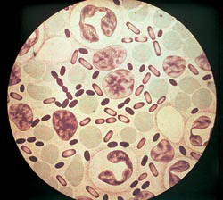
Rice. 3. The photo shows the causative agents of the plague. The intensity of coloration with aniline dyes is greatest at the poles of bacteria.
Php?post=4145&action=edit#

Rice. 4. In the photo, the pathogens of the plague are growing on a dense colony medium. At first, the colonies look like broken glass. Next, their central part becomes denser, and the periphery resembles lace.
Rodents (tarbagans, marmots, gerbils, gophers, rats and house mice) and animals (camels, cats, foxes, hares, hedgehogs, etc.) are easily susceptible to the plague bacillus. Among laboratory animals, white mice, guinea pigs, rabbits and monkeys are susceptible to infection.
Dogs never get plague, but they transmit the pathogen through the bites of blood-sucking insects - fleas. An animal that dies from a disease ceases to be a source of infection. If rodents infected with plague bacilli hibernate, their disease becomes latent, and after hibernation they again become distributors of pathogens. In total, there are up to 250 species of animals that are sick, and therefore are a source and reservoir of infection.

Rice. 5. Rodents are the reservoir and source of the plague pathogen.
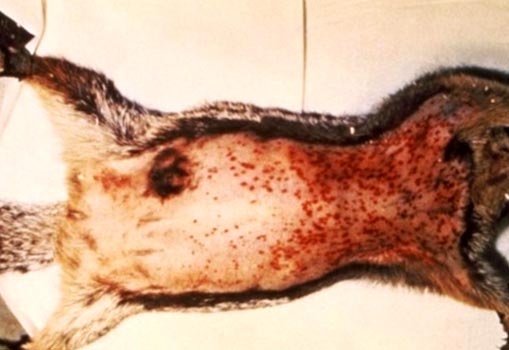
Rice. 6. The photo shows signs of plague in rodents: enlarged lymph nodes and multiple hemorrhages under the skin.

Rice. 7. In the photo, the small jerboa is a carrier of the plague in Central Asia.

Rice. 8. In the photo, the black rat is a carrier not only of plague, but also of leptospirosis, leishmaniasis, salmonellosis, trichinosis, etc.
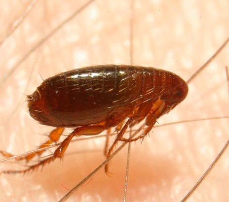
Rice. 9. The photo shows a flea on human skin.
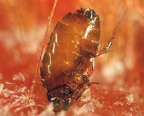
Rice. 10. The photo shows the moment of a flea bite.

Rice. 11. The moment of a flea bite.
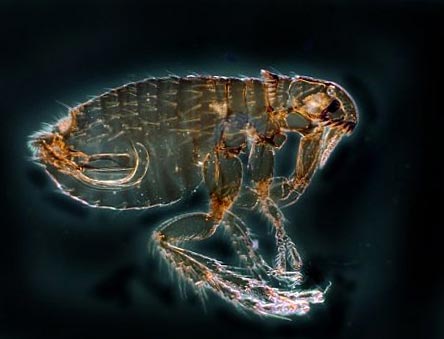
Rice. 12. In the photo, the flea is the main carrier of the plague. There are more than 100 species of these insects in nature.
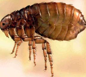
Rice. 13. In the photo, the gopher flea is the main carrier of plague.
Infection occurs through an insect bite and rubbing of its feces and intestinal contents when regurgitating during feeding. When bacteria multiply in the intestinal tube of a flea under the influence of coagulase (an enzyme secreted by pathogens), a “plug” is formed that prevents human blood from entering its body. As a result, the flea regurgitates a clot onto the skin of the bitten person. Infected fleas remain highly infective for 7 weeks to 1 year.

Rice. 14. In the photo, the appearance of a flea bite is pulicotic irritation.
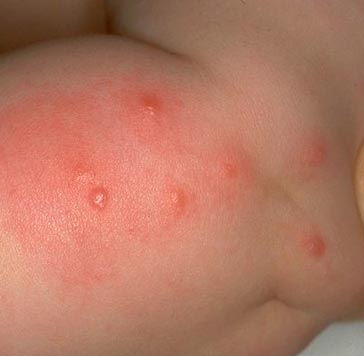
Rice. 15. The photo shows a characteristic series of flea bites.

Rice. 16. View of the lower leg with flea bites.
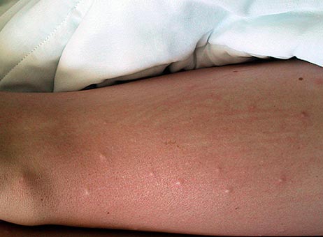
Rice. 17. Appearance of the thigh with flea bites.
The ability of the plague bacillus to form a capsule and antiphagocytic mucus does not allow macrophages and leukocytes to actively fight it, as a result of which the pathogen quickly multiplies in the organs and tissues of humans and animals.

Rice. 18. The photo shows the bubonic plague. Typical enlargement of the lymph node in the axillary area.
The disease manifests itself after the pathogen enters the body on days 3–6 (rarely, but there have been cases of the disease manifesting itself on days 9). When infection enters the blood, the incubation period is several hours.
Clinical picture of the initial period

Rice. 19. In the photo, assistance to a plague patient is provided by doctors dressed in anti-plague suits.
At the site of a flea bite or contact with an infected animal, a papule appears on the skin, which quickly ulcerates. Next, a black scab and scar appear. Most often, skin manifestations are the first signs of more severe manifestations of the plague.
The most common form of manifestation of the disease. Enlarged lymph nodes appear near the site of the insect bite (inguinal, axillary, cervical). More often one lymph node becomes inflamed, less often several. When several lymph nodes become inflamed at once, a painful bubo is formed. Initially, the lymph node has a hard consistency, painful on palpation. Gradually it softens, acquiring a dough-like consistency. Next, the lymph node either resolves or becomes ulcerated and sclerosed. From the affected lymph node, the infection can enter the bloodstream, with the subsequent development of bacterial sepsis. The acute phase of bubonic plague lasts about a week.

Rice. 20. The photo shows the affected cervical lymph nodes (buboes). Multiple hemorrhages of the skin.

Rice. 21. In the photo, the bubonic form of plague affects the cervical lymph nodes. Multiple hemorrhages in the skin.

Rice. 22. The photo shows the bubonic form of plague.
When the pathogen enters the bloodstream, widespread (generalized) forms of plague develop.
If the infection, bypassing the lymph nodes, immediately enters the blood, then the primary septic form of the disease develops. Intoxication develops at lightning speed. With the massive proliferation of pathogens in the patient’s body, a huge number of inflammatory mediators are produced. This leads to the development of disseminated intravascular coagulation syndrome (DIC), which affects all internal organs. Hemorrhages in the heart muscle and adrenal glands pose a particular danger to the body. The developed infectious-toxic shock causes the death of the patient.
When the infection spreads beyond the affected lymph nodes and pathogens enter the bloodstream, infectious sepsis develops, which is manifested by a sharp deterioration in the patient’s condition, increased symptoms of intoxication and the development of DIC syndrome. The developed infectious-toxic shock causes the death of the patient.

Rice. 23. In the photo, the septic form of plague is the consequences of disseminated intravascular coagulation syndrome.

Rice. 24. In the photo, the septic form of plague is the consequences of disseminated intravascular coagulation syndrome.

Rice. 25. 59-year-old Paul Gaylord (resident of Portland, Oregon, USA). Plague bacteria entered his body from a stray cat. As a result of the development of a secondary septic form of the disease, his fingers and toes were amputated.

Rice. 26. Consequences of DIC syndrome.
The pneumonic form of plague is the most severe and dangerous form of the disease. The infection penetrates the alveoli through airborne droplets. Damage to the lung tissue is accompanied by cough and shortness of breath. An increase in body temperature occurs with severe chills. The sputum at the beginning of the disease is thick and transparent (vitreous), then it becomes liquid and foamy, mixed with blood. The scant data from physical examinations do not correspond to the severity of the disease. DIC syndrome develops. Internal organs are affected. Hemorrhages in the heart muscle and adrenal glands pose a particular danger to the body. The patient's death occurs from infectious-toxic shock.
When the lungs are affected, patients become highly contagious. They form around themselves a focus of a particularly dangerous infectious disease.
It is an extremely dangerous and severe form of the disease. Pathogens penetrate the lung tissue from affected lymph nodes or through the bloodstream during bacterial sepsis. The clinical picture and outcome of the disease are the same as in the primary pulmonary form.
The existence of this form of the disease is considered controversial. It is assumed that infection occurs through the consumption of contaminated products. Initially, against the background of intoxication syndrome, abdominal pain and vomiting appear. Then comes diarrhea and numerous urges (tenesmus). The stool is copious, mucous-bloody.
Rice. 27. Photo of an anti-plague suit - special equipment for medical workers when eliminating an outbreak of a particularly dangerous infectious disease.
The basis for diagnosing plague is the rapid detection of the plague bacillus. First, bacterioscopy of smears is performed. Next, a culture of the pathogen is isolated, which infects experimental animals.
The material for research is the contents of the bubo, sputum, blood, feces, pieces of tissue from the organs of deceased animals and corpses.
The causative agent of plague (Yersinia pestis) is a rod-shaped bipolar coccobacilli. Analysis for the detection of plague bacillus by direct bacterioscopy is the simplest and fastest method. The waiting time for the result is no more than 2 hours.
The culture of the plague pathogen is isolated in specialized high-security laboratories designed to work with. The growth time of the pathogen culture is two days. Next, an antibiotic sensitivity test is performed.
The use of serological methods makes it possible to determine the presence and growth of antibodies in the patient’s blood serum to the plague pathogen. The time to receive results is 7 days.

Rice. 28. Diagnosis of plague is carried out in special sensitive laboratories.

Rice. 29. The photo shows the causative agents of the plague. Fluorescence microscopy.
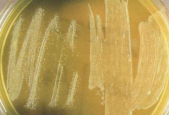
Rice. 30. The photo shows the culture of Yersinia pestis.
Antibodies against the introduction of the plague pathogen are formed quite late in the development of the disease. Immunity after an illness is not long-lasting or intense. There are repeated cases of the disease, which are as severe as the first.
Before treatment begins, the patient is hospitalized in a separate room. Medical personnel serving the patient wear a special anti-plague suit.
Antibacterial treatment begins at the first signs and manifestations of the disease. Among antibiotics, preference is given to antibacterial drugs of the aminoglycoside group (streptomycin), the tetracycline group (vibromycin, morphocycline), the fluoroquinolone group (ciprofloxacin), and the ansamycin group (rifampicin). An antibiotic of the amphenicol group (cortrimoxazole) has proven itself well in the treatment of the skin form of the disease. For septic forms of the disease, a combination of antibiotics is recommended. The course of antibacterial therapy is at least 7–10 days.
The goal of pathogenetic therapy is to reduce intoxication syndrome by removing toxins from the patient’s blood.
Symptomatic treatment is aimed at suppressing and eliminating the manifestations (symptoms) of the plague and, as a result, alleviating the suffering of the patient. It is aimed at eliminating pain, cough, shortness of breath, suffocation, tachycardia, etc.
The patient is considered healthy if all symptoms of the disease have disappeared and 3 negative bacteriological test results have been obtained.
Identification of a plague patient is a signal for immediate action, which includes:
After vaccination with an anti-plague vaccine, immunity lasts for a year. Re-vaccinate after 6 months. persons at risk of re-infection: shepherds, hunters, agricultural workers and employees of anti-plague institutions.

Rice. 31. In the photo, the medical team is dressed in anti-plague suits.
The prognosis of plague depends on the following factors:
The most favorable prognosis is for patients with lymph node involvement. The mortality rate for this form of the disease reaches 5%. In the septic form of the disease, the mortality rate reaches 95%.
The plague is, and even with the use of all the necessary medications and manipulations, the disease often ends in the death of the patient. Plague pathogens constantly circulate in nature and cannot be completely destroyed and controlled. Symptoms of plague are varied and depend on the form of the disease. The bubonic form of plague is the most common.
Articles in the section "Particularly dangerous infections"
Most popularThere are different diseases in the world. But none of them caused such horror and fear as the plague. This disease has had no mercy since ancient times. It claimed millions of lives, regardless of gender, age and welfare of people. Today, the disease no longer brings a huge amount of death and grief. Thanks to the wonders of modern medicine, the plague has been transformed into a less dangerous disease. However, it was not possible to completely eradicate the disease. The plague bacillus (Yersinia pestis), which causes the disease, continues to exist in this world and infect people.
Several years ago, microbiologists began conducting research to study the evolution of pathogens. The plague wand was also studied. Among the existing microorganisms, a genetically similar bacterium was found - Yersinia pseudotuberculosis. It is the causative agent of pseudotuberculosis.
The conducted research allowed scientists to draw one conclusion. When life began to emerge on the planet, there was no plague stick yet. About 15-20 thousand years ago there was a pathogen called pseudotuberculosis. It was a consumer of dead organic matter and multiplied in animal excrement and around corpses buried in the ground. Several factors provoked its further evolution. Some pathogens of pseudotuberculosis transformed into the plague bacillus.
In those places where the primary foci of plague arose, the causative agent of pseudotuberculosis lived in the burrows of marmots (tarbagans). Its evolution, that is, the appearance of the plague wand, was facilitated by certain factors:
When the animals hibernated, they covered their muzzles with their paws. The pathogens of pseudotuberculosis got into the wounds formed due to flea bites. This bacterium would not be able to survive in the circulatory system of active animals. She would have been killed instantly by macrophages. But in sleeping marmots there were no threats to Yersinia pseudotuberculosis. The blood was cooled to favorable temperatures, and the immune system was “turned off.” Of course, temperature rises occurred, but they were rare and short. They created ideal conditions for the natural selection of pathogen forms. All these processes ultimately led to the birth of the plague bacillus.

Modern scientists cannot say whether the plague has always haunted people. According to the surviving information, only three major epidemics are known. The first of these, the so-called Justinian Plague, began around the 540s in Egypt. Over the course of several decades, the plague bacillus devastated almost all Mediterranean states.
The second epidemic, called the “Black Death,” was recorded in the middle of the 14th century. The plague spread from a natural source in the Gobi Desert due to sudden climate change. The pathogen subsequently spread to Asia, Europe, and North Africa. The island of Greenland was also affected by the disease. The second epidemic greatly affected the population. The plague wand claimed approximately 60 million lives.
The third plague epidemic began at the end of the 19th century. An outbreak of the disease was recorded in China. In 6 months, 174 thousand people died in this country. The next outbreak occurred in India. Between 1896 and 1918, the causative agent of a dangerous disease killed 12.5 million people.

Currently, scientists, analyzing the consequences of epidemics and studying important historical sources, call the plague “the queen of diseases.” At the same time, it no longer causes such fear and horror, because no more major outbreaks have been recorded in the world, claiming millions of lives.
Statistics are kept on the manifestations of plague in the modern period. The World Health Organization notes that between 2010 and 2015, 3,248 people fell ill with the plague. There were deaths in 584 cases. This means that 82% of people recovered.
The plague wand has become less dangerous for several reasons. Firstly, people began to observe the rules of hygiene and cleanliness. For example, we can compare the modern period with the Middle Ages. Several centuries ago in Western Europe, people threw all their food waste and feces directly onto the streets. Due to environmental pollution, the townspeople suffered from various diseases and died from the plague.
Secondly, modern people live far from the only people most often encountering infected rodents and fleas are hunters and tourists.
Thirdly, today medicine knows effective methods of treating and preventing a dangerous disease. Experts have created vaccines and identified drugs that can kill the plague bacillus.

If we talk about the structure of the plague bacillus, then Yersinia pestis is a gram-negative small bacterium. It is distinguished by pronounced polymorphism. This is confirmed by the occurring forms - granular, thread-like, flask-shaped, oblong, etc.
Yersinia pestis is a zoonotic bacterium belonging to the Enterobacteriaceae family. The generic name Yersinia was given to this microorganism in honor of the French bacteriologist Alexandre Yersin. It was this specialist who, in 1894, during a study of biological materials of people who died from a dangerous disease, was able to identify the pathogen.
A microorganism capable of causing epidemics with a high mortality rate has always been of interest to microbiologists after its discovery. Since the discovery of Yersinia pestis, specialists have been studying the structure of the bacterium (plague bacillus) and its features. The result of some research conducted by domestic scientists was the compilation in 1985 of a classification of the pathogen isolated in the territory of the USSR and Mongolia.
The causative agent of plague lives in the body of small mammals. The rod multiplies in the circulatory system. When a flea bites an infected animal, it becomes a carrier of infection. In the insect's body, the bacterium settles in the crop and begins to multiply intensively. Due to the increase in the number of rods, the goiter becomes clogged. The flea begins to experience severe hunger. To satisfy it, she jumps from one owner to another, spreading the infection between animals.
The stick enters the human body in several ways:

Depending on the methods of penetration of the plague bacillus into the body, 3 forms of the disease are distinguished. The first of them is bubonic. With such a plague, the pathogen enters the human lymphatic system after a flea bite. Due to the disease, the lymph nodes become inflamed and become so-called buboes. In the later stages of the plague, they turn into festering wounds.
The second form of the disease is septic. With it, the pathogen enters directly into the circulatory system. Buboes are not formed. The septic form occurs when the plague bacillus enters the human body in two ways - after the bite of an infected flea, as well as after contact with infected materials (entry of the pathogen through skin lesions).
The third form is pulmonary. It is transmitted from infected patients through airborne droplets. The pneumonic form of plague is considered the most dangerous. Without treatment, the progression of the disease in most cases is death.

For a long time, humanity did not know about the methods of penetration of the plague bacillus, and had no idea how to stop the deadly disease. Doctors came up with various bizarre methods that did not lead to a cure. For example, in the Middle Ages, healers prepared strange potions from plants and crushed snakes, and advised people to quickly and for a long time escape from the contaminated area.
Today, plague is treated with antibiotics from the aminoglycoside group (streptomycin, amikacin, gentamicin), tetracyclines, rifampicin, chloramphenicol. Lethal outcomes occur in cases where the disease occurs in a lightning-fast form, and specialists are unable to identify the pathogenic bacterium in a timely manner.

The plague bacillus, despite the achievements of modern medicine, is still an insidious pathogen. Foci of the disease in nature occupy about 7% of the land. They are located on desert and steppe plains, in highlands. People who have visited natural plague foci should pay attention to their health. When the pathogen enters the body, the incubation period lasts from several hours to 9 days. Then the first symptoms appear - body temperature suddenly rises to 39 degrees or higher, convulsions, chills, severe headache and muscle pain occur, and breathing becomes difficult. Such symptoms require immediate medical attention.
All iLive content is reviewed by medical experts to ensure it is as accurate and factual as possible.
We have strict sourcing guidelines and only link to reputable sites, academic research institutions and, where possible, proven medical studies. Please note that the numbers in parentheses (, etc.) are clickable links to such studies.
If you believe that any of our content is inaccurate, out of date, or otherwise questionable, please select it and press Ctrl + Enter.

Yersinia pestis is 1-2 µm long and 0.3-0.7 µm thick. In smears from the patient’s body and from the corpses of people and rodents who died from the plague, it looks like a short ovoid (ovoid) rod with a bipolar color. In smears from broth culture, the rod is located in a chain, in smears from agar cultures - randomly. Bipolar coloring is preserved in both cases, but in smears from agar cultures it is somewhat weaker. The causative agent of plague stains negatively according to Gram, stains better with alkaline and carbolic dyes (Leffler's blue), does not form spores, and has no flagella. The G + C content in DNA is 45.8-46.0 mol% (for the entire genus). At a temperature of 37 °C it forms a delicate capsule of a protein nature, which is detected in moist and slightly acidic nutrient media.
Yersinia pestis is an aerobe and grows well on regular nutrient media. The optimal temperature for growth is 27-28 °C (range - from 0 to 45 °C), pH = 6.9-7.1. The plague bacillus exhibits characteristic growth on liquid and solid nutrient media: on broth it manifests itself in the formation of a loose film, from which threads descend in the form of icicles resembling stalactites; at the bottom there is a loose sediment, the broth remains transparent. The development of colonies on solid media goes through three stages: after 10-12 hours under a microscope, growth in the form of colorless plates (the “broken glass” stage); after 18-24 hours - the stage of “lace scarves”; when microscopying, a light lace zone is noticeable, located around the protruding central part, yellowish or slightly brownish in color. After 40-48 hours, the “adult colony” stage begins - a brownish-outlined center with a pronounced peripheral zone. Yersinia pseudotuberculosis and Yersinia enterocolitica do not have the “broken glass” stage. On media with blood, colonies of Yersinia pestis are granular with a weakly defined peripheral zone. In order to quickly obtain growth characteristic of Yersinia pestis on media, it is recommended to add growth stimulants to them: sodium sulfite, blood (or its preparations) or sarcina culture lysate. The plague bacillus is characterized by pronounced polymorphism, especially in media with high concentrations of NaCl, in old cultures, and in the organs of decomposed plague corpses.
The plague bacillus does not have oxidase, does not form indole and H2S, has catalase activity and ferments glucose, maltose, galactose, mannitol with the formation of acid without gas.
, , , , , , ,
Up to 18 similar somatic antigens have been found in Yersinia pestis, Yersinia pseudotuberculosis and Yersinia enterocolitica. Yersinia pestis is characterized by the presence of capsular antigen (fraction I), T, V-W antigens, plasmacoagulase proteins, fibrinolysin, outer membrane proteins and pHb antigen. However, unlike Yersinia pseudotuberculosis and Yersinia enterocolitica, Yersinia pestis is antigenically more homogeneous; There is no serological classification for this species.
, , , , , , , ,
The plague bacillus can persist in sputum for up to 10 days; on linen and clothes stained with the patient’s secretions, it persists for weeks (protein and mucus protect it from the harmful effects of drying). It survives in the corpses of people and animals that died from the plague from early autumn until winter; low temperature, freezing and thawing do not kill it. Sun, drying, and high temperatures are destructive for Yersinia pestis. Heating to 60 °C kills in 1 hour, at a temperature of 100 °C it dies in a few minutes; 70% alcohol, 5% phenol solution, 5% Lysol solution and some other chemical disinfectants kill in 5-10-20 minutes.
Yersinia pestis is the most pathogenic and aggressive among bacteria, and therefore causes the most severe disease. In all sensitive animals and humans, the plague pathogen suppresses the protective function of the phagocytic system. It penetrates phagocytes, suppresses the “oxidative explosion” in them and multiplies unhindered. The inability of phagocytes to carry out their killer function against Yersinia pestis is the main reason for susceptibility to plague. High invasiveness, aggressiveness, toxigenicity, toxicity, allergenicity and the ability to suppress phagocytosis are due to the presence of a whole arsenal of pathogenicity factors in U. pestis, which are listed below.
A significant part of the pathogenicity factors of Yersinia pestis is controlled by genes, the carriers of which are the following 3 classes of plasmids, usually found together in all pathogenic strains:
It determines the dependence of the growth of Y. pestis at 37 °C on the presence of Ca2+ ions in the medium, therefore it has another name - Lcr-plasmid (low calcium response). The genes of this particularly important plasmid also encode the synthesis of antigens V and W and the heat-inducible proteins Yop. Their synthesis is carried out under complex genetic control at a temperature of 37 °C and in the absence of Ca2+ in the environment. All types of Yop proteins, except YopM and YopN, are hydrolyzed due to the activity of the plasminogen activator (pla gene of the pYP plasmid). Yop proteins largely determine the virulence of Yersinia pestis. YopE protein has antiphagocytic and cytotoxic effects. YopD ensures entry of YopE into the target cell; YopH has antiphagocytic and protein tyrosine phosphatase activities; protein YopN - properties of a calcium sensor; YopM binds to human blood atrombin.
, , ,
Post-infectious immunity is strong and lifelong. Repeated cases of plague are extremely rare. The nature of immunity is cellular. Although antibodies appear and play a role in acquired immunity, it is mediated primarily by T lymphocytes and macrophages. In persons who have recovered from the plague or have been vaccinated, phagocytosis is complete. It determines acquired immunity.
The range of warm-blooded carriers of the plague microbe is extremely extensive and includes more than 200 species of 8 orders of mammals. The main sources of plague in nature are rodents and lagomorphs. Natural infestation has been established in more than 180 of their species, over 40 of them are part of the Fauna of Russia and adjacent territories (within the former USSR). Of the 60 species of fleas for which the possibility of transmitting the plague pathogen has been established under experimental conditions, 36 live in this territory.
The plague microbe multiplies in the lumen of the digestive tube of fleas. A plug (“plague block”) forms in its anterior section, containing a large number of microbes. When a mammal is bitten, some of the microbes are washed away from the plug with reverse blood flow into the wound, which leads to infection. In addition, excrement released by a flea when feeding, if it gets into the wound, can also cause infection.
The main carriers of Y. pestis in Russia and Central Asia are ground squirrels, gerbils and marmots, and in some areas also pikas and voles. The existence of the following plague foci is associated with them.
Different classifications of Yersinia pestis are based on different groups of characteristics - biochemical characteristics (glycerol-positive and glycerol-negative variants), area of distribution (oceanic and continental variants), types of main carriers (rat and gopher variants). According to one of the most common classifications, proposed in 1951 by the French plague researcher R. Devignat, depending on the geographic distribution of the pathogen and its biochemical properties, three intraspecific forms (biovars) of Yersinia pestis are distinguished.
According to the classification of domestic scientists (Saratov, 1985), the species Yersinia pestis is divided into 5 subspecies: Yersinia pestis subsp. pestis (main subspecies; it includes all three biovars of R. Devigna’s classification), Y. pestis subsp. altaica (Altai subspecies), Yersinia pestis subsp. caucasica (Caucasian subspecies), Y. pestis subsp. hissarica (Gissar subspecies) and Yersinia pestis subsp. ulegeica (Udege subspecies).
Human infection occurs through a flea bite, direct contact with infectious material, airborne droplets, and rarely through nutrition (for example, by consuming the meat of camels with plague). In 1998-1999 30,534 people worldwide were infected with the plague, of which 2,234 died.
, , , , , ,
Depending on the method of infection, bubonic, pneumonic, and intestinal forms of plague are distinguished; rarely septic and skin (purulent blisters at the site of a flea bite). The incubation period for plague varies from several hours to 9 days. (in persons subjected to seroprophylaxis, up to 12 days). The causative agent of plague penetrates through the smallest damage to the skin (flea bite), sometimes through the mucous membrane or by airborne droplets, reaches regional lymph nodes, where it begins to rapidly multiply. The disease begins suddenly: severe headache, high temperature with chills, the face is hyperemic, then it darkens, dark circles under the eyes (“black death”). A bubo (an enlarged, inflamed lymph node) appears on the second day. Sometimes the plague develops so rapidly that the patient dies before the bubo appears. Pneumonic plague is especially severe. It can occur as a result of complications of bubonic plague, and during infection by airborne droplets. The disease also develops very rapidly: chills, high temperature, and already in the first hours pain in the side, cough, initially dry, and then with bloody sputum; delirium, cyanosis, collapse appear, and death occurs. A patient with pneumonic plague poses an exceptional danger to others, as he secretes a huge amount of the pathogen with sputum. In the development of the disease, the main role is played by the suppression of the activity of phagocytes: neutrophilic leukocytes and macrophages. Uncontrolled reproduction and spread of the pathogen through the blood throughout the body completely suppresses the immune system and leads (in the absence of effective treatment) to the death of the patient.
Bacterioscopic, bacteriological, serological and biological methods are used, as well as an allergy test with pestin (for retrospective diagnosis). The materials for the study are: punctate from the bubo (or its discharge), sputum, blood, and in the intestinal form - feces. Yersinia pestis is identified based on morphology, cultural, biochemical characteristics, a test with a plague phage and using a biological test.
A simple and reliable method for determining the antigens of the plague bacillus in the test material is the use of RPGA, especially using an erythrocyte diagnosticum sensitized with monoclonal antibodies to the capsular antigen, and IFM. The same reactions can be used to detect antibodies in the serum of patients.
Despite the presence of natural foci, since 1930 in Russia there has not been a single case of human plague. For specific prevention of plague, a plague vaccination is used - a live attenuated vaccine from the EV strain. It is administered cutaneously, intradermally or subcutaneously. In addition, a dry tablet vaccine for oral administration has been proposed. Post-vaccination immunity is formed by the 5-6th day after vaccination and persists for 11-12 months. For its assessment and retrospective diagnosis of plague, an intradermal allergy test with pestin has been proposed. The reaction is considered positive if, after 24-48 hours, a compaction of at least 10 mm in diameter forms at the site of pestin injection and redness appears. The allergy test is also positive in persons with post-infectious immunity.
Russian scientists made a great contribution to the study of the plague and the organization of the fight against it: D. S. Samoilovich (the first “hunter” of the plague microbe back not only in Russia, but also in Europe back in the 18th century, he was the first to propose vaccinations against the plague ), D.K. Zabolotny, N.P. Klodnitsky, I.A. Deminsky (study of natural foci of plague, carriers of its pathogen in foci, etc.), etc.
It is well known how quickly infectious diseases can spread, which means that equally rapid methods of detecting infections literally in the field should exist and be as accessible as possible, which is critical in the fight against epidemics.
Plague stick(Yersinia pestis) - a type of gram-negative sporogenous bacteria, facultative anaerobes. The causative agent of bubonic plague, pneumonia (pneumonic plague) and septicemic plague.
 The mortality rate for plague if untreated ranges from 63% to 93%. When treated with modern antibiotics - approximately 16%. Timely treatment with antimicrobial drugs such as aminoglycosides, fluoroquinolones or doxycycline significantly increases the likelihood of a favorable outcome.
 In the XIV-XVII centuries in Europe, from the bubonic plague, according to various estimates, from 50 to 75 million people died. The last outbreak of plague was recorded in Madagascar in the summer of 2015.
 that it includes an infection caused by Yersinia pestis. This category includes four-character categories:
 From the moment of its appearance, a person is exposed to bacterial infections. Various pathogenic microorganisms have contributed to the history of mankind, but the bloodiest trace was left by the plague pathogen. The bacterium Yersinia pestis, the causative agent of the plague, was isolated only at the end of the 19th century. And before that, it wasn’t even epidemics, but pandemics that claimed millions of lives.
Long before scientists discovered the pathogen, it was known that the disease was highly contagious. In the Middle Ages, in order to prevent the spread of infection, strict quarantine measures were applied to people and things that fell into the area of infection. The first plague quarantine was introduced in Venice in 1422.
Attempts to identify the causes that provoke the development of plague have been made by doctors at all times. However, only with the advent of developed microbiological research techniques, scientists were able to discover the microorganism that is the causative agent of the disease. Russian doctors Samoilovich D.S., Skvortsov I.P. began to look for the causative agent of the disease using microscopes. But poor technique for working with microspecimens and the lack of microbiological research methods did not allow us to identify the cause of the infection.
Only in 1894 was the causative agent of the plague discovered - scientists were working in Hong Kong, where the third pandemic began. Having examined tissue samples taken from corpses and infected people, Japanese bacteriologist Kitasato Shibasaburo identified identical microorganisms in the form of short rods. He managed to grow a pure culture of the plague pathogen using nutrient media. Laboratory animals infected with the grown culture died, and autopsies revealed characteristic pathological changes. Kitasato reported the results of the study - identifying the cause of the plague - in Hong Kong on July 7, 1894.
At the same time as Kitasato, the French bacteriologist Alexandre Yersin, examining the corpses of those infected with the plague, isolated the microorganism that caused the disease and grew a pure culture. He published the results of his research on July 30, 1894. But only in 1926 did Khavkin V.A. succeeded in creating an effective vaccine against the plague. Today, only isolated cases of infection are recorded in natural foci of infection.
Although Kitasato was the first to report the discovery of the microorganism that causes plague, the honor of discovering the plague bacillus belongs to the French bacteriologist and physician Alexandre Yersin. While studying the isolated bacterium, Kitasato made mistakes when staining smears and incorrectly assessed the mobility of the microorganism. As a result, Kitasato erroneously characterized the isolated microorganism as gram-positive and weakly motile. Initially, the plague bacterium was assigned to the genus Bacterium, then to Pasteurella. In 1967, this genus, in honor of A. Yersin, was renamed Yersinia.
The causative agent of plague is the non-spore-forming coccobacillus Yersinia pestis. The bacillus is immobile and has a mucous capsule.
Taxonomy of the plague pathogen:
In Yersinia, microbiology includes 18 species (as of May 2015), among which only three are dangerous to humans, being infectious agents:
All Yersinia are gram-negative rods, but, unlike pseudotuberculosis and yersinia, the prokaryotic plague bacillus does not have a flagellum.
The morphology of the plague causative agent has been studied quite fully. The causative agent of bubonic plague is a coccobacilli in cell shape and looks like a motionless short ovoid rod. Yersinia pestis is characterized by polymorphism - elongated, filamentous, spherical and granular varieties have been found. Due to the peculiarity of the structure of Yersinia (heterogeneous distribution of cytoplasm in the cell with an increase in concentration in the terminal areas), the plague bacillus is characterized by bipolar staining. It colors better at the poles than at the center. Like all prokaryotes, the nucleus is what Yersinia pestis cells do not have.
The bacterium appears blue when stained with Leffler's methylene blue or stained with Romanowsky-Giemsa (blue) with pronounced bipolarity.
The causative agent of plague easily tolerates low temperatures, even freezing. At low temperatures it can be stored for quite a long time:
At room temperature, microorganisms that cause plague can remain viable for up to 4 months. In the secretions of sick people, which get on clothes and underwear, bacteria live for weeks. Microorganisms are protected by a mucous capsule from drying out, which is detrimental to them.
The coccobacilli Yersinia pestis is sensitive to UV irradiation and heat, during which it quickly dies:
When treated with disinfectant solutions, the plague pathogen quickly dies - just a 5-minute exposure to a 5% solution of Acidum carbolicum (carbolic acid) is enough.
Bacteria - the causative agents of plague - have a complex antigenic structure. It consists of about 10 different antigens, including:
The causative agent of plague is one of the most aggressive and pathogenic bacteria, so the disease is always extremely severe.
The coccobacilli Yersinia pestis is a facultative anaerobe in its form of existence; it grows well on meat peptone agar and broth. The optimal temperature for cultivating the plague pathogen is considered to be 25-30°C, and reproduction begins at +5°C. Yersinia pestis bacilli placed in nutrient media grow in the form of specific colonies, which can be of two forms:
Plague bacteria sown on agar form a light gray coating. After 48 hours, a loose film is formed on the nutrient broth, from which icicles descend. The bacterium Yersinia pestis is not capable of liquefying gelatin and does not curdle milk. Decomposes a number of sugars into acid.
Jpg" alt="dead from the bubonic plague" width="500" height="372" srcset="" data-srcset="https://probakterii.ru/wp-content/uploads/2018/01/vozbuditel-chumy-4-500x372..jpg 300w, https://probakterii.ru/wp-content/uploads/2018/01/vozbuditel-chumy-4.jpg 528w" sizes="(max-width: 500px) 100vw, 500px"> !}
A burial pit excavated in France contained numerous human remains. Research has proven that people died from the bubonic plague
The toxins secreted by the plague bacillus are a specific protein that has the properties of an endo- and exotoxin. The protein consists of two fractions (A and B), which have different compositions and have different antigenic properties. One part is responsible for fixation to the cell wall, and the second is responsible for the production of toxin. The plague toxin is called “mouse”, and its synthesis in the bacterial cell is carried out under the control of a plasmid. The toxicity of the plague bacillus is due to the ability to have a destructive effect on cell mitochondria, and leads to:
Plague is a naturally occurring, vector-borne zoonosis. Transmissible diseases are human infectious diseases whose pathogens are transmitted by blood-sucking insects and ticks. Zoonoses are infections common to humans and animals. The main source and carrier of the pathogen were and remain wild rodents (about 300 species) living everywhere. The causative agent of anthropozoonotic plague, the coccobacilli Yersinia pestis, infects wild animals, forming cases of irregular plague (sporadic).
In natural conditions, the natural carriers of the plague pathogen are most often mice, gophers and similar rodents, with each territorial focus retaining its own specific keeper of the infection. Infection with plague coccobacilli occurs through contact of infected animals with healthy ones. As a result of the development of an acute form of the disease, infected animals die, and the epizootic may end. Others, during hibernation, carry the plague in a sluggish form and, waking up in the spring, are a natural source of the disease, maintaining a natural infectious focus in a given area.
The bacterium Yersinia pestis, despite the similarity of the name of the disease, has nothing to do with rinderpest (rinderpest). Its infectious agent is an RNA virus that is closest to the causative agent of canine distemper. In June 2011, the UN declared that rinderpest had been completely eradicated from the planet.
If in the wild rodents are carriers of the bacilli, then in cities the main reservoir of the plague bacillus is synanthropic rats (that is, those whose lifestyle is associated with humans). The main types of rats responsible for the spread of plague are:
When a person becomes infected from an infected animal, the following routes of transmission are available:
The high virulence and pathogenicity of the plague bacillus is due to its significant penetrating ability and the presence of a protein toxin. The pathogenicity factors of Yersinia pestis are encoded in the plasmid and chromosome of the bacterium.
Plague is an acute infectious disease and is considered especially dangerous. This is a strictly quarantine infection, which is characterized by:
The plague bacillus enters the body through a wound from an insect bite or through intact epidermis and mucous membranes of the respiratory tract or gastrointestinal tract. The disease has affected people at all times - it is reliably known about three plague pandemics that covered vast territories:
During the last pandemic, it was possible to identify the causative agent of the plague - the bacterium Yersinia pestis. An effective vaccine against these microorganisms was created only in 1926.
The latent period of the disease can last up to 9 days, and for the pulmonary form - no more than 1-2 days. The plague begins acutely, the temperature rises sharply to 40°C, accompanied by chills, signs of intoxication are always pronounced. As the disease develops, the lymph nodes, lungs, liver, and heart are quickly affected. Regardless of the form, typical complaints from patients of muscle pain and constant headache are typical for the plague. Psychomotor agitation is often present, and hallucinations are possible.
External manifestation of plague on the patient’s face:
Such symptoms of the initial stage are typical for plague of any form. Based on their symptoms of the disease, Rudnev G.P. A clinical classification of plague was proposed, which is still used today:
The symptoms of the disease vary depending on the type of plague:
Data-lazy-type="image" data-src="https://probakterii.ru/wp-content/uploads/2018/01/vozbuditel-chumy8-500x381.jpg" alt=" Finger necrosis" width="500" height="381" srcset="" data-srcset="https://probakterii.ru/wp-content/uploads/2018/01/vozbuditel-chumy8-500x381..jpg 300w, https://probakterii.ru/wp-content/uploads/2018/01/vozbuditel-chumy8.jpg 600w" sizes="(max-width: 500px) 100vw, 500px">!}
Laboratory diagnosis of plague is carried out using modern methods of microbiology, immunoserology and genetics. The use of modern methods for diagnosing the disease, which is caused by plague bacteria, is fully justified when examining patients with an abnormally high temperature who were in the source of the infection.
After extensive research, microbiologists were able to establish that plague in humans is caused by the bacteria Yersinia pestis. Plague is a particularly dangerous infectious disease, so its treatment is carried out exclusively in a specialized hospital. Patients are prescribed etiotropic therapy and symptomatic treatment. Drugs, dosage and regimens are selected according to the form of infection. At the same time, deep detoxification is carried out, antipyretic, cardiac, respiratory and vascular analeptics, as well as symptomatic drugs, are prescribed.
Although immunity is formed after suffering from the disease, it is extremely weak and short-lived. Cases of re-infection were often observed, and the disease was as severe as the first time. Anti-plague vaccination provides immunity to the disease only for 1 year and is not 100% guaranteed.
If there is a threat of infection, people at risk - shepherds, agricultural workers, hunters, employees of anti-plague institutions - are re-vaccinated after 6 months.

Contents Dietary supplement based on an extract obtained from the Spanish beetle (or Spanish beetle...

Ekaterina MirimanovaSystem minus 60. RevolutionSystem minus 60 with Ekaterina Mirimanova"System minus 60. Revolution"...

Heaviness and bloating are the causes of both ordinary overeating and more serious problems with the digestive tract....

The second blood group, Rh-negative, appeared many years ago, when a person stopped being a hunter, and...

PSYCHOLOGICAL ASPECTS OF ANOREXIA PHENOMENON (EXPERIMENTAL STUDY) T. V. Tarasova, E. V. Arsentieva V...

Contents Since the skin in this area is thin, it is more prone to the appearance of various types of spots. Redness under...

vseslav Sat, 10/17/2015 - 20:50 Vasileostrovskaya station is one of the oldest stations...

Most often, the goat mushroom (Suillus bovinus) is called a goat mushroom in the common parlance of mushroom pickers. This tubular...

Every person has had dreams at least once in their life. Many do not attach any importance to them, but some...

Daedalus (crater). Diameter: 93 km Depth: 3 km (NASA photo) The moon has attracted the attention of people since ancient times. In II...

Presidents' Day is celebrated on the third Monday of February in the United States. Until the 70s, celebrations were dedicated to...

On the day of the last call, I want to say just one word to my beloved head teacher: “Thank you.” But how much is this...

Watermelon is considered a tasty and healthy berry; its seeds are especially useful. Small, dark brown or...

“Speech development in 6-year-old children” A child is not born with developed speech. It is impossible to definitively answer the question about...

Ekaterina MirimanovaSystem minus 60. RevolutionSystem minus 60 with Ekaterina Mirimanova“System minus 60....

Heaviness and bloating are the causes of both ordinary overeating and more serious digestive problems...