Spanish Fly for two - how they affect libido in women and men
Contents Biologically active additive based on an extract obtained from a beetle with a fly (or fly...
Human blood is a liquid substance consisting of plasma and formed elements, or blood cells, that are in suspension in it, which make up approximately 40-45% of the total volume. They are small and can only be seen under a microscope.
There are several types of blood cells that perform specific functions. Some of them function only inside the circulatory system, others go beyond it. What they all have in common is that they are all formed in the bone marrow from stem cells, the process of their formation is continuous, and their life span is limited.
All blood cells are divided into red and white. The first are erythrocytes, which make up most of all cells, the second are leukocytes.
Platelets are also considered to be blood cells. These small platelets are not actually complete cells. They are small fragments separated from large cells - megakaryocytes.
Erythrocytes are called red blood cells. This is the largest group of cells. They carry oxygen from the respiratory organs to the tissues and take part in the transport of carbon dioxide from the tissues to the lungs.
The place of formation of red blood cells is the red bone marrow. They live 120 days and are destroyed in the spleen and liver.
They are formed from precursor cells - erythroblasts, which, before turning into an erythrocyte, go through different stages of development and divide several times. Thus, up to 64 red blood cells are formed from an erythroblast.

Erythrocytes are devoid of a nucleus and in shape resemble a disc concave on both sides, the average diameter of which is about 7-7.5 microns, and the thickness along the edges is 2.5 microns. This shape helps to increase the plasticity required for passage through small vessels and the surface area for diffusion of gases. Old red blood cells lose their plasticity, which is why they linger in the small vessels of the spleen and are destroyed there.
Most of the erythrocytes (up to 80%) have a biconcave spherical shape. The remaining 20% may have a different one: oval, cup-shaped, simple spherical, crescent-shaped, etc. Violation of the shape is associated with various diseases (anemia, vitamin B 12 deficiency, folic acid, iron, etc.).

Most of the cytoplasm of the erythrocyte is occupied by hemoglobin, consisting of protein and heme iron, which gives the blood a red color. The non-protein part consists of four heme molecules with an Fe atom in each. It is thanks to hemoglobin that the erythrocyte is able to carry oxygen and remove carbon dioxide. In the lungs, an iron atom binds to an oxygen molecule, hemoglobin is converted to oxyhemoglobin, which gives the blood a scarlet color. In tissues, hemoglobin gives off oxygen and attaches carbon dioxide, turning into carbohemoglobin, as a result, the blood becomes dark. In the lungs, carbon dioxide is separated from hemoglobin and excreted by the lungs to the outside, and the incoming oxygen again binds to iron.
In addition to hemoglobin, the cytoplasm of the erythrocyte contains various enzymes (phosphatase, cholinesterases, carbonic anhydrase, etc.).
The erythrocyte membrane has a fairly simple structure compared to the membranes of other cells. It is an elastic thin mesh, which ensures rapid gas exchange.
On the surface of red blood cells are different types of antigens that determine the Rh factor and blood type. The Rh factor can be positive or negative depending on the presence or absence of the Rh antigen. The blood type depends on which antigens are on the membrane: 0, A, B (the first group is 00, the second is 0A, the third is 0B, the fourth is AB).
In the blood of a healthy person, there may be small amounts of immature red blood cells called reticulocytes. Their number increases with significant blood loss, when replacement of red cells is required and the bone marrow does not have time to produce them, therefore it releases immature ones, which, nevertheless, are able to perform the functions of red blood cells for transporting oxygen.
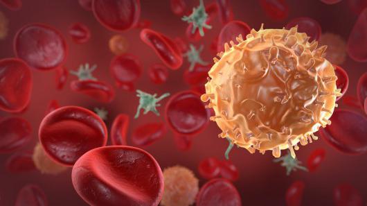
Leukocytes are white blood cells whose main task is to protect the body from internal and external enemies.
They are usually divided into granulocytes and agranulocytes. The first group is granular cells: neutrophils, basophils, eosinophils. The second group does not have granules in the cytoplasm, it includes lymphocytes and monocytes.

This is the most numerous group of leukocytes - up to 70% of the total number of white cells. Neutrophils got their name due to the fact that their granules are stained with dyes with a neutral reaction. Its granularity is fine, the granules have a purple-brownish tint.

The main task of neutrophils is phagocytosis,
which consists in capturing pathogenic microbes and tissue decay products and destroying them inside the cell with the help of lysosomal enzymes located in granules. These granulocytes fight mainly bacteria and fungi and, to a lesser extent, viruses. Pus consists of neutrophils and their residues. Lysosomal enzymes are released during the breakdown of neutrophils and soften nearby tissues, thus forming a purulent focus.A neutrophil is a round-shaped nuclear cell, reaching a diameter of 10 microns. The core may be rod-shaped or consist of several segments (from three to five) connected by strands. An increase in the number of segments (up to 8-12 or more) indicates pathology. Thus, neutrophils can be stab or segmented. The first are young cells, the second are mature. Cells with a segmented nucleus make up to 65% of all leukocytes, stab cells in the blood of a healthy person - no more than 5%.

In the cytoplasm there are about 250 varieties of granules containing substances due to which the neutrophil performs its functions. These are protein molecules that affect metabolic processes (enzymes), regulatory molecules that control the work of neutrophils, substances that destroy bacteria and other harmful agents.
These granulocytes are formed in the bone marrow from neutrophilic myeloblasts. A mature cell stays in the brain for 5 days, then enters the bloodstream and lives here for up to 10 hours. From the vascular bed, neutrophils enter the tissues, where they stay for two or three days, then they enter the liver and spleen, where they are destroyed.
There are very few of these cells in the blood - no more than 1% of the total number of leukocytes. They have a rounded shape and a segmented or rod-shaped nucleus. Their diameter reaches 7-11 microns. Inside the cytoplasm are dark purple granules of various sizes. The name was given due to the fact that their granules are stained with dyes with an alkaline, or basic (basic) reaction. Basophil granules contain enzymes and other substances involved in the development of inflammation.

Their main function is the release of histamine and heparin and participation in the formation of inflammatory and allergic reactions, including the immediate type (anaphylactic shock). In addition, they can reduce blood clotting.

Formed in the bone marrow from basophilic myeloblasts. After maturation, they enter the blood, where they stay for about two days, then go into the tissues. What happens next is still unknown.
These granulocytes make up approximately 2-5% of the total white cells. Their granules are stained with an acidic dye - eosin.
They have a rounded shape and a weakly colored core, consisting of segments of the same size (usually two, less often three). In diameter, eosinophils reach 10-11 microns. Their cytoplasm stains pale blue and is almost invisible among a large number of large round yellow-red granules.

These cells are formed in the bone marrow, their precursors are eosinophilic myeloblasts. Their granules contain enzymes, proteins and phospholipids. A mature eosinophil lives in the bone marrow for several days, after entering the blood it stays in it for up to 8 hours, then it moves to tissues that have contact with the external environment (mucous membranes).

These are round cells with a large nucleus that occupies most of the cytoplasm. Their diameter is 7 to 10 microns. The kernel is round, oval or bean-shaped, has a rough structure. It consists of lumps of oxychromatin and basiromatin, resembling lumps. The nucleus may be dark purple or light purple, sometimes there are light blotches in the form of nucleoli. The cytoplasm is stained light blue, around the nucleus it is lighter. In some lymphocytes, the cytoplasm has an azurophilic granularity that turns red when stained.

Two types of mature lymphocytes circulate in the blood:
Of the atypical lymphocytes in the blood, one can detect:
Lymphocytes are formed in the bone marrow from lymphoblasts and in the process of maturation they go through several stages of division. Its full maturation occurs in the thymus, lymph nodes and spleen. Lymphocytes are immune cells that provide immune responses. There are T-lymphocytes (80% of the total) and B-lymphocytes (20%). The first passed maturation in the thymus, the second - in the spleen and lymph nodes. B-lymphocytes are larger in size than T-lymphocytes. The life span of these leukocytes is up to 90 days. Blood for them is a transport medium through which they enter the tissues where their help is required.
The actions of T-lymphocytes and B-lymphocytes are different, although both are involved in the formation of immune responses.
The former are engaged in the destruction of harmful agents, usually viruses, by phagocytosis. The immune reactions in which they participate are non-specific resistance, since the actions of T-lymphocytes are the same for all harmful agents.
According to the actions performed, T-lymphocytes are divided into three types:
B-lymphocytes act differently: against pathogens, they produce antibodies - immunoglobulins. This happens as follows: in response to the actions of harmful agents, they interact with monocytes and T-lymphocytes and turn into plasma cells that produce antibodies that recognize the corresponding antigens and bind them. For each type of microbes, these proteins are specific and are able to destroy only a certain type, so the resistance that these lymphocytes form is specific, and it is directed mainly against bacteria.

These cells provide the body's resistance to certain harmful microorganisms, which is commonly called immunity. That is, having met with a harmful agent, B-lymphocytes create memory cells that form this resistance. The same thing - the formation of memory cells - is achieved by vaccinations against infectious diseases. In this case, a weak microbe is introduced so that the person can easily endure the disease, and as a result, memory cells are formed. They can remain for life or for a certain period, after which the vaccination is required to be repeated.
Monocytes are the largest of the white blood cells. Their number is from 2 to 9% of all white blood cells. Their diameter reaches 20 microns. The monocyte nucleus is large, occupies almost the entire cytoplasm, can be round, bean-shaped, have the shape of a mushroom, a butterfly. When stained, it becomes red-violet. The cytoplasm is smoky, bluish-smoky, rarely blue. It usually has an azurophilic fine grain. It may contain vacuoles (voids), pigment grains, phagocytosed cells.

Monocytes are produced in the bone marrow from monoblasts. After maturation, they immediately appear in the blood and stay there for up to 4 days. Some of these leukocytes die, some move to tissues, where they mature and turn into macrophages. These are the largest cells with a large round or oval nucleus, blue cytoplasm and a large number of vacuoles, which makes them appear foamy. The life span of macrophages is several months. They can constantly be in one place (resident cells) or move (wandering).

Monocytes form regulatory molecules and enzymes. They are able to form an inflammatory reaction, but they can also slow it down. In addition, they are involved in the process of wound healing, helping to speed it up, contribute to the restoration of nerve fibers and bone tissue. Their main function is phagocytosis. Monocytes destroy harmful bacteria and inhibit the reproduction of viruses. They are able to follow commands but cannot distinguish between specific antigens.
These blood cells are small non-nucleated plates and may be round or oval in shape. During activation, when they are at the damaged vessel wall, they form outgrowths, so they look like stars. Platelets contain microtubules, mitochondria, ribosomes, specific granules containing substances necessary for blood clotting. These cells are equipped with a three-layer membrane.

Platelets are produced in the bone marrow, but in a completely different way than other cells. Platelets are formed from the largest brain cells - megakaryocytes, which, in turn, were formed from megakaryoblasts. Megakaryocytes have a very large cytoplasm. After cell maturation, membranes appear in it, dividing it into fragments, which begin to separate, and thus platelets appear. They leave the bone marrow into the blood, stay in it for 8-10 days, then die in the spleen, lungs, and liver.

Blood platelets can have different sizes:
Platelets perform a very important function - they are involved in the formation of a blood clot, which closes the damage in the vessel, thereby preventing blood from flowing out. In addition, they maintain the integrity of the vessel wall, contribute to its fastest recovery after damage. When bleeding begins, platelets stick to the edge of the lesion until the hole is completely closed. Adhering plates begin to break down and release enzymes that act on blood plasma. As a result, insoluble fibrin strands are formed, tightly covering the injury site.
Blood cells have a complex structure, and each type performs a specific job: from transporting gases and substances to producing antibodies against foreign microorganisms. Their properties and functions are not fully understood to date. For normal human life, a certain amount of each type of cell is necessary. According to their quantitative and qualitative changes, physicians have the opportunity to suspect the development of pathologies. The composition of the blood is the first thing that the doctor studies when the patient is contacted.
Thank you
The site provides reference information for informational purposes only. Diagnosis and treatment of diseases should be carried out under the supervision of a specialist. All drugs have contraindications. Expert advice is required!
Blood is a liquid connective tissue that fills the entire human cardiovascular system. Its amount in the body of an adult reaches 5 liters. It consists of a liquid part called plasma and formed elements such as leukocytes, platelets and erythrocytes. In this article, we will talk specifically about erythrocytes, their structure, functions, method of formation, etc.
2. Enzymatic:
are carriers of various enzymes ( specific protein catalysts);
3. Respiratory:
this function is carried out by hemoglobin, which is able to attach to itself and give off both oxygen and carbon dioxide;
4. Protective:
bind toxins due to the presence of special substances of protein origin on their surface.
With an increase in indicators, we are talking about violations of the body. There is an opinion that in most cases, ESR increases against the background of an increase in the ratio of large and small protein particles in the blood plasma. As soon as fungi, viruses or bacteria enter the body, the level of protective antibodies immediately increases, which leads to changes in the ratio of blood proteins. From this it follows that especially often ESR increases against the background of inflammatory processes such as inflammation of the joints, tonsillitis, pneumonia, etc. The higher this indicator, the more pronounced the inflammatory process. With a mild course of inflammation, the rate increases to 15 - 20 mm / h. If the inflammatory process is severe, then it jumps up to 60-80 mm/hour. If during the course of therapy the indicator begins to decrease, then the treatment was chosen correctly.
In addition to inflammatory diseases, an increase in ESR is also possible with some non-inflammatory ailments, namely:
Modern experts distinguish the following types of hemolysis:
1. By the nature of the flow:
 The total number of these cells in human blood is simply enormous. So, for example, if your weight is about 60 kg, then there are at least 25 trillion red blood cells in your blood. The figure is very large, so for practicality and convenience, experts do not calculate the total level of these cells, but their number in a small amount of blood, namely in its 1 cubic millimeter. It is important to note that the norms for the content of these cells are determined immediately by several factors - the age of the patient, his gender and place of residence.
 The most common causes of this condition are:
An erythrocyte is called capable of transporting oxygen to the tissues due to hemoglobin, and carbon dioxide to the lungs. This is a cell of simple structure, which is of great importance for the life of mammals and other animals. The erythrocyte is the most numerous organism: about a quarter of all body cells are red blood cells.
An erythrocyte is a cell that originated from a red germ of hematopoiesis. About 2.4 million of these cells are produced per day, they enter the bloodstream and begin to perform their functions. During the experiments, it was determined that in an adult, erythrocytes, the structure of which is significantly simplified compared to other cells of the body, live 100-120 days.
In all vertebrates (with rare exceptions), oxygen is transported from the respiratory organs to the tissues through the hemoglobin of erythrocytes. There are exceptions: all members of the white-blooded fish family exist without hemoglobin, although they can synthesize it. Since, at the temperature of their habitat, oxygen dissolves well in water and blood plasma, these fish do not need its more massive carriers, which are erythrocytes.

A cell such as an erythrocyte has a different structure depending on the class of chordates. For example, in fish, birds and amphibians, the morphology of these cells is similar. They differ only in size. The shape of red blood cells, volume, size, and the absence of some organelles distinguish mammalian cells from others found in other chordates. There is also a pattern: mammalian erythrocytes do not contain extra organelles and they are much smaller, although they have a large contact surface.

Considering the structure and the person, common features can be identified immediately. Both cells contain hemoglobin and are involved in oxygen transport. But human cells are smaller, they are oval and have two concave surfaces. The erythrocytes of a frog (as well as birds, fish and amphibians, except salamander) are spherical, they have a nucleus and cell organelles that can be activated when necessary.
In human erythrocytes, as in the red blood cells of higher mammals, there are no nuclei and organelles. The size of erythrocytes in a goat is 3-4 microns, in humans - 6.2-8.2 microns. In amphium, the cell size is 70 microns. Clearly, size is an important factor here. The human erythrocyte, although smaller, has a large surface due to two concavities.
The small size of the cells and their large number made it possible to greatly increase the ability of the blood to bind oxygen, which is now little dependent on external conditions. And such structural features of human erythrocytes are very important, because they allow you to feel comfortable in a certain habitat. This is a measure of adaptation to life on land, which began to develop even in amphibians and fish (unfortunately, not all fish in the process of evolution were able to populate the land), and reached its peak in higher mammals.
The structure of blood cells depends on the functions that are assigned to them. It is described from three angles:
Outwardly, in profile, the erythrocyte looks like a biconcave disk, and in full face - like a round cell. The diameter is normally 6.2-8.2 microns.
More often in the blood serum there are cells with small differences in size. With a lack of iron, the run-up decreases, and anisocytosis is recognized in the blood smear (many cells with different sizes and diameters). With a deficiency of folic acid or vitamin B 12, the erythrocyte increases to a megaloblast. Its size is approximately 10-12 microns. The volume of a normal cell (normocyte) is 76-110 cubic meters. µm.
The structure of red blood cells in the blood is not the only feature of these cells. Much more important is their number. The small size allowed to increase their number and, consequently, the area of the contact surface. Oxygen is more actively captured by human erythrocytes than frogs. And most easily it is given in tissues from human erythrocytes.
The quantity really matters. In particular, an adult has 4.5-5.5 million cells per cubic millimeter. A goat has about 13 million red blood cells per milliliter, while reptiles have only 0.5-1.6 million, and fish have 0.09-0.13 million per milliliter. In a newborn child, the number of red blood cells is about 6 million per milliliter, while in an elderly child it is less than 4 million per milliliter.

Red blood cells - erythrocytes, the number, structure, functions and developmental features of which are described in this publication, are very important for humans. They implement some very important features:
Let us continue the consideration of such a cell as an erythrocyte, its structure is maximally optimized for the implementation of the above functions. It is as light and mobile as possible, has a large contact surface for gaseous diffusion and chemical reactions with hemoglobin, and also quickly divides and replenishes losses in peripheral blood. This is a highly specialized cell, the functions of which cannot yet be replaced.

A cell such as an erythrocyte has a very simple structure, which does not apply to its membrane. It is 3 layers. The mass fraction of the membrane is 10% of the cell. It contains 90% proteins and only 10% lipids. This makes erythrocytes special cells in the body, since in almost all other membranes, lipids predominate over proteins.
The volumetric shape of erythrocytes can change due to the fluidity of the cytoplasmic membrane. Outside the membrane itself is a layer of surface proteins with a large number of carbohydrate residues. These are glycopeptides, under which there is a bilayer of lipids, with their hydrophobic ends facing in and out of the erythrocyte. Under the membrane, on the inner surface, there is again a layer of proteins that do not have carbohydrate residues.
The function of the membrane is to ensure the deformability of the erythrocyte, which is necessary for capillary passage. At the same time, the structure of human erythrocytes provides additional opportunities - cellular interaction and electrolyte current. Proteins with carbohydrate residues are receptor molecules, thanks to which erythrocytes are not "hunted" by CD8 leukocytes and macrophages of the immune system.
Red blood cells exist thanks to receptors and are not destroyed by their own immunity. And when, due to repeated pushing through the capillaries or due to mechanical damage, erythrocytes lose some receptors, spleen macrophages "extract" them from the bloodstream and destroy them.
What is an erythrocyte? Its structure is no less interesting than its functions. This cell is similar to a bag of hemoglobin bounded by a membrane on which receptors are expressed: clusters of differentiation and various blood groups (according to Landsteiner, according to Rhesus, according to Duffy and others). But inside the cell is special and very different from other cells in the body.
The differences are as follows: erythrocytes in women and men do not contain a nucleus, they do not have ribosomes and an endoplasmic reticulum. All of these organelles were removed after filling with hemoglobin. Then the organelles turned out to be unnecessary, because a cell with a minimum size was required to push through the capillaries. Therefore, inside it contains only hemoglobin and some auxiliary proteins. Their role has not yet been clarified. But due to the lack of an endoplasmic reticulum, ribosomes and a nucleus, it has become light and compact, and most importantly, it can easily deform along with a fluid membrane. And these are the most important structural features of erythrocytes.
The main features of erythrocytes are their short life. They cannot divide and synthesize protein due to the nucleus removed from the cell, and therefore structural damage to their cells accumulates. As a result, erythrocytes tend to age. However, the hemoglobin that is captured by splenic macrophages at the time of RBC death will always be sent to form new oxygen carriers.
The life cycle of an erythrocyte begins in the bone marrow. This organ is present in the lamellar substance: in the sternum, in the wings of the ilium, in the bones of the base of the skull, and also in the cavity of the femur. Here, a precursor of myelopoiesis with a code (CFU-GEMM) is formed from a blood stem cell under the action of cytokines. After division, she will give the ancestor of hematopoiesis, denoted by the code (BOE-E). From it, a precursor of erythropoiesis is formed, which is indicated by the code (CFU-E).
This same cell is called a colony-forming red blood cell. It is sensitive to erythropoietin, a hormonal substance secreted by the kidneys. An increase in the amount of erythropoietin (according to the principle of positive feedback in functional systems) accelerates the processes of division and production of red blood cells.
The sequence of cellular bone marrow transformations of CFU-E is as follows: an erythroblast is formed from it, and from it - a pronormocyte, giving rise to a basophilic normoblast. As the protein accumulates, it becomes a polychromatophilic normoblast and then an oxyphilic normoblast. After the nucleus is removed, it becomes a reticulocyte. The latter enters the bloodstream and differentiates (matures) to a normal erythrocyte.
Approximately 100-125 days the cell circulates in the blood, constantly carries oxygen and removes metabolic products from tissues. It transports carbon dioxide bound to hemoglobin and sends it back to the lungs, filling its protein molecules with oxygen along the way. And as it gets damaged, it loses phosphatidylserine molecules and receptor molecules. Because of this, the erythrocyte falls "under the sight" of the macrophage and is destroyed by it. And the heme obtained from all the digested hemoglobin is sent again for the synthesis of new red blood cells.
The erythrocyte, the structure and functions of which we will consider in our article, is the most important component of the blood. It is these cells that carry out gas exchange, providing respiration at the cellular and tissue level.
The circulatory system of humans and mammals is characterized by the most perfect structure compared to other organisms. It consists of a four-chambered heart and a closed system of blood vessels through which blood circulates continuously. This tissue consists of a liquid component - plasma, and a number of cells: erythrocytes, leukocytes and platelets. Every cell has a role to play. The structure of a human erythrocyte is determined by the functions performed. This concerns the size, shape and number of these blood cells.
Erythrocytes have the shape of a biconcave disc. They are not able to move independently in the bloodstream, like leukocytes. They reach the tissues and internal organs thanks to the work of the heart. Erythrocytes are prokaryotic cells. This means that they do not contain a decorated core. Otherwise, they could not carry oxygen and carbon dioxide. This function is performed due to the presence of a special substance inside the cells - hemoglobin, which also determines the red color of human blood.

The structure and functions of erythrocytes are largely due to the characteristics of this particular substance. Hemoglobin has two components. This is an iron-containing component called heme, and a protein called globin. For the first time, the English biochemist Max Ferdinand Perutz managed to decipher the spatial structure of this chemical compound. For this discovery, he was awarded the Nobel Prize in 1962. Hemoglobin is a member of the group of chromoproteins. These include complex proteins consisting of a simple biopolymer and a prosthetic group. For hemoglobin, this group is heme. This group also includes plant chlorophyll, which ensures the flow of the process of photosynthesis.

In humans and other chordates, hemoglobin is located inside the red blood cells, while in invertebrates it is dissolved directly in the blood plasma. In any case, the chemical composition of this complex protein allows the formation of unstable compounds with oxygen and carbon dioxide. Oxygenated blood is called arterial blood. It is enriched with this gas in the lungs.
From the aorta, it goes to the arteries, and then to the capillaries. These smallest vessels are suitable for every cell of the body. Here, red blood cells give off oxygen and attach the main product of respiration - carbon dioxide. With the blood flow, which is already venous, they enter the lungs again. In these organs, gas exchange occurs in the smallest bubbles - alveoli. Here, hemoglobin removes carbon dioxide, which is removed from the body through exhalation, and the blood is again saturated with oxygen.
Such chemical reactions are due to the presence of ferrous iron in the heme. As a result of the connection and decomposition, oxy- and carbhemoglobin are sequentially formed. But the complex protein of erythrocytes can also form stable compounds. For example, incomplete combustion of fuel releases carbon monoxide, which forms carboxyhemoglobin with hemoglobin. This process leads to the death of red blood cells and poisoning of the body, which can lead to death.

Shortness of breath, noticeable weakness, tinnitus, noticeable pallor of the skin and mucous membranes may indicate an insufficient amount of hemoglobin in the blood. The norm of its content varies depending on the gender. In women, this figure is 120 - 140 g per 1000 ml of blood, and in men it reaches 180 g / l. The content of hemoglobin in the blood of newborns is the highest. It exceeds this figure in adults, reaching 210 g / l.
Lack of hemoglobin is a serious condition called anemia or anemia. It can be caused by a lack of vitamins and iron salts in foodstuffs, an addiction to alcohol, the effect of radiation pollution on the body and other negative environmental factors.
A decrease in the amount of hemoglobin may also be due to natural factors. For example, in women, anemia can be caused by the menstrual cycle or pregnancy. Subsequently, the amount of hemoglobin is normalized. A temporary decrease in this indicator is also observed in active donors who often donate blood. But an increased number of red blood cells is also quite dangerous and undesirable for the body. It leads to an increase in blood density and the formation of blood clots. Often an increase in this indicator is observed in people living in high mountainous areas.
It is possible to normalize the level of hemoglobin by eating foods containing iron. These include liver, tongue, meat of cattle, rabbit, fish, black and red caviar. Plant products also contain the necessary trace element, but the iron in them is much more difficult to digest. These include legumes, buckwheat, apples, molasses, red peppers and herbs.
The structure of blood erythrocytes is characterized primarily by their shape, which is quite unusual. It really resembles a disk concave on both sides. This form of red blood cells is not accidental. It increases the surface of red blood cells and ensures the most efficient penetration of oxygen into them. This unusual shape also contributes to an increase in the number of these cells. So, normally, 1 cubic mm of human blood contains about 5 million red blood cells, which also contributes to the best gas exchange.

Scientists have long established that human red blood cells have structural features that provide the most efficient gas exchange. This applies to form, quantity, and internal content. This is especially evident when comparing the structure of human and frog erythrocytes. In the latter, red blood cells are oval in shape and contain a nucleus. This significantly reduces the content of respiratory pigments. Frog erythrocytes are much larger than human ones, and therefore their concentration is not so high. For comparison: if a person has more than 5 million of them in a cubic mm, then in amphibians this figure reaches 0.38.
The structure of human and frog erythrocytes allows us to draw conclusions about the evolutionary transformations of such structures. Respiratory pigments are also found in the simplest ciliates. In the blood of invertebrates, they are found directly in the plasma. But this significantly increases the density of the blood, which can lead to the formation of blood clots inside the vessels. Therefore, over time, evolutionary transformations went towards the appearance of specialized cells, the formation of their biconcave shape, the disappearance of the nucleus, a decrease in their size and an increase in concentration.
The erythrocyte, the structure of which has a number of characteristic features, remains viable for 120 days. This is followed by their destruction in the liver and spleen. The main hematopoietic organ in humans is the red bone marrow. It continuously produces new red blood cells from stem cells. Initially, they contain a nucleus, which, as it matures, is destroyed and replaced by hemoglobin.

In a person's life, there are often situations in which a blood transfusion is required. For a long time, such operations led to the death of patients, and the real reasons for this remained a mystery. Only at the beginning of the 20th century it was established that the erythrocyte was to blame. The structure of these cells determines the blood groups of a person. There are four of them in total, and they are distinguished according to the AB0 system.
Each of them is distinguished by a special type of protein substances contained in red blood cells. They are called agglutinogens. They are absent in people with the first blood group. From the second - they have agglutinogens A, from the third - B, from the fourth - AB. At the same time, agglutinin proteins are contained in the blood plasma: alpha, beta, or both at the same time. The combination of these substances determines the compatibility of blood groups. This means that the simultaneous presence of agglutinogen A and agglutinin alpha in the blood is impossible. In this case, red blood cells stick together, which can lead to the death of the body.

The structure of a human erythrocyte determines the performance of another function - the determination of the Rh factor. This sign is also necessarily taken into account during blood transfusion. In Rh-positive people, a special protein is located on the erythrocyte membrane. The majority of such people in the world - more than 80%. Rh-negative people do not have this protein.
What is the danger of mixing blood with red blood cells of different types? During the pregnancy of an Rh-negative woman, fetal proteins can enter her bloodstream. In response, the mother's body will begin to produce protective antibodies that neutralize them. During this process, the RBCs of the Rh-positive fetus are destroyed. Modern medicine has created special drugs that prevent this conflict.
Erythrocytes are red blood cells whose main function is to carry oxygen from the lungs to cells and tissues and carbon dioxide in the opposite direction. This role is possible due to the biconcave shape, small size, high concentration and the presence of hemoglobin in the cell.
Erythrocytes are one of the very important elements of the blood. Filling organs with oxygen (O 2) and removing carbon dioxide (CO 2) from them is the main function of the formed elements of the blood fluid.
Other properties of blood cells are also significant. Knowing what red blood cells are, how long they live, where other data are destroyed, allows a person to monitor health and correct it in time.
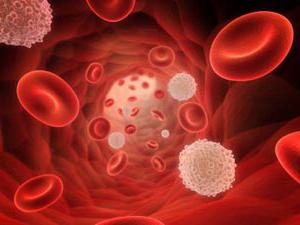
If you look at blood under a scanning electron microscope, you can see what shape and size red blood cells have.

 Human blood under a microscope
 Healthy (intact) cells are small discs (7-8 microns), concave on both sides. They are also called red blood cells.
The number of erythrocytes in the blood fluid exceeds the level of leukocytes and platelets. One drop of human blood contains about 100 million of these cells.
A mature erythrocyte is covered with a membrane. It does not have a nucleus and organelles, except for the cytoskeleton. The inside of the cell is filled with a concentrated fluid (cytoplasm). It is rich in the pigment hemoglobin.
The chemical composition of the cell, in addition to hemoglobin, includes:
Hemoglobin is a protein made up of heme and globin. Heme contains iron atoms. Iron in hemoglobin, binding oxygen in the lungs, stains the blood in a light red color. It turns dark when oxygen is released into the tissues.
Blood cells have a large surface due to their shape. The increased plane of the cells improves the exchange of gases.
The red blood cell is elastic. The very small size of the erythrocyte and flexibility allow it to easily pass through the smallest vessels - capillaries (2-3 microns).
The lifespan of erythrocytes is 120 days. During this time, they perform all their functions. Then they are destroyed. The place of death is the liver, spleen.
Red blood cells decompose faster if their shape changes. When bulges appear in them, echinocytes are formed, depressions - stomatocytes. Poikilocytosis (change in shape) leads to cell death. Disk shape pathology arises from damage to the cytoskeleton.
Video -blood functions. red blood cells
The life path of erythrocytes begins in the red bone marrow of all human bones (up to the age of five).
In an adult, after 20 years, red blood cells are produced in:


Their formation takes place under the influence of erythropoietin, a renal hormone.
With age, erythropoiesis, that is, the process of formation of red blood cells, decreases.
The formation of a blood cell begins with a proerythroblast. As a result of repeated division, mature cells are created.
From the unit that forms the colony, the erythrocyte goes through the following stages:
The primordial cell has a nucleus, which first becomes smaller and then leaves the cell altogether. Its cytoplasm is gradually filled with hemoglobin.
If there are reticulocytes in the blood along with mature red blood cells, this is normal. Earlier types of red blood cells in the blood indicate pathology.
Red blood cells realize their main purpose in the body - they are carriers of respiratory gases - oxygen and carbon dioxide.
This process is carried out in a certain order:

In addition to gas exchange, shaped elements perform other functions:

Normally, each red blood cell in the bloodstream is a free cell in movement. With an increase in blood acidity pH and other negative factors, gluing of red blood cells occurs. Their bonding is called agglutination.
Such a reaction is possible and very dangerous when blood is transfused from one person to another. In this case, to prevent agglutination of red blood cells, you need to know the blood group of the patient and his donor.
The agglutination reaction served as the basis for dividing people's blood into four groups. They differ from each other by the combination of agglutinogens and agglutinins.
The following table will introduce the features of each blood group:
In determining the blood type, it is impossible to make mistakes in any case. Knowing the group affiliation of blood is especially important when it is transfused. Not everyone suits a certain person.
Extremely important! Before a blood transfusion, it is imperative to determine its compatibility. It is impossible to inject incompatible blood into a person. It's life-threatening.
With the introduction of incompatible blood, agglutination of red blood cells occurs. This occurs with this combination of agglutinogens and agglutinins: Aα, Bβ. In this case, the patient has signs of hemotransfusion shock.
They can be:
Agglutination ends with hemolysis, that is, the destruction of red blood cells occurs in the body.
A small amount of blood or red blood cells can be transfused as follows:
Important! If it becomes necessary to transfuse a large amount of fluid, only the blood of the same group is infused.
The number of red blood cells in the blood is determined during a laboratory analysis and counted in 1 mm 3 of blood.
Reference. For any disease, a clinical blood test is prescribed. It gives an idea of the hemoglobin content, the level of erythrocytes and their sedimentation rate (ESR). Blood is given in the morning, on an empty stomach.
Normal hemoglobin value:
The presence of red pigment in excess of the norm may indicate:
The inhabitants of the highlands, lovers of frequent smoking, hemoglobin is also elevated. Low hemoglobin levels occur with anemia (anemia).
Number of non-core drives:

A decrease in the number of red cells or its increase (erythrocytosis) show that disturbances are possible in the activity of the body.
So, with anemia, blood loss, a decrease in the rate of formation of red cells in the bone marrow, their rapid death, and an increased water content, the level of red blood cells decreases.
An increased number of red cells can be detected while taking certain medications, such as corticosteroids, diuretics. A consequence of insignificant erythrocytosis is a burn, diarrhea.
Erythrocytosis also occurs in conditions such as:
Important! In pregnant women, normal blood cell counts change. This is most often associated with the birth of the fetus, the appearance of the child's own circulatory system, and not with the disease.
An indicator of a malfunction in the body is the erythrocyte sedimentation rate (ESR).
It is not recommended to make diagnoses on the basis of tests. Only a specialist after a thorough examination using various techniques can draw the right conclusions and prescribe an effective treatment.

Contents Biologically active additive based on an extract obtained from a beetle with a fly (or fly...

Ekaterina MirimanovaSystem minus 60. Revolution System minus 60 with Ekaterina Mirimanova"System minus 60. Revolution"...

Heaviness and bloating are the causes of both ordinary overeating and more serious problems with the digestive tract....

The second blood type, Rh-negative, appeared many years ago, when a person stopped being a hunter, and ...

PSYCHOLOGICAL ASPECTS OF THE PHENOMENON OF ANOREXIA (EXPERIMENTAL STUDY) T. V. Tarasova, E. V. Arsent'eva V...

Contents Due to the fact that the skin in this area is thin, it is more prone to the appearance of various kinds of spots. Redness under...

vseslav Sat, 10/17/2015 - 20:50 Vasileostrovskaya station is one of the oldest stations...

Most often, the goat mushroom (Suillus bovinus) is referred to in the everyday speech of mushroom pickers as a goat. This tubular...

Every person has had dreams at least once in their life. Many do not attach any importance to them, but some ...

Daedalus (crater). Diameter: 93 km Depth: 3 km (NASA photo) The moon has attracted people's attention since ancient times. In II...

The United States celebrates President's Day on the third Monday in February. Until the 70s, the celebrations were timed to...

On the day of the last call, I want to say just one word to my beloved head teacher: “Thank you.” But how much is...

Watermelon is considered a tasty and healthy berry, its seeds are especially useful. Small, dark brown or...

“The development of speech in children of 6 years old” A child is not born with an established speech. It is not possible to give a definitive answer to...

Although obesity in itself is not a specific disease, it becomes a breeding ground for ...

Parsley is a green plant with a spicy and rich aroma. The most common...