Azan and Iqamat (detailed analysis)
While reading the adhan, the muezzin's hands should touch his earlobes and his gaze should be directed towards the Kaaba. After...
The sensory organization of a personality is the level of development of individual sensitivity systems and the possibility of their unification. Human sensory systems are his sense organs, like receivers of his sensations, in which the transformation of sensation into perception occurs.
The main feature of a person’s sensory organization is that it develops as a result of his entire life path. A person’s sensitivity is given to him at birth, but its development depends on the circumstances, desires and efforts of the person himself. Feeling – lower mental process of reflecting individual properties of objects or phenomena of the internal and external world through direct contact.
It is obvious that the primary cognitive process occurs in the human sensory systems and, on its basis, cognitive processes that are more complex in structure arise: perceptions, ideas, memory, thinking. No matter how simple the primary cognitive process may be, it is precisely it that is the basis of mental activity; only through the “inputs” of sensory systems does the surrounding world penetrate into our consciousness. The physiological mechanism of sensations is the activity of the nervous apparatus - analyzers, consisting of 3 parts:
· receptor- the perceiving part of the analyzer (carries out the transformation of external energy into a nervous process)
· central section of the analyzer- afferent or sensory nerves
· cortical sections of the analyzer, in which nerve impulses are processed.
Each type of sensation is characterized not only by specificity, but also has common properties with other types: quality, intensity, duration, spatial localization. The minimum magnitude of the stimulus at which the sensation appears is absolute threshold of sensation. The value of this threshold characterizes absolute sensitivity, which is numerically equal to a value inversely proportional to the absolute threshold of sensations. Sensitivity to changes in stimulus is called relative or difference sensitivity. The minimum difference between two stimuli that causes a slightly noticeable difference in sensation is called difference threshold.
Classification of sensations
A widespread classification is based on the modality of sensations (specificity of the sense organs) - this is the division of sensations into visual, auditory, vestibular, tactile, olfactory, gustatory, motor, visceral. There are intermodal sensations - synesthesia. The main and most significant group of sensations brings information from the outside world to a person and connects him with the external environment. These are exteroceptive - contact and distant sensations; they occur in the presence or absence of direct contact of the receptor with the stimulus. Vision, hearing, and smell are distant sensations. These types of sensations provide orientation in the immediate environment. Taste, pain, tactile sensations are contact. According to the location of the receptors on the surface of the body, in muscles and tendons or inside the body, they are distinguished accordingly:
– exteroceptive sensations (arising from the influence of external stimuli on receptors located on the surface of the body, externally) visual, auditory, tactile;
– proprioceptive(kinesthetic) sensations (reflecting the movement and relative position of body parts with the help of receptors located in muscles, tendons, joint capsules);
– interoceptive(organic) sensations - arising from the reflection of metabolic processes in the body with the help of specialized receptors, hunger and thirst.
In order for a sensation to arise, it is necessary that the stimulus reaches a certain value, which is called threshold of perception.
Relative threshold- the magnitude that the stimulus must reach for us to feel this change.
Absolute thresholds– these are the upper and lower limits of the organ’s resolution. Threshold research methods:
Bounds method
consists in gradually increasing the stimulus from subthreshold, then the reverse procedure
Installation method
the subject independently distinguishes the magnitude of the stimulus
Sensory systems are considered components of the nervous system, which is involved in the perception of information from the outside world, its transmission into the brain and analysis. Receiving data from the environment and one’s body is a necessary factor for an individual’s life.
This analyzer is one of the most important components of the central nervous system, which involves sensory receptors, nerve fibers that carry information to the brain and its parts. Next, they begin to process and analyze the data.
Each analyzer implies the presence of peripheral receptors, conducting ducts and switching nuclei. In addition, they have a special hierarchy and have several levels of step-by-step data processing. At the lowest level of such perception, primary sensory neurons located in special sensory organs or ganglia are involved. They help conduct excitation from peripheral receptors to the central nervous system. Peripheral receptors are receptive, highly specialized neoplasms that are capable of perceiving, converting and transmitting external energy to primary sensory neurons.
To understand how the sensory system functions, you need to learn about its structure. There are 3 components:
The beginning of the analyzer is receptors, and the end is neurons. Analyzers should not be confused with . The former lack the effector part.
General rules for the operation of analyzers:

Vision is a multi-element process that begins with the projection of an image onto the retina. After the photoreceptors are excited, they are then transformed in the neural layer and finally a decision is made about the sensory image.
The visual analyzer involves certain departments:
The auditory analyzer provides encoding of acoustic images and makes it possible to orient in space thanks to the assessment of the stimulus. The peripheral areas of this analyzer represent the hearing organs and phonoreceptors located in the inner ear. Based on the formation of analyzers, the nominative purpose of speech appears - the association of things and names.
The auditory analyzer is considered one of the most important because it becomes a means of communication between people.
The external passage of the ear helps conduct sound impulses into the eardrum, which separates the outer ear from the middle ear. It is a thin partition and looks like a funnel oriented inward. After exposure to sound impulses through the outer ear, the membrane vibrates.
 It contains 3 bones: the malleus, the incus and the stirrup, which gradually transform the vibrational impulses of the eardrum into the inner ear. The handle of the malleus is woven into the membrane itself, and part 2 is connected to the anvil, which in turn directs the impulse of the stapes. It transmits impulses of smaller amplitude, but more intense. There are 2 muscles located inside the middle ear. The stirrup secures the stirrup, preventing it from moving, and the tensioner contracts and increases tension. By contracting after approximately 10 ms, these muscles prevent overload in the inner ear.
The inner ear contains the cochlea, which is a bony spiral with dimensions of 0.04 mm in width and 0.5 mm at the top. This channel is divided by 2 membranes. At the top of the cochlea, each of these membranes is connected. The upper one will overlap with the lower canal through the foramen ovale using the scala tympani. They are filled with perilymph, similar in consistency to cerebrospinal fluid. In the middle of the 2 channels there is a membranous one, which is filled with endolymph. In it, on the main membrane, there is an apparatus that perceives sounds and includes receptor cells that convert mechanical impulses.
This analyzer perceives and analyzes chemical stimuli that are located in the surrounding world and act on the olfactory system. The process itself is the perception through special organs of any characteristics (flavors) of various substances.
The olfactory system in an individual is expressed by the epithelium, which is located at the top of the nasal cavity and includes sections of the lateral concha and septum on each side. It is enveloped in olfactory mucus and includes special chemoreceptors, supporting and basal cells. The respiratory area has free endings of sensory fibers that react to aromatic substances.
Contains the following departments:
The somatosensory analyzer involves the neural processes that process sensory data throughout the body. Somatic perception is opposed to specific sensations that involve visual and auditory function, aroma, taste and coordination.
There are 3 physiological types of such sensations:
There are other criteria for dividing such sensations:
Visceral sensations are associated with the state of the body. Deep feelings come from deep tissues. These include mainly “deep” pressure, pain and vibration.
It is a more confusing psycho-emotional process regarding sensation. Perception is a holistic image of objects and events that arise as a result of the synthesis of sensations. During this process, the identification of the most significant and important characteristics of an object is noted, with separation from those that are insignificant for such a case, and the correlation of what is perceived with the experience experienced. Any perception presupposes an active functional component (palpation, eye activity when examining, etc.) and complex analytical work of the brain.
Perception can manifest itself in the following forms: conscious, subliminal and extrasensory.
Specialists mainly study the study of the conscious, having made great progress in understanding the mechanisms and patterns of this process. Its study is based on data from psychophysiological studies.
The sensory system is a complex of peripheral and central parts of the central nervous system, which are responsible for receiving impulses of various images from the outside world or one’s own body.
This structure suggests the presence of receptors, neural ducts and sections in the brain. They are responsible for converting outgoing signals. The most famous are the visual, auditory, olfactory, and somatosensory analyzers. Thanks to them, it is possible to differentiate various physical characteristics (temperature, taste, sound vibrations or pressure). Sensory analyzers are the most important elements of the individual’s nervous system. They take an active part in processing data from the external environment, its transformation and analysis. Reception of information from the environment will become a necessary condition for life.
Students, graduate students, young scientists who use the knowledge base in their studies and work will be very grateful to you.
Posted on http://www.allbest.ru/
The sensory information that we receive with the help of our sense organs (analyzers) is important not only for organizing the activities of internal organs and behavior in accordance with the requirements of the environment, but also for the full development of a person.
Sense organs are “windows” through which the outside world enters our consciousness. Without this information, the optimal organization of both the most primitive, “animal” functions of our body, and the higher cognitive mental processes of a person would be impossible.
However, a person does not perceive all changes in the environment; he is not able, for example, to sense the effects of ultrasound, X-rays or radio waves. The range of human sensory perception is limited by the sensory systems available to him, each of which processes information about stimuli of a certain physical nature.
The human sensory system consists of the following subsystems: visual system, auditory system, somatosensory system, gustatory system, olfactory system. Types of analyzers are shown in Appendix 1.
3. The central, or cortical section of the analyzer, consists of two parts: the central part - the “core” - represented by specific neurons that process afferent information from receptors, and the peripheral part - “scattered elements” - neurons dispersed throughout the cerebral cortex. The cortical ends of the analyzers are also called “sensory zones”, which are not strictly limited areas; they overlap each other. These structural features of the central department ensure the process of compensation for impaired functions. At the level of the cortical region, higher analysis and synthesis of afferent excitations are carried out, which provide a complete picture of the environment.
Each sensory system forms connections with various structures of the motor and integrative systems of the brain. Sensory systems are a necessary link for the formation of responses to environmental influences. The sensory system is characterized by the presence of feedback addressed to the receptor or first central section. Activating them makes it possible to regulate the process of perceiving information and its conduction along ascending pathways in the brain.
1. The principle of multi-channel (duplication in order to increase the reliability of the system).
2. The principle of multi-level information transfer.
3. The principle of convergence (the terminal branches of one neuron contact several neurons of the previous level; Sherrington’s funnel).
4. The principle of divergence (animation; contact with several neurons of a higher level).
5. The principle of feedback (all levels of the system have both an ascending and descending path; feedback has an inhibitory value as part of the signal processing process).
6. The principle of corticalization (all sensory systems are represented in the new cortex; therefore, the cortex is functionally multivalued, and there is no absolute localization).
7. The principle of bilateral symmetry (exists to a relative extent).
8. The principle of structural-functional correlations (corticalization of different sensory systems has different degrees).
Basic functions of sensory systems: Bezrukikh M.M. Psychophysiology. Dictionary / M.M. Bezrukikh, D.A. Faber - M.: PER SE, 2006. - signal detection; signal discrimination; transmission and transformation; coding and detection of features; pattern recognition. This sequence is observed in all sensory systems, reflecting the hierarchical principle of their organization. At the same time, detection and primary discrimination of signals is provided by receptors, and detection and identification of signals by neurons of the cerebral cortex. Transmission, transformation and coding of signals is carried out by neurons of all layers of sensory systems.
1. Detection of signals begins in a receptor - a specialized cell, evolutionarily adapted to perceive a stimulus of a certain modality from the external or internal environment and convert it from a physical or chemical form into a form of nervous excitation.
2. An important characteristic of the sensory system is the ability to notice differences in the properties of simultaneously or sequentially acting stimuli. Discrimination begins in the receptors, but this process involves neurons throughout the sensory system. It characterizes the minimum difference between stimuli that the sensory system can notice (differential, or difference, threshold).
3. The processes of transformation and transmission of signals in the sensory system convey to the higher centers of the brain the most important (essential) information about the stimulus in a form convenient for its reliable and quick analysis. Signal transformations can be conditionally divided into spatial and temporal. Among spatial transformations, changes in the ratio of different parts of the signal are distinguished.
4. Information coding is the transformation of information into a conditional form - a code - carried out according to certain rules. In a sensory system, signals are encoded with a binary code, i.e., the presence or absence of an electrical impulse at a given time. Information about stimulation and its parameters is transmitted in the form of individual impulses, as well as groups or “packs” of impulses (“volleys” of impulses). The amplitude, duration and shape of each pulse are the same, but the number of pulses in a burst, their repetition rate, the duration of the bursts and the intervals between them, as well as the temporal “pattern” of the burst are different and depend on the characteristics of the stimulus. Sensory information is also encoded by the number of simultaneously excited neurons, as well as the location of excitation in the neural layer.
5. Signal detection is the selective selection by a sensory neuron of one or another sign of a stimulus that has behavioral significance. This analysis is carried out by detector neurons that selectively respond only to certain stimulus parameters. Thus, a typical neuron in the visual cortex responds with a discharge to only one specific orientation of a dark or light strip located in a certain part of the visual field. At other inclinations of the same strip, other neurons will respond. Detectors of complex features and entire images are concentrated in the higher parts of the sensory system.
6. Pattern recognition is the final and most complex operation of the sensory system. It consists in assigning an image to one or another class of objects that the organism has previously encountered, i.e., in the classification of images. By synthesizing signals from detector neurons, the higher department of the sensory system forms an “image” of the stimulus and compares it with many images stored in memory. Identification ends with a decision about what object or situation the organism encountered. As a result of this, perception occurs, i.e. we realize whose face we see in front of us, whom we hear, what smell we smell. Recognition often occurs regardless of signal variability. Thus, we reliably identify objects under different illumination, color, size, angle, orientation and position in the field of view. This means that the sensory system forms an (invariant) sensory image independent of changes in a number of signal features.
Thus, the sensory system (analyzer) is a functional system consisting of a receptor, an afferent pathway and a zone of the cerebral cortex where this type of sensitivity is projected.
The cortical analyzers of the human brain and their functional connections with various organs are clearly shown in the figure in Appendix 3.
Human sensory systems provide:
1) formation of sensations and perception of current stimuli;
2) control of voluntary movements;
3) control of the activities of internal organs;
4) the level of brain activity necessary for a person to be awake.
The process of transmitting sensory signals (they are often called sensory messages) is accompanied by their multiple transformations and recoding at all levels of the sensory system and ends with the recognition of a sensory image. Sensory information entering the brain is used to organize simple and complex reflex acts, as well as to form mental activity. The entry of sensory information into the brain may be accompanied by awareness of the presence of a stimulus (sensation of the stimulus). Sensation is a subjective sensory response to an actual sensory stimulus (for example, the sensation of light, heat or cold, touch, etc.). as mentioned earlier, the totality of sensations provided by any one analyzer is denoted by the term “modality,” which can include various qualitative types of sensations. Independent modalities are touch, vision, hearing, smell, taste, feeling of cold or heat, pain, vibration, sensation of limb position and muscle load. Within modalities there are different qualities, or submodalities; For example, the taste modality distinguishes between sweet, salty, sour and bitter tastes.
Based on the totality of sensations, sensory perception is formed, i.e., comprehension of sensations and readiness to describe them. Perception is not a simple reflection of the current stimulus; it depends on the distribution of attention at the moment of its action, the memory of past sensory experience and the subjective attitude to what is happening, expressed in emotional experiences.
Thus, the sensory system enters information into the brain and analyzes it. The work of any sensory system begins with the perception by receptors of physical or chemical energy external to the brain, transforming it into nerve signals and transmitting them to the brain through chains of neurons. The process of transmitting sensory signals is accompanied by their repeated transformation and recoding and ends with higher analysis and synthesis (image recognition), after which the body’s response is formed.
2. Characteristics of the main sensory systems
In physiology, it is customary to divide analyzers into external and internal. External human analyzers react to those stimuli that come from the external environment. Human internal analyzers are those structures that respond to changes within the body. For example, muscle tissue has specific receptors that respond to pressure and other indicators that change inside the body.
External analyzers are divided into contact (in direct contact with the stimulus) and distant, which respond to remote stimuli:
1) contact: taste and touch;
2) distant: vision, hearing and smell.
The activity of each of the sense organs represents an elementary mental process - sensation. Sensory information from external stimuli enters the central nervous system in 2 ways:
1) Characteristic sensory pathways:
a) vision - through the retina, lateral geniculate body and superior colliculus into the primary and secondary visual cortex;
b) hearing - through the nuclei of the cochlea and quadrigeminal, the medial geniculate body into the primary auditory cortex;
c) taste - through the medulla oblongata and thalamus to the somatosensory cortex;
d) smell - through the olfactory bulb and piriform cortex to the hypothalamus and limbic system;
e) touch - passes through the spinal cord, brain stem and thalamus to the somatosensory cortex.
2) Nonspecific sensory pathways: pain and temperature sensations located in the nuclei of the thalamus and brain stem.
The visual sensory system provides the brain with more than 90% of sensory information. Vision is a multi-link process that begins with the projection of an image onto the retina. Then the photoreceptors are excited, the transmission and transformation of visual information occurs in the neural layers of the visual system, and visual perception ends with the decision about the visual image being made by the higher cortical parts of this system.
The adaptation of the eye to clearly seeing objects at different distances is called accommodation; the main role here is played by the lens, which changes its curvature and, consequently, refractive power.
The peripheral part of the visual sensory system is the eye (Fig. 1). It consists of the eyeball and supporting structures: lacrimal glands, ciliary muscle, blood vessels and nerves. Characteristics of the membranes of the eyeball in Appendix 4.
The conductive section of the visual sensory system is the optic nerve, the nuclei of the superior colliculus of the midbrain, and the nuclei of the external geniculate body of the diencephalon.
The central section of the visual analyzer is located in the occipital lobe.
The eyeball has a spherical shape, which makes it easier to rotate to point at the object in question. The amount of light that enters the retina is regulated by the pupil, which is capable of dilating and contracting. The pupil is the hole in the center of the iris through which light rays pass into the eye. The pupil sharpens the image on the retina, increasing the depth of field of the eye.
The light beam breaks on the cornea, lens and vitreous body. Thus, the image falls on the retina, which contains many nerve receptors - rods and cones. Thanks to chemical reactions, an electrical impulse is formed here, which follows the optic nerve and is projected in the occipital lobes of the cerebral cortex.
Figure 1 - Organ of vision:
1 - tunica albuginea; 2 - cornea; 3 - lens; 4 - ciliary body; 5 - iris; 6 - choroid; 7 - retina; 8 - blind spot; 9 - vitreous body; 10 - posterior chamber of the eye; 11 - anterior chamber of the eye; 12 - optic nerve
The retina is the inner light-sensitive layer of the eye. There are two types of photoreceptors here (rods and cones: cones function in high light conditions, they provide daytime and color vision; much more photosensitive rods are responsible for twilight vision) and several types of nerve cells. All of the listed retinal neurons with their processes form the nervous apparatus of the eye, which not only transmits information to the visual centers of the brain, but also participates in its analysis and processing. Therefore, the retina is called the part of the brain located in the periphery. From the retina, visual information travels along the optic nerve fibers to the brain.
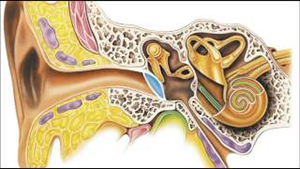
The auditory sensory system is one of the most important distant sensory systems in humans. The receptor here is the ear. Like any other analyzer, the auditory one also consists of three parts: the auditory receptor, the auditory nerve with its pathways and the auditory zone of the cerebral cortex, where the analysis and assessment of sound stimulation occurs (Fig. 2).
The peripheral auditory sensory system consists of three parts: the outer, middle and inner ear.
Wiring department. The hair cells are covered by the nerve fibers of the cochlear branch of the auditory nerve, which carries the nerve impulse to the medulla oblongata, then, crossing with the second neuron of the auditory tract, it is directed to the posterior colliculus and the nuclei of the internal geniculate bodies of the diencephalon, and from them to the temporal region of the cortex, where the central part of the auditory analyzer is located.
Figure 2 - Hearing organ:
A - general view: 1 - external auditory canal; 2 - eardrum; 3 - middle ear;
4 - hammer; 5 - anvil; 6 - stirrup; 7 - auditory nerve; 8 - snail; 9 - auditory (Eustachian) tube; B - section of the cochlea; B - cross section of the cochlear canal: 10 - bone labyrinth; 11 - membranous labyrinth; 12 - spiral (Corti) organ; 13 - main (basal) plate
The central section of the auditory analyzer is located in the temporal lobe. The primary auditory cortex occupies the upper edge of the superior temporal gyrus and is surrounded by the secondary cortex. The meaning of what is heard is interpreted in associative zones. In humans, in the central nucleus of the auditory analyzer, Wernicke's area, located in the posterior part of the superior temporal gyrus, is of particular importance. This zone is responsible for understanding the meaning of words; it is the center of sensory speech. With prolonged exposure to strong sounds, the excitability of the sound analyzer decreases, and with prolonged exposure to silence it increases. This adaptation is observed in the zone of higher sounds.
Acoustic (sound) signals are air vibrations with different frequencies and strengths. They stimulate the auditory receptors located in the cochlea of the inner ear. The receptors activate the first auditory neurons, after which sensory information is transmitted to the auditory area of the cerebral cortex through a number of sequential sections:
Outer ear - the auditory canal conducts sound vibrations to the eardrum. The eardrum, which separates the outer ear from the tympanic cavity, or middle ear, is a thin (0.1 mm) partition shaped like an inward funnel. The membrane vibrates under the action of sound vibrations coming to it through the external auditory canal.
In the middle ear, filled with air, there are three bones: the hammer, the incus and the stapes, which sequentially transmit vibrations of the eardrum to the inner ear. The hammer is woven into the eardrum with a handle; its other side is connected to the anvil, which transmits vibrations to the stapes. Due to the peculiarities of the geometry of the auditory ossicles, vibrations of the eardrum of reduced amplitude but increased strength are transmitted to the stapes.
There are two muscles in the middle ear: the tensor tympani and the stapedius. The first of them, contracting, increases the tension of the eardrum and thereby limits the amplitude of its vibrations during strong sounds, and the second fixes the stapes and thereby limits its movements. This automatically protects the inner ear from overload;
The inner ear contains the cochlea, which contains auditory receptors. The cochlea is a bony spiral canal forming 2.5 turns. Inside the middle canal of the cochlea, on the main membrane, there is a sound-perceiving apparatus - a spiral organ containing receptor hair cells. These cells transform mechanical vibrations into electrical potentials.
Comparative characteristics of the parts of the hearing organ in Appendix 5.
The mechanisms of auditory reception are as follows. Sound, which is vibrations of air, enters the external auditory canal in the form of air waves through the auricle and acts on the eardrum. Vibrations of the eardrum are transmitted to the auditory ossicles, the movements of which cause vibration of the oval window membrane. These vibrations are transmitted to the perilymph and endolymph, then perceived by the fibers of the main membrane. High sounds cause vibrations of short fibers, low sounds cause vibrations of longer ones located at the top of the cochlea. These vibrations excite the receptor hair cells of the organ of Corti. Next, the excitation is transmitted along the auditory nerve to the temporal lobe of the cerebral cortex, where the final synthesis and synthesis of sound signals occurs.
The taste sensory system is a collection of sensitive chemical receptors that respond to certain chemicals. Taste, like smell, is based on chemoreception. Chemoreceptors - taste cells - are located at the bottom of the taste bud. They are covered with microvilli that come into contact with substances dissolved in water.
Taste buds carry information about the nature and concentration of substances entering the mouth. Their excitation triggers a complex chain of reactions in different parts of the brain, leading to different functioning of the digestive organs or to the removal of substances harmful to the body that enter the mouth with food.
The peripheral section of this system is represented by taste buds - taste receptors - located in the epithelium of the grooved, leaf-shaped and mushroom-shaped papillae of the tongue and in the mucous membrane of the palate, pharynx and epiglottis. Most of them are on the tip, edges and back of the tongue. Each of the approximately 10,000 human taste buds consists of several (2-6) receptor cells and, in addition, supporting cells. The taste bud is flask-shaped; in humans its length and width are about 70 microns. The taste bud does not reach the surface of the mucous membrane of the tongue and is connected to the oral cavity through the taste pore.
The conduction section of this analyzer is represented by the trigeminal nerve, the chorda tympani, the glossopharyngeal nerve, the nuclei of the medulla oblongata, and the nuclei of the thalamus.
The central section (cortical end) of the taste analyzer is located in the evolutionarily ancient formations of the cerebral hemispheres, located on their medial (middle) and lower surfaces. This is the cortex of the hippocampus (Ammon's horn), parahippocampus and uncinate, as well as the lateral part of the postcentral gyrus (Fig. 5.3).
Rice. 5.3. Fornix and hippocampus:
1 - hook; 9 - dentate gyrus; 2 - parahippocampal gyrus; 3 - hippocampal peduncle; 4 - hippocampus; 5 - corpus callosum; 6 - central groove; 7 - occipital lobe; 8 - parietal lobe; 9 - temporal lobe
The conductors of all types of taste sensitivity are the chorda tympani and the glossopharyngeal nerve, the nuclei of which in the medulla oblongata contain the first neurons of the taste system. Many of the fibers coming from taste buds are distinguished by a certain specificity, since they respond by increasing the frequency of pulse discharges only to the action of salt, acid and quinine. Other fibers respond to sugar. The most convincing hypothesis is that information about the 4 main taste sensations: bitter, sweet, sour and salty is encoded not by impulses in single fibers, but by different distributions of discharge frequencies in a large group of fibers, differently excited by the taste substance.
Taste afferent signals enter the nucleus of the solitary fasciculus of the brainstem. From the nucleus of the solitary fasciculus, the axons of the second neurons ascend as part of the medial lemniscus to the arcuate nucleus of the thalamus, where third neurons are located, the axons of which are sent to the cortical taste center. The research results do not yet allow us to assess the nature of the transformations of taste afferent signals at all levels of the taste system.
Olfactory analyzer. The peripheral section of the olfactory sensory system is located in the upper posterior nasal cavity - this is the olfactory epithelium, which contains olfactory cells that interact with molecules of odorant substances.
The conduction section is represented by the olfactory nerve, olfactory bulb, olfactory tract, and the nuclei of the amygdala complex.
The central, cortical section is the uncus, the hippocampal gyrus, the septum pellucidum and the olfactory gyrus.
The nuclei of the taste and olfactory analyzers are closely connected with each other, as well as with the brain structures responsible for the formation of emotions and long-term memory. From here it is clear how important the normal functional state of the taste and olfactory analyzer is.
The olfactory receptor cell is a bipolar cell, at the apical pole of which there are cilia, and an unmyelinated axon extends from its basal part. The receptor axons form the olfactory nerve, which penetrates the base of the skull and enters the olfactory bulb.
Molecules of odorous substances enter the mucus produced by the olfactory glands with a constant flow of air or from the oral cavity during eating. Sniffing accelerates the flow of odorous substances to the mucus.
Each olfactory cell has only one type of membrane receptor protein. This protein itself is capable of binding many odorous molecules of various spatial configurations. The rule “one olfactory cell - one olfactory receptor protein” greatly simplifies the transmission and processing of information about odors in the olfactory bulb - the first nerve center for switching and processing chemosensory information in the brain.
The peculiarity of the olfactory system is, in particular, that its afferent fibers do not switch in the thalamus and do not move to the opposite side of the cerebrum. The olfactory tract emerging from the bulb consists of several bundles that are sent to different parts of the forebrain: the anterior olfactory nucleus, the olfactory tubercle, the prepiriform cortex, the periamygdala cortex and part of the nuclei of the amygdala complex. The connection of the olfactory bulb with the hippocampus, piriform cortex and other parts of the olfactory brain occurs through several switches. It has been shown that the presence of a significant number of centers of the olfactory brain is not necessary for the recognition of odors, therefore, most of the nerve centers into which the olfactory tract is projected can be considered as associative centers that ensure the connection of the olfactory sensory system with other sensory systems and the organization on this basis of a number of complex forms behavior - food, defensive, sexual, etc.
The sensitivity of the human olfactory system is extremely high: one olfactory receptor can be excited by one molecule of an odorant, and the stimulation of a small number of receptors leads to the appearance of sensation. Adaptation in the olfactory system occurs relatively slowly (tens of seconds or minutes) and depends on the speed of air flow over the olfactory epithelium and on the concentration of the odorous substance.
The somatosensory system (musculocutaneous sensory system) includes the skin sensitivity system and the sensitive system of the musculoskeletal system, which are corresponding receptors located in different layers of the skin. The receptor surface of the skin is huge (1.4-2.1 m2). There are many receptors concentrated in the skin. They are localized at different depths of the skin and are distributed unevenly over its surface.
The peripheral part of this important sensory system is represented by a variety of receptors, which, according to their location, are divided into skin receptors, proprioceptors (receptors of muscles, tendons and joints) and visceral receptors (receptors of internal organs). Based on the nature of the perceived stimulus, mechanoreceptors, thermoreceptors, chemoreceptors and pain receptors - nociceptors - are distinguished.
The role of a sense organ here, in fact, is the entire surface of the human body, its muscles, joints, and, to a certain extent, internal organs.
The conduction section is represented by numerous afferent fibers, centers of the dorsal horns of the spinal cord, nuclei of the medulla oblongata, and thalamic nuclei.
The central section is located in the parietal lobe: the primary cortex is in the posterior central gyrus, the secondary cortex is in the superior parietal lobule.
The skin has several analyzer systems: tactile (touch sensations), temperature (sensations of cold and heat), pain. The tactile sensitivity system is unevenly distributed throughout the body. But most of all, the accumulation of tactile cells is observed in the palm of the hand, on the tips of the fingers and on the lips. Tactile sensations of the hand, combining with muscle-joint sensitivity, form the sense of touch - a specifically human system of cognitive activity of the hand, developed through labor.
If you touch the surface of the body and then press on it, the pressure can cause pain. Thus, tactile sensitivity provides knowledge about the qualities of an object, and painful sensations signal the body about the need to move away from the stimulus and have a pronounced emotional tone.
The third type of skin sensitivity - temperature sensations - is associated with the regulation of heat exchange between the body and the environment. The distribution of heat and cold receptors on the skin is uneven. The back is most sensitive to cold, the chest is the least sensitive.
The position of the body in space is signaled by static sensations. Static sensitivity receptors are located in the vestibular apparatus of the inner ear. Sudden and frequent changes in body position relative to the plane of the earth can lead to dizziness.
Mechanisms of excitation of skin receptors: the stimulus leads to deformation of the receptor membrane, as a result of which the electrical resistance of the membrane decreases. An ionic current begins to flow through the receptor membrane, leading to the generation of a receptor potential. When the receptor potential increases to a critical level, impulses are generated in the receptor, propagating along the fiber to the central nervous system.
Conclusion
Thus, information about the surrounding world is perceived by a person through the senses, called sensory systems (analyzers) in physiology.
The activity of analyzers is associated with the emergence of five senses - vision, hearing, taste, smell and touch, through which the body communicates with the external environment.
Sense organs are complex sensory systems (analyzers), including perceptive elements (receptors), nerve pathways and corresponding sections in the brain, where the signal is converted into sensation. The main characteristic of the analyzer is sensitivity, which is characterized by the value of the sensation threshold.
The main functions of the sensory system: detection and discrimination of signals; transmission and conversion of signals; information coding; signal detection and pattern recognition.
Each sensory system includes three sections: 1) peripheral or receptor, 2) conductive, 3) cortical.
Sensory systems perceive signals from the outside world and carry to the brain the information necessary for the body to navigate the external environment and to assess the state of the body itself. These signals arise in perceptive elements - sensory receptors that receive stimuli from the external or internal environment, nerve pathways, and are transmitted from the receptors to the brain and those parts of the brain that process this information - through chains of neurons and the nerve fibers of the sensory system connecting them.
Signal transmission is accompanied by multiple transformations and recoding at all levels of the sensory system and ends with the recognition of a sensory image.
Bibliography
1. Atlas of human anatomy: textbook. allowance for medical textbook establishments / ed. T.S. Artemyev, A.A. Vlasova, N.T. Shindina. - M.: RIPOL CLASSIC, 2007. - 528 p.
2. Fundamentals of psychophysiology: Textbook / Rep. ed. Yu.I. Alexandrov. - St. Petersburg: Peter, 2003. - 496 p.
3. Ostrovsky M.A. Human physiology. Textbook. In 2 vols. T. 2 / M.A. Ostrovsky, I.A. Shevelev; Ed. V.M. Pokrovsky, G.F. Briefly. - M. - 368 p. - P. 201-259.
4. Rebrova N.P. Physiology of sensory systems: Educational manual / N.P. Rebrova. - St. Petersburg: NP “Strategy of the Future”, 2007. - 106 p.
5. Serebryakova T.A. Physiological foundations of mental activity: Textbook. - N.-Novgorod: VGIPU, 2008. - 196 p.
6. Smirnov V.M. Physiology of sensory systems and higher nervous activity: Proc. allowance / V.M. Smirnov, S.M. Budylina. - M.: Academy, 2009. - 336 p. - pp. 178-196.
7. Titov V.A. Psychophysiology. Lecture notes / V.A. Titov. - M.: Prior-izdat, 2003. - 176 p.
8. Physiology of sensory systems and higher nervous activity: textbook. In 2 vols. T. 1. / Ed. Ya.A. Altman, G.A. Kulikova. - M. Academy, 2009. - 288 p.
9. Human physiology / Ed. V.M. Smirnova - M.: Academy, 2010. - p.364-370, 372-375,377-378, 370-371,381-386.
Annex 1
Types of analyzers
|
Analyzer |
Functions (what stimuli it perceives) |
Peripheral department |
Wiring department |
Central department |
|
|
Visual |
Light |
Retinal photoreceptors |
Optic nerve |
Visual area in the occipital lobe of the cerebral cortex |
|
|
Auditory |
Sound |
Auditory receptors of the organ of Corti |
Auditory nerve |
Auditory zone in the temporal lobe of the CBP |
|
|
Vestibular (gravitational) |
Mechanical |
Receptors of the semicircular canals and otolithic apparatus |
Vestibular, then auditory nerve |
Vestibular zone in the temporal lobe of the CBP |
|
|
Sensorimotor sensitive (somatosensory) |
Mechanical, temperature, pain. |
Touch receptors in the skin |
Spinothalamic tract: cutaneous sensory nerves |
Somatosensory area in the posterior central gyrus of the GBP |
|
|
Sensorimotor motor (motor) |
Mechanical |
Proprioceptors of muscles and joints |
Sensory nerves of the musculoskeletal system |
Somatosensory area and motor area in the anterior central gyrus of the GBP |
|
|
Olfactory |
Gaseous chemicals |
Olfactory receptors in the nasal cavity |
Olfactory nerve |
Olfactory nuclei and olfactory centers of the temporal lobe of the CBP |
|
|
Flavoring |
Chemical solutes |
Taste buds in the mouth |
Facial glossopharyngeal nerve |
Taste zone in the parietal lobe of the KBP |
|
|
Visceral (internal environment) |
Mechanical |
Interoreceptors of internal organs |
Vagus, splanchnic and pelvic nerves |
Limbic system and sensorimotor area KBP |
Appendix 2
Comparative characteristics of the peripheral section of analyzers
|
Analyzers |
Sensitive organ |
Quality |
Receptors |
|
|
Visual analyzer |
Retina |
Brightness, contrast, motion, size, color |
Rods and cones |
|
|
Hearing analyzer |
Height, timbre of sound |
Hair cells |
||
|
Vestibular analyzer |
Vestibular organ |
Force of gravity |
Vestibular cells |
|
|
Vestibular analyzer |
Vestibular organ |
Rotation |
Vestibular cells |
|
|
Skin analyzer |
Touch |
Touch, cold and heat receptors |
||
|
Taste analyzer |
Sweet and sour taste |
Taste buds on the tip of the tongue |
||
|
Taste analyzer |
Bitter and salty taste |
Taste buds at the base of the tongue |
||
|
Olfactory analyzer |
Olfactory nerves |
Olfactory receptors |
Comparative characteristics of the conductor and central sections of the analyzers
|
Analyzers |
Switching levels: primary |
Switching levels secondary |
Switching levels: tertiary |
Central department |
|
|
Visual analyzer |
Retina |
Primary and secondary visual cortex |
Occipital lobes of the brain |
||
|
Hearing analyzer |
Cochlear nuclei |
Primary auditory cortex |
Temporal lobe of the brain |
||
|
Vestibular analyzer |
Vestibular nuclei |
Somatosensory cortex |
Parietal and temporal lobes of the brain |
||
|
Skin analyzer |
Spinal cord |
Somatosensory cortex |
Superior portion of the posterior central gyrus of the brain |
||
|
Olfactory analyzer |
Olfactory bulb |
Piriform bark |
Limbic system, hypothalamus |
Temporal lobe (seahorse cortex) of the brain |
|
|
Taste analyzer |
Medulla |
Somatosensory cortex |
Inferior portion of the posterior central gyrus of the brain |
Appendix 3
Cortical analyzers of the human brain and their functional connection with various organs
1 - peripheral link; 2 - conductive; 3 - central, or cortical; 4 - interoreceptive; 5 - motor; 6 - gustatory and olfactory; 7 - cutaneous, 8 - auditory, 9 - visual)
Appendix 4
Comparative characteristics of the membranes of the eyeball
|
Shells |
Structural features |
|||
|
Sclera (albuginea) |
Supportive, protective |
|||
|
Fibrous casing (outer casing) |
Cornea |
Transparent, connective tissue, convex in shape |
Transmits and refracts light rays |
|
|
The choroid itself |
Contains many blood vessels |
Uninterrupted power supply to the eyes |
||
|
Choroid (tunica media) |
Ciliary body |
Contains ciliary muscle |
Change in lens curvature |
|
|
Choroid (tunica media) |
Contains the pupil, muscles and melanin pigment |
Transmits light rays and determines eye color |
||
|
Retina (inner layer) |
Two layers: outer pigment (contains fuscin pigment) and inner photosensitive (contains rods, cones) |
Converts light stimulation into a nerve impulse, primary processing of the visual signal |
||
|
Shells |
Structural features |
|||
|
Fibrous casing (outer casing) |
Sclera (albuginea) |
Opaque, connective tissue |
Supportive, protective |
Appendix 5
Comparative characteristics of the parts of the hearing organ
|
Structural features |
|||
|
Outer ear |
Auricle, external auditory canal |
Protective (hairs, earwax), conductive, resonator |
|
|
Middle ear |
Tympanic cavity, tympanic membrane, auditory ossicles (hammer, incus, stapes), auditory (eustachian) tube |
Conductive, increasing vibration power, protective (from strong sound vibrations) |
|
|
Inner ear |
The cochlea of the membranous labyrinth, which contains the spiral organ of Corti |
Conductive, sound-receiving (spiral organ) |
Posted on Allbest.ru
Sensory organization of personality as the level of development of individual sensitivity systems and the possibility of their unification. Analyzers of sensory systems. Activity of sensory receptors. General principles of the design of sensory systems. The work of the senses.
abstract, added 05/24/2012
General characteristics of the sense organs. Receptors and their functional characteristics. Processing of sensory stimuli at the level of the spinal cord, thalamus and cerebral cortex. Auscultation as a diagnostic method. General principle of the structure of sensory systems.
presentation, added 09/26/2013
Violations of sensory systems in an adult attract attention and are considered by others as a pathology. Accessory organs of the eye. Organ of hearing and balance. Methods for studying each sensory system. Methods of unconditioned reflexes.
course work, added 04/14/2009
General physiology of sensory systems. Somatosensory, gustatory and olfactory analyzers. Identifying touch points. Determination of spatial thresholds of tactile reception and localization of pain receptors. Determination of taste sensations and thresholds.
training manual, added 02/07/2013
The structure of the cerebral cortex. Characteristics of the cortical projection zones of the brain. Voluntary regulation of human mental activity. The main disorders in damage to the structure of the functional part of the brain. Tasks of the programming and control unit.
presentation, added 04/01/2015
Processing of somatosensory and auditory signals. Features of the organization of fine touch receptors. Properties of responses of cortical neurons. Parallel processing of sensory modalities. Pain and temperature pathways. Central pathways of pain.
abstract, added 10/27/2009
Characteristics of the brain, the most important human organ that regulates all processes, reflexes and movements in the body. The membranes of the brain: soft, arachnoid, hard. Functions of the medulla oblongata. The main meaning of the cerebellum. Gray matter of the spinal cord.
presentation, added 10/28/2013
The concept and principles of the structure of human analytical systems, study from the point of view of neurophysiology. Causes and types of disorders of the analytical systems, their clinical signs and ways of elimination. Structure, role of the visual analyzer.
test, added 09/18/2009
Higher nervous activity. The work of the reception apparatus and the higher levels of the brain. The problem of reflection adequacy. Differentiation of irritations, their fractional analysis. The energy of external irritation. Afferent impulses from muscle-articular receptors.
abstract, added 06/16/2013
Regulation of body functions, coordinated activity of organs and systems, connection of the body with the external environment are the main functions of the nervous system. Properties of nervous tissue - excitability and conductivity. The structure of the brain and its zones.
General information
Adhering to the cognitive approach to describing the psyche, we imagine a person as a kind of system that processes symbols when solving its problems, then we can imagine the most important feature of a person’s individuality - the sensory organization of the personality.
Sensory organization of personality
The sensory organization of a personality is the level of development of individual sensitivity systems and the possibility of their unification. Human sensory systems are his sense organs, like receivers of his sensations, in which the transformation of sensation into perception occurs.
Any receiver has a certain sensitivity. If we turn to the animal world, we will see that the predominant level of sensitivity of any species is a generic characteristic. For example, bats have developed sensitivity to the perception of short ultrasonic pulses, and dogs have olfactory sensitivity.
The main feature of a person’s sensory organization is that it develops as a result of his entire life path. A person’s sensitivity is given to him at birth, but its development depends on the circumstances, desires and efforts of the person himself.
What do we know about the world and ourselves? Where do we get this knowledge? How? The answers to these questions come from the depths of centuries from the cradle of all living things.
Feel
Sensation is a manifestation of a general biological property of living matter - sensitivity. Through sensation there is a psychic connection with the external and internal world. Thanks to sensations, information about all phenomena of the external world is delivered to the brain. In the same way, a loop is closed through sensations to receive feedback about the current physical and partly mental state of the body.
Through sensations we learn about taste, smell, color, sound, movement, the state of our internal organs, etc. From these sensations, holistic perceptions of objects and the whole world are formed.
It is obvious that the primary cognitive process occurs in the human sensory systems and, on its basis, cognitive processes that are more complex in structure arise: perceptions, ideas, memory, thinking.
No matter how simple the primary cognitive process may be, it is precisely it that is the basis of mental activity; only through the “inputs” of sensory systems does the surrounding world penetrate into our consciousness.
Processing sensations
After the brain receives information, the result of its processing is the development of a response action or strategy aimed, for example, at improving physical tone, focusing more attention on the current activity, or setting up an accelerated involvement in mental activity.
Generally speaking, the response or strategy developed at any given time is the best choice of the options available to a person at the time of decision making. However, it is clear that the number of options available and the quality of choice varies from person to person and depends, for example, on:
mental properties of the individual,
strategies for relationships with others,
partly physical condition,
experience, the presence of the necessary information in memory and the ability to retrieve it.
degree of development and organization of higher nervous processes, etc.
For example, a baby goes out undressed into the cold, his skin feels cold, perhaps a chill appears, he becomes uncomfortable, a signal about this goes to the brain and a deafening roar is heard. An adult's reaction to cold (stimulus) may be different; he will either rush to get dressed, or jump into a warm room, or try to warm up in another way, for example, by running or jumping.
Improving higher mental functions of the brain
Over time, children improve their reactions, greatly increasing the effectiveness of the results achieved. But after growing up, opportunities for improvement do not disappear, despite the fact that an adult’s sensitivity to them decreases. This is exactly what “Effecton” sees as part of its mission: increasing the efficiency of intellectual activity by training the higher mental functions of the brain.
Effecton's software products allow you to measure various indicators of the human sensorimotor system (in particular, the Jaguar package contains time tests for simple audio and visual-motor reactions, complex visual-motor reactions, and accuracy of perception of time intervals). Other packages of the Effecton complex evaluate the properties of cognitive processes at higher levels.
Therefore, it is necessary to develop the child’s perception, and using the “Jaguar” package can help you with this.
Physiology of sensations
Analyzers
The physiological mechanism of sensations is the activity of nervous apparatus - analyzers, consisting of 3 parts:
receptor - the perceiving part of the analyzer (converts external energy into a nervous process)
central section of the analyzer - afferent or sensory nerves
cortical sections of the analyzer, in which nerve impulses are processed.
Certain receptors correspond to their own areas of cortical cells.
The specialization of each sense organ is based not only on the structural features of the analyzer-receptors, but also on the specialization of the neurons that are part of the central nervous apparatus, which receive signals perceived by the peripheral sense organs. The analyzer is not a passive receiver of energy; it reflexively adapts under the influence of stimuli.
Movement of a stimulus from the external to the internal world
According to the cognitive approach, the movement of a stimulus during its transition from the external world to the internal world occurs as follows:
the stimulus causes certain energy changes in the receptor,
energy is converted into nerve impulses,
information about nerve impulses is transmitted to the corresponding structures of the cerebral cortex.
Sensations depend not only on the capabilities of the human brain and sensory systems, but also on the characteristics of the person himself, his development and condition. When sick or tired, a person's sensitivity to certain influences changes.
There are also cases of pathologies when a person is deprived, for example, of hearing or vision. If this problem is congenital, then there is a disruption in the flow of information, which can lead to mental development delays. If these children were taught special techniques that compensate for their deficiencies, then some redistribution within the sensory systems is possible, thanks to which they will be able to develop normally.
Properties of sensations
Each type of sensation is characterized not only by specificity, but also has common properties with other types:
quality,
intensity,
duration,
spatial localization.
But not every irritation causes a sensation. The minimum magnitude of the stimulus at which sensation appears is the absolute threshold of sensation. The value of this threshold characterizes absolute sensitivity, which is numerically equal to a value inversely proportional to the absolute threshold of sensations. And sensitivity to changes in the stimulus is called relative or difference sensitivity. The minimum difference between two stimuli that causes a slightly noticeable difference in sensation is called the difference threshold.
Based on this, we can conclude that it is possible to measure sensations. And once again you are amazed by the amazing, finely working instruments - human sense organs or human sensory systems.
Effecton's software products allow you to measure various indicators of the human sensory system (for example, the Jaguar package contains speed tests for simple audio and visual-motor reactions, complex visual-motor reactions, accuracy of time perception, accuracy of space perception and many others). Other packages of the Effecton complex also evaluate the properties of cognitive processes at higher levels.
Classification of sensations
Five main types of sensations: vision, hearing, touch, smell and taste - were already known to the ancient Greeks. Currently, ideas about the types of human sensations have been expanded; about two dozen different analyzer systems can be distinguished, reflecting the impact of the external and internal environment on receptors.
The classification of sensations is carried out according to several principles. The main and most significant group of sensations brings information from the outside world to a person and connects him with the external environment. These are exteroceptive - contact and distant sensations; they occur in the presence or absence of direct contact of the receptor with the stimulus. Vision, hearing, and smell are distant sensations. These types of sensations provide orientation in the immediate environment. Taste, pain, tactile sensations are contact.
According to the location of the receptors on the surface of the body, in muscles and tendons or inside the body, they are distinguished accordingly:
exteroception - visual, auditory, tactile and others;
proprioception - sensations from muscles, tendons;
interoception - sensations of hunger, thirst.
During the evolution of all living things, sensitivity has undergone changes from the most ancient to the modern. Thus, distant sensations can be considered more modern than contact ones, but in the structure of the contact analyzers themselves it is also possible to identify more ancient and completely new functions. For example, pain sensitivity is more ancient than tactile sensitivity.
Such classification principles help to group all types of sensations into systems and see their interactions and connections.
Types of sensations
Vision, hearing
Let's look at the different types of sensations, keeping in mind that vision and hearing are the most well studied.
The idea of sensory systems was formulated by I.P. Pavlov in the doctrine of analyzers in 1909 during his study of higher nervous activity. Analyzer- a set of central and peripheral formations that perceive and analyze changes in the external and internal environments of the body. Concept sensory system, which appeared later, replaced the concept of analyzer, including the mechanisms of regulation of its various departments with the help of direct and feedback connections. Along with this, the concept still exists sense organ as a peripheral formation that perceives and partially analyzes environmental factors. The main part of the sensory organ is the receptors, equipped with auxiliary structures that ensure optimal perception. Thus, the organ of vision consists of the eyeball, the retina, which contains visual receptors, and a number of auxiliary structures: eyelids, muscles, lacrimal apparatus. The organ of hearing consists of the outer, middle and inner ear, where in addition to the spiral (corti) organ and its hair (receptor) cells there are also a number of auxiliary structures. The tongue can be considered an organ of taste. When directly exposed to various environmental factors with the participation of analyzers in the body, Feel, which are reflections of the properties of objects in the objective world. The peculiarity of sensations is their modality, those. a set of sensations provided by any one analyzer. Within each modality, in accordance with the type (quality) of the sensory impression, different qualities can be distinguished, or valence. Modalities are, for example, vision, hearing, taste. Qualitative types of modality (valence) for vision are different colors, for taste - the sensation of sour, sweet, salty, bitter.
The activity of analyzers is usually associated with the emergence of five senses - vision, hearing, taste, smell and touch, through which the body communicates with the external environment. However, in reality there are much more of them. For example, the sense of touch in a broad sense, in addition to the tactile sensations arising from touch, includes the feeling of pressure and vibration. The temperature sense includes sensations of warmth or cold, but there are also more complex sensations, such as sensations of hunger, thirst, sexual need (libido), due to the special (motivational) state of the body. The sense of body position in space is associated with the activity of the vestibular and motor analyzers and their interaction with the visual analyzer. The sensation of pain occupies a special place in sensory function. In addition, we can, although “vaguely,” perceive other changes, not only in the external, but also in the internal environment of the body, and in this case emotionally charged sensations are formed. Thus, coronary spasm in the initial stage of the disease, when pain does not yet occur, can cause a feeling of melancholy and despondency. Thus, there are actually much more structures that perceive irritation from the living environment and the internal environment of the body than is commonly believed.
The classification of analyzers can be based on various characteristics: the nature of the current stimulus, the nature of the sensations that arise, the level of receptor sensitivity, the speed of adaptation, and much more.
But the most significant is the classification of analyzers, which is based on their purpose (role). In this regard, there are several types of analyzers.
External analyzers perceive and analyze changes in the external environment. This should include visual, auditory, olfactory, gustatory, tactile and temperature analyzers, the excitation of which is perceived subjectively in the form of sensations.
Internal (visceral) analyzers, perceiving and analyzing changes in the internal environment of the body, indicators of homeostasis. Fluctuations in indicators of the internal environment within the physiological norm in a healthy person are usually not perceived subjectively in the form of sensations. Thus, we cannot subjectively determine the value of blood pressure, especially if it is normal, the state of the sphincters, etc. However, information coming from the internal environment plays an important role in regulating the functions of internal organs, ensuring the body’s adaptation to various conditions of its life. The significance of these analyzers is studied as part of a physiology course (adaptive regulation of the activity of internal organs). But at the same time, changes in some constants of the internal environment of the body can be perceived subjectively in the form of sensations (thirst, hunger, sexual desire) formed on the basis of biological needs. To satisfy these needs, behavioral responses are activated. For example, when a feeling of thirst arises due to stimulation of osmo- or volume receptors, behavior is formed aimed at searching for and receiving water.
Body position analyzers perceive and analyze changes in the position of the body in space and body parts relative to each other. These include the vestibular and motor (kinesthetic) analyzers. As we evaluate the position of our body or its parts relative to each other, this impulse reaches our consciousness. This is evidenced, in particular, by the experiment of D. McLosky, which he performed on himself. Primary afferent fibers from muscle receptors were stimulated by threshold electrical stimuli. An increase in the frequency of impulses of these nerve fibers caused the subject to have subjective sensations of a change in the position of the corresponding limb, although its position did not actually change.
Pain analyzer should be highlighted separately due to its special significance for the body - it carries information about damaging actions. Painful sensations can occur when both extero- and interoreceptors are irritated.
Structural and functional organization of analyzers
According to the presentation of I.P. Pavlov (1909), any analyzer has three sections: peripheral, conductive and central, or cortical. The peripheral section of the analyzer is represented by receptors. Its purpose is the perception and primary analysis of changes in the external and internal environments of the body. In the receptors, the energy of the stimulus is transformed into a nerve impulse, as well as the signal is amplified due to the internal energy of metabolic processes. Receptors are characterized by specificity (modality), i.e. the ability to perceive a certain type of stimulus to which they have adapted in the process of evolution (adequate stimuli), on which the primary analysis is based. Thus, the receptors of the visual analyzer are adapted to the perception of light, and the auditory receptors are adapted to perceive sound, etc. That part of the receptor surface from which one afferent fiber receives the signal is called its receptive field. Receptive fields can have a different number of receptor formations (from 2 to 30 or more), among which there is a leader receptor, and overlap each other. The latter ensures greater reliability of the function and plays a significant role in compensation mechanisms.
Receptors are characterized by great diversity.
In classification receptors, the central place is occupied by their division depending on the type of perceived stimulus. There are five types of such receptors.
1. Mechanoreceptors are excited by mechanical deformation and are located in the skin, blood vessels, internal organs, musculoskeletal system, auditory and vestibular systems.
2. Chemoreceptors perceive chemical changes in the external and internal environment of the body. These include taste and olfactory receptors, as well as receptors that respond to changes in the composition of blood, lymph, intercellular and cerebrospinal fluid (changes in O 2 and CO 2 tension, osmolarity and pH, glucose levels and other substances). Such receptors are found in the mucous membrane of the tongue and nose, carotid and aortic bodies, hypothalamus and medulla oblongata.
3. Thermoreceptors perceive temperature changes. They are divided into heat and cold receptors and are found in the skin, mucous membranes, blood vessels, internal organs, hypothalamus, midbrain, medulla oblongata and spinal cord.
4. Photoreceptors in the retina of the eye perceive light (electromagnetic) energy.
5. Nociceptors, the excitation of which is accompanied by painful sensations (pain receptors). The irritants of these receptors are mechanical, thermal and chemical (histamine, bradykinin, K +, H +, etc.) factors. Painful stimuli are perceived by free nerve endings, which are found in the skin, muscles, internal organs, dentin, and blood vessels.
From a psychophysiological point of view receptors are divided according to the sense organs and the sensations generated into visual, auditory, gustatory, olfactory and tactile.
By location in the body receptors are divided into extero- and interoreceptors.
Exteroceptors include receptors of the skin, visible mucous membranes and sensory organs: visual, auditory, gustatory, olfactory, tactile, pain and temperature. Interoreceptors include receptors of internal organs (visceroreceptors), blood vessels and the central nervous system. A variety of interoreceptors are receptors of the musculoskeletal system (proprioceptors) and vestibular receptors. If the same type of receptors (for example, chemoreceptors sensitive to CO 3) is localized both in the central nervous system (in the medulla oblongata) and in other places (vessels), then such receptors are divided into central and peripheral.
By speed of adaptation receptors are divided into three groups: rapidly adapting (phasic), slowly adapting (tonic) and mixed (phasotonic), adapting at an average speed. An example of rapidly adapting receptors are the vibration (Pacini corpuscles) and touch (Meissner corpuscles) receptors on the skin. Slowly adapting receptors include proprioceptors, lung stretch receptors, and pain receptors. Retinal photoreceptors and skin thermoreceptors adapt at an average speed.
According to structural and functional organization distinguish between primary and secondary receptors. Primary receptors are the sensory endings of the dendrite of the afferent neuron. The neuron body is located in the spinal ganglion or cranial nerve ganglion. In the primary receptor, the stimulus acts directly on the endings of the sensory neuron. Primary receptors are phylogenetically more ancient structures; they include olfactory, tactile, temperature, pain receptors and proprioceptors.
In secondary receptors there is a special cell that is synaptically connected to the end of the dendrite of the sensory neuron. This is a cell, such as a photoreceptor, of epithelial nature or neuroectodermal origin.
This classification allows us to understand how receptor excitation occurs.
Mechanism of receptor excitation. When a stimulus acts on a receptor cell, a change in the spatial configuration of protein receptor molecules occurs in the protein-lipid layer of the membrane. This leads to a change in the permeability of the membrane to certain ions, most often sodium ions, but in recent years the role of potassium in this process has also been discovered. Ionic currents arise, the membrane charge changes, and a receptor potential (RP) is generated. And then the process of excitation occurs in different receptors in different ways. In the primary sensory receptors, which are the free bare ends of a sensitive neuron (olfactory, tactile, proprioceptive), the RP acts on the adjacent, most sensitive areas of the membrane, where an action potential (AP) is generated, which then spreads in the form of impulses along the nerve fiber. The conversion of external stimulus energy into AP in primary receptors can occur both directly on the membrane and with the participation of some auxiliary structures. This, for example, happens in the Pacinian corpuscle. The receptor here is represented by a bare axon ending, which is surrounded by a connective tissue capsule. When the Pacinian corpuscle is compressed, RP is recorded, which is further converted into an impulse response of the afferent fiber. In secondary sensory receptors, which are represented by specialized cells (visual, auditory, gustatory, vestibular), RP leads to the formation and release of a transmitter from the presynaptic section of the receptor cell into the synaptic cleft of the receptor-afferent synapse. This transmitter acts on the postsynaptic membrane of the sensitive neuron, causing its depolarization and the formation of a postsynaptic potential, which is called the generator potential (GP). GP, acting on extrasynaptic areas of the membrane of a sensitive neuron, causes the generation of APs. GP can be both de- and hyperpolarizing and, accordingly, cause excitation or inhibit the impulse response of the afferent fiber.
Properties and features of receptor and generator potentials
Receptor and generator potentials are bioelectric processes that have the properties of a local or local response: they spread with decrement, i.e. with attenuation; the magnitude depends on the strength of irritation, since they obey the “law of force”; the value depends on the rate of increase in the stimulus amplitude over time; can be summed up when applying rapidly successive irritations.
So, the transformation of stimulus energy into a nerve impulse occurs in the receptors, i.e. primary coding of information, transformation of information into sensory code.
Most receptors have so-called background activity, i.e. excitation occurs in them in the absence of any stimuli.
Conductor section of the analyzer includes afferent (peripheral) and intermediate neurons of the stem and subcortical structures of the central nervous system (CNS), which constitute a chain of neurons located in different layers at each level of the CNS. The conduction section ensures the conduction of excitation from receptors to the cerebral cortex and partial processing of information. The conduction of excitation through the conduction section is carried out by two afferent pathways:
1) a specific projection path (direct afferent paths) from the receptor along strictly designated specific paths with switching at different levels of the central nervous system (at the level of the spinal and medulla oblongata, in the visual thalamus and in the corresponding projection zone of the cerebral cortex);
2) in a nonspecific way, with the participation of the reticular formation. At the level of the brain stem, collaterals extend from a specific pathway to the cells of the reticular formation, to which various afferent excitations can converge, ensuring the interaction of analyzers. In this case, afferent excitations lose their specific properties (sensory modality) and change the excitability of cortical neurons. Excitation is carried out slowly through a large number of synapses. Due to collaterals, the hypothalamus and other parts of the limbic system of the brain, as well as motor centers, are included in the excitation process. All this provides the autonomic, motor and emotional components of sensory reactions.
Central, or cortical, analyzer department, according to I.P. Pavlov, consists of two parts: the central part, i.e. “core”, represented by specific neurons that process afferent impulses from receptors, and the peripheral part, i.e. “scattered elements” - neurons dispersed throughout the cerebral cortex. The cortical ends of the analyzers are also called “sensory zones”, which are not strictly limited areas; they overlap each other. Currently, in accordance with cytoarchitectonic and neurophysiological data, projection (primary and secondary) and associative tertiary zones of the cortex are distinguished. Excitation from the corresponding receptors to the primary zones is directed along fast-conducting specific pathways, while activation of the secondary and tertiary (associative) zones occurs along polysynaptic nonspecific pathways. In addition, the cortical zones are interconnected by numerous associative fibers. Neurons are distributed unevenly throughout the thickness of the cortex and usually form six layers. The main afferent pathways to the cortex end on the neurons of the upper layers (III - IV). These layers are most strongly developed in the central parts of the visual, auditory and skin analyzers. Afferent impulses with the participation of stellate cells of the cortex (IV layer) are transmitted to pyramidal neurons (III layer), from here the processed signal leaves the cortex to other brain structures.
In the cortex, input and output elements, together with stellate cells, form so-called columns - functional units of the cortex, organized in the vertical direction. The column has a diameter of about 500 μm and is determined by the distribution zone of collaterals of the ascending afferent thalamocortical fiber. Adjacent columns have relationships that organize the participation of multiple columns to carry out a particular reaction. Excitation of one of the columns leads to inhibition of neighboring ones.
Cortical projections of sensory systems have a topical principle of organization. The volume of the cortical projection is proportional to the receptor density. Due to this, for example, the central fovea of the retina in the cortical projection is represented by a larger area than the periphery of the retina.
To determine the cortical representation of various sensory systems, the method of recording evoked potentials (EP) is used. EP is a type of evoked electrical activity in the brain. Sensory EPs are recorded during stimulation of receptor formations and are used to characterize such an important function as perception.
Among the general principles of analyzer organization, multi-level and multi-channel systems should be highlighted.
Multilevelness provides the possibility of specialization of different levels and layers of the central nervous system for processing certain types of information. This allows the body to more quickly respond to simple signals that are analyzed at individual intermediate levels.
The existing multichannel nature of analyzer systems is manifested in the presence of parallel neural channels, i.e. in each of the layers and levels there are many nerve elements connected with many nerve elements of the next layer and level, which in turn transmit nerve impulses to elements of a higher level, thereby ensuring the reliability and accuracy of the analysis of the influencing factor.
At the same time existing hierarchical principle the construction of sensory systems creates conditions for fine regulation of perception processes through influences from higher levels to lower ones.
These structural features of the central department ensure the interaction of various analyzers and the process of compensation for impaired functions. At the level of the cortical region, higher analysis and synthesis of afferent excitations are carried out, providing a complete picture of the environment.
The main properties of the analyzers are the following.
1. High sensitivity to an adequate stimulus. All parts of the analyzer, and especially the receptors, are highly excitable. Thus, the photoreceptors of the retina can be excited by the action of only a few quanta of light, and the olfactory receptors inform the body about the appearance of single molecules of odorous substances. However, when considering this property of analyzers, it is preferable to use the term “sensitivity” rather than “excitability”, since in humans it is determined by the occurrence of sensations.
Sensitivity is assessed using a number of criteria.
Threshold of sensation(absolute threshold) - the minimum force of irritation that causes such excitation of the analyzer, which is perceived subjectively in the form of a sensation.
Discrimination threshold(differential threshold) - a minimal change in the strength of the current stimulus, perceived subjectively in the form of a change in the intensity of sensation. This pattern was established by E. Weber in an experiment with the determination of the force of pressure on the palm by the test subject’s sensation. It turned out that when a load of 100 g was applied, it was necessary to add a load of 3 g to feel an increase in pressure, when a load of 200 g was applied, it was necessary to add 6 g, 400 g - 12 g, etc. In this case, the ratio of the increase in the strength of stimulation (L) to the strength of the active stimulus (L) is a constant value (C):
This value is different for different analyzers, in this case it is equal to approximately 1/30 of the strength of the current stimulus. A similar pattern is observed when the strength of the current stimulus decreases.
Intensity of sensations with the same stimulus strength can be different, since it depends on the level of excitability of various structures of the analyzer at all its levels. This pattern was studied by G. Fechner, who showed that the intensity of sensation is directly proportional to the logarithm of the strength of stimulation. This position is expressed by the formula:
where E is the intensity of sensations,
K - constant,
L is the strength of the current stimulus,
L 0 - sensation threshold (absolute threshold).
Weber's and Fechner's laws are not accurate enough, especially when the strength of irritation is low. Psychophysical research methods, although they suffer from some inaccuracy, are widely used in studies of analyzers in practical medicine, for example, in determining visual acuity, hearing, smell, tactile sensitivity, and taste.
2. Inertia- relatively slow onset and disappearance of sensations. The latent time for the occurrence of sensations is determined by the latent period of excitation of receptors and the time required for the transition of excitation in synapses from one neuron to another, the time of excitation of the reticular formation and generalization of excitation in the cerebral cortex. The persistence of sensations for a certain period after the stimulus is turned off is explained by the phenomenon of aftereffects in the central nervous system - mainly by the circulation of excitation. Thus, a visual sensation does not arise and disappear instantly. The latent period of visual sensation is 0.1 s, the aftereffect time is 0.05 s. Light stimuli (flickers) quickly following one after another can give a feeling of continuous light (the phenomenon of “flickering fusion”). The maximum frequency of light flashes, which are perceived separately, is called the critical flickering frequency, which is greater, the stronger the brightness of the stimulus and the higher the excitability of the central nervous system, and is about 20 flickers per second. Along with this, if two stationary stimuli are projected sequentially with an interval of 20-200 ms onto different parts of the retina, a sensation of object movement arises. This phenomenon is called the “Phi Phenomenon.” This effect is observed even when one stimulus is slightly different in shape from the other. These two phenomena: “flicker fusion” and “Phi-phenomenon” are the basis of cinematography. Due to the inertia of perception, the visual sensation from one frame lasts until the appearance of another, which is why the illusion of continuous movement arises. Typically, this effect occurs when still images are presented on the screen in rapid succession at a speed of 18-24 frames per second.
3. Ability sensory system to adaptation with a constant strength of a long-acting stimulus, it mainly consists of a decrease in absolute and an increase in differential sensitivity. This property is inherent in all sections of the analyzer, but it is most clearly manifested at the level of receptors and consists in a change not only in their excitability and impulses, but also in indicators of functional mobility, i.e. in changing the number of functioning receptor structures (P.G. Snyakin). Based on the speed of adaptation, all receptors are divided into quickly and slowly adapting, and sometimes a group of receptors with an average speed of adaptation is also distinguished. In the conductive and cortical sections of the analyzers, adaptation is manifested in a decrease in the number of activated fibers and nerve cells.
An important role in sensory adaptation is played by efferent regulation, which is carried out through descending influences that change the activity of the underlying structures of the sensory system. Thanks to this, the phenomenon of “tuning” sensory systems to optimal perception of stimuli in a changed environment arises.
4. Interaction of analyzers. With the help of analyzers, the body learns the properties of objects and phenomena in the environment, the beneficial and negative aspects of their impact on the body. Therefore, dysfunction of external analyzers, especially visual and auditory, makes it extremely difficult to understand the outside world (the outside world is very poor for a blind or deaf person). However, only analytical processes in the central nervous system cannot create a real picture of the environment. The ability of analyzers to interact with each other provides a figurative and holistic view of objects in the external world. For example, we evaluate the quality of a lemon slice using visual, olfactory, tactile and taste analyzers. At the same time, an idea is formed both about individual qualities - color, consistency, smell, taste, and about the properties of the object as a whole, i.e. a certain holistic image of the perceived object is created. The interaction of analyzers when assessing phenomena and objects also underlies compensation for impaired functions when one of the analyzers is lost. Thus, in blind people the sensitivity of the auditory analyzer increases. Such people can determine the location of large objects and walk around them if there is no extraneous noise. This is done by reflecting sound waves from an object in front. American researchers observed a blind man who quite accurately determined the location of a large cardboard plate. When the subject's ears were covered with wax, he could no longer determine the location of the cardboard.
Interactions of sensory systems can manifest themselves in the form of the influence of excitation of one system on the state of excitability of another according to the dominant principle. Thus, listening to music can cause pain relief during dental procedures (audioanalgesia). Noise impairs visual perception; bright light increases the perception of sound volume. The process of interaction between sensory systems can manifest itself at various levels. The reticular formation of the brain stem, the cerebral cortex, plays a particularly important role in this. Many cortical neurons have the ability to respond to complex combinations of signals from different modalities (multisensory convergence), which is very important for cognition of the environment and the evaluation of new stimuli.
Encoding information in analyzers
Concepts. Coding- the process of converting information into a conditional form (code) convenient for transmission over a communication channel. Any transformation of information in the analyzer departments is coding. In the auditory analyzer, the mechanical vibration of the membrane and other sound-conducting elements is at the first stage converted into a receptor potential, the latter ensures the release of the transmitter into the synaptic cleft and the emergence of a generator potential, as a result of which a nerve impulse arises in the afferent fiber. The action potential reaches the next neuron, at the synapse of which the electrical signal again turns into a chemical signal, i.e. the code changes many times. It should be noted that at all levels of analyzers there is no restoration of the stimulus in its original form. This physiological coding differs from most technical communication systems, where the message, as a rule, is restored in its original form.
Nervous system codes. IN Computer technology uses binary code, when two symbols are always used to form combinations - 0 and 1, which represent two states. Encoding of information in the body is carried out on the basis of non-binary codes, which makes it possible to obtain a larger number of combinations with the same code length. The universal code of the nervous system is nerve impulses that travel along nerve fibers. In this case, the content of information is determined not by the amplitude of the pulses (they obey the “All or nothing” law), but by the frequency of the pulses (time intervals between individual pulses), their combination into bursts, the number of pulses in a burst, and the intervals between bursts. The transmission of a signal from one cell to another in all sections of the analyzer is carried out using a chemical code, i.e. various mediators. To store information in the central nervous system, encoding is carried out using structural changes in neurons (memory mechanisms).
Coded characteristics of the stimulus. The analyzers encode the qualitative characteristics of the stimulus (for example, light, sound), the strength of the stimulus, the time of its action, as well as space, i.e. the place of action of the stimulus and its localization in the environment. All sections of the analyzer take part in encoding all the characteristics of the stimulus.
In the peripheral section of the analyzer coding of the quality of the stimulus (type) is carried out due to the specificity of the receptors, i.e. the ability to perceive a stimulus of a certain type to which it is adapted in the process of evolution, i.e. to an adequate stimulus. Thus, a light beam excites only the receptors of the retina; other receptors (smell, taste, tactile, etc.) usually do not respond to it.
The strength of the stimulus can be encoded by a change in the frequency of impulses generated by the receptors when the strength of the stimulus changes, which is determined by the total number of impulses per unit time. This is the so-called frequency coding. Moreover, with increasing stimulus strength, the number of impulses arising in the receptors usually increases, and vice versa. When the strength of the stimulus changes, the number of excited receptors may also change; in addition, the strength of the stimulus can be encoded by varying the latency period and reaction time. A strong stimulus reduces the latency period, increases the number of impulses and lengthens the reaction time. Space is encoded by the size of the area over which the receptors are excited; this is spatial encoding (for example, we can easily determine whether a pencil touches the surface of the skin with a sharp or blunt end). Some receptors are more easily excited when a stimulus acts on them at a certain angle (Pacinian corpuscles, retinal receptors), which is an assessment of the direction of action of the stimulus on the receptor. The localization of the action of the stimulus is encoded by the fact that receptors in different parts of the body send impulses to certain areas of the cerebral cortex.
The time of action of the stimulus on the receptor is encoded by the fact that it begins to be excited with the onset of the stimulus and stops being excited immediately after the stimulus is turned off (temporal coding). It should be noted that the time of action of the stimulus in many receptors is not encoded accurately enough due to their rapid adaptation and cessation of excitation with a constant strength of the stimulus. This inaccuracy is partially compensated by the presence of on-, off- and on-off receptors, which are excited respectively when the stimulus is turned on, off, and also when the stimulus is turned on and off. With a long-acting stimulus, when adaptation of the receptors occurs, a certain amount of information about the stimulus (its strength and duration) is lost, but sensitivity increases, i.e., sensitization of the receptor to changes in this stimulus develops. An increase in stimulus acts on the adapted receptor as a new stimulus, which is also reflected in a change in the frequency of impulses coming from the receptor.
In the conductor section of the analyzer, coding is carried out only at “switching stations,” that is, when transmitting a signal from one neuron to another, where the code changes. Information is not encoded in nerve fibers; they act as wires through which information encoded in receptors and processed in the centers of the nervous system is transmitted.
There can be different intervals between impulses in a separate nerve fiber, impulses are formed into packets with different numbers, and there can also be different intervals between individual packets. All this reflects the nature of the information encoded in the receptors. In this case, the number of excited nerve fibers in the nerve trunk can also change, which is determined by a change in the number of excited receptors or neurons at the previous signal transition from one neuron to another. At switching stations, for example in the thalamus, information is encoded, firstly, by changing the volume of impulses at the input and output, and secondly, by spatial coding, i.e. due to the connection of certain neurons with certain receptors. In both cases, the stronger the stimulus, the more neurons are excited.
In the overlying parts of the central nervous system, a decrease in the frequency of neuronal discharges and the transformation of long-term impulses into short bursts of impulses are observed. There are neurons that are excited not only when a stimulus appears, but also when it is turned off, which is also associated with the activity of receptors and the interaction of the neurons themselves. Neurons, called “detectors,” respond selectively to one or another stimulus parameter, for example, to a stimulus moving in space, or to a light or dark strip located in a certain part of the visual field. The number of such neurons, which only partially reflect the properties of the stimulus, increases at each subsequent level of the analyzer. But at the same time, at each subsequent level of the analyzer there are neurons that duplicate the properties of the neurons of the previous section, which creates the basis for the reliability of the analyzer function. In the sensory nuclei, inhibitory processes occur that filter and differentiate sensory information. These processes provide control of sensory information. This reduces noise and changes the ratio of spontaneous and evoked neuronal activity. This mechanism is realized through types of inhibition (lateral, recurrent) in the process of ascending and descending influences.
At the cortical end of the analyzer frequency-spatial coding occurs, the neurophysiological basis of which is the spatial distribution of ensembles of specialized neurons and their connections with certain types of receptors. Impulses arrive from receptors in certain areas of the cortex at different time intervals. Information arriving in the form of nerve impulses is recoded into structural and biochemical changes in neurons (memory mechanisms). The cerebral cortex carries out the highest analysis and synthesis of incoming information.
Analysis consists in the fact that, with the help of the sensations that arise, we distinguish between the current stimuli (qualitatively - light, sound, etc.) and determine the strength, time and place, i.e. the space on which the stimulus acts, as well as its localization (source of sound, light, smell).
Synthesis is realized in the recognition of a known object, phenomenon or in the formation of an image of an object or phenomenon encountered for the first time.
There are cases where blind people from birth began to see only in adolescence. Thus, a girl who gained sight only at the age of 16 could not use her vision to recognize objects that she had used many times before. But as soon as she took the object in her hands, she happily named it. Thus, she had to practically re-learn the world around her with the participation of the visual analyzer, reinforced by information from other analyzers, in particular from the tactile one. In this case, tactile sensations turned out to be decisive. This is evidenced, for example, by the long-standing experience of Strato. It is known that the image on the retina is reduced and inverted. A newborn sees the world exactly like this. However, in early ontogenesis, the child touches everything with his hands, compares and compares visual sensations with tactile ones. Gradually, the interaction of tactile and visual sensations leads to the perception of the location of objects as they appear in reality, although the image on the retina remains inverted. Straton put on glasses with lenses that turned the image on the retina to a position corresponding to reality. The observed world around us turned upside down. However, within 8 days, by comparing tactile and visual sensations, he again began to perceive all things and objects as usual. When the experimenter took off his glasses, the world “turned upside down” again, and normal perception returned after 4 days.
If information about an object or phenomenon enters the cortical section of the analyzer for the first time, then an image of a new object or phenomenon is formed due to the interaction of several analyzers. But even at the same time, incoming information is compared with traces of memory about other similar objects or phenomena. Information received in the form of nerve impulses is encoded using long-term memory mechanisms.
So, the process of transmitting a sensory message is accompanied by repeated recoding and ends with higher analysis and synthesis, which occurs in the cortical section of the analyzers. After this, the choice or development of a program for the body’s response takes place.
sensory receptor visual analyzer
General plan of the structure of sensory systems
|
Analyzer name |
Nature of the stimulus |
Peripheral department |
Wiring department |
Central hotel |
|
visual |
Electromagnetic vibrations reflected or emitted by objects in the external world and perceived by the organs of vision. |
Rod and cone neurosensory cells, the outer segments of which are rod-shaped ("rods") and cone-shaped ("cones"), respectively. Rods are receptors that perceive light rays in low light conditions, i.e. colorless, or achromatic, vision. Cones, on the other hand, function in bright light conditions and are characterized by different sensitivity to the spectral properties of light (color or chromatic vision) |
The first neuron of the conduction section of the visual analyzer is represented by bipolar cells of the retina. The axons of the bipolar cells in turn converge on the ganglion cells (the second neuron). Bipolar and ganglion cells interact with each other due to numerous lateral connections formed by collaterals of dendrites and axons of the cells themselves, as well as with the help of amacrine cells |
Located in the occipital lobe. There are complex and super complex receptive fields of the detector type. This feature allows you to isolate from a whole image only individual parts of lines with different locations and orientations, and the ability to selectively respond to these fragments is manifested. |
|
auditory |
Sounds, i.e. oscillatory movements of particles of elastic bodies, propagating in the form of waves in a wide variety of media, including air, and perceived by the ear |
Converting the energy of sound waves into the energy of nervous excitation, it is represented by the receptor hair cells of the organ of Corti (organ of Corti), located in the cochlea. The inner ear (sound-receiving apparatus), as well as the middle ear (sound-transmitting apparatus) and the outer ear (sound-receiving apparatus) are combined into the concept organ of hearing |
Represented by a peripheral bipolar neuron located in the spiral ganglion of the cochlea (first neuron). The fibers of the auditory (or cochlear) nerve, formed by the axons of the neurons of the spiral ganglion, end on the cells of the nuclei of the cochlear complex of the medulla oblongata (second neuron). Then, after partial decussation, the fibers go to the medial geniculate body of the metathalamus, where switching occurs again (third neuron), from here the excitation enters the cortex (fourth neuron). In the medial (internal) geniculate bodies, as well as in the lower tuberosities of the quadrigeminal, there are centers of reflex motor reactions that occur when exposed to sound. |
Located in the upper part of the temporal lobe of the cerebrum. The transverse temporal gyrus (Heschl's gyrus) is important for the function of the auditory analyzer. |
|
Vestibular |
Provides the so-called acceleration feeling, i.e. a sensation that occurs during linear and rotational acceleration of body movement, as well as during changes in head position. The vestibular analyzer plays a leading role in the spatial orientation of a person and maintaining his posture. |
Represented by hair cells of the vestibular organ, located, like the cochlea, in the labyrinth of the pyramid of the temporal bone. The vestibular organ (organ of balance, organ of gravity) consists of three semicircular canals and the vestibule. The vestibule consists of two sacs: a round one (sacculus), located closer to the cochlea, and an oval one (utriculus), located closer to the semicircular canals. For the hair cells of the vestibule, adequate stimuli are acceleration or deceleration of the rectilinear movement of the body, as well as tilting of the head. For the hair cells of the semicircular canals, an adequate stimulus is the acceleration or deceleration of rotational movement in any plane |
The peripheral fibers of the bipolar neurons of the vestibular ganglion located in the internal auditory canal (the first neuron) approach the receptors. The axons of these neurons as part of the vestibular nerve are directed to the vestibular nuclei of the medulla oblongata (second neuron). The vestibular nuclei of the medulla oblongata (upper - Bechterew's nucleus, medial - Schwalbe's nucleus, lateral - Deiters' nucleus and lower - Roller's nucleus) receive additional information on afferent neurons from muscle proprioceptors or from the articular joints of the cervical spine. These nuclei of the vestibular analyzer are closely connected with various parts of the central nervous system. Thanks to this, control and management of effector reactions of a somatic, vegetative and sensory nature are ensured. The third neuron is located in the nuclei of the visual thalamus, from where excitation is sent to the cerebral cortex. |
The central section of the vestibular analyzer is localized in the temporal region of the cerebral cortex, somewhat anterior to the auditory projection zone (Brodmann fields 21 - 22, fourth neuron). |
|
Motor |
Provides the formation of the so-called muscle feeling when the tension of muscles, their membranes, joint capsules, ligaments, and tendons changes. In the muscular sense, three components can be distinguished: a sense of position, when a person can determine the position of his limbs and their parts relative to each other; a sense of movement, when, by changing the angle of flexion in a joint, a person is aware of the speed and direction of movement; a sense of strength in which a person can estimate the muscle strength required to move or hold joints in a certain position when lifting or moving a load. Along with the cutaneous, visual, and vestibular motor analyzers, the motor analyzer evaluates the position of the body in space, posture, and is involved in the coordination of muscle activity. |
It is represented by proprioceptors located in muscles, ligaments, tendons, joint capsules, and fascia. These include muscle spindles, Golgi bodies, Pacinian bodies, and free nerve endings. The muscle spindle is a collection of thin, short, striated muscle fibers that are surrounded by a connective tissue capsule. The muscle spindle with intrafusal fibers is located parallel to the extrafusal ones, therefore they are excited when the skeletal muscle relaxes (lengthens). Golgi bodies are found in tendons. These are grape-shaped sensory endings. Golgi corpuscles, located in the tendons, are connected in series relative to the skeletal muscle, so they are excited when it contracts due to tension in the muscle tendon. Golgi receptors control the force of muscle contraction, i.e. voltage. Panin's corpuscles are encapsulated nerve endings, localized in the deep layers of the skin, in tendons and ligaments, and respond to pressure changes that occur during muscle contraction and tension in tendons, ligaments and skin. |
Represented by neurons that are located in the spinal ganglia (first neuron). The processes of these cells, as part of the Gaulle and Burdach bundles (posterior columns of the spinal cord), reach the tender and wedge-shaped nuclei of the medulla oblongata, where the second neurons are located. From these neurons, the fibers of muscle-articular sensitivity, having crossed, as part of the medial loop, reach the visual thalamus, where third neurons are located in the ventral posterolateral and posteromedial nuclei. |
The central section of the motor analyzer is the neurons of the anterior central gyrus. |
|
Internal (visceral) |
They analyze and synthesize information about the state of the internal environment of the body and participate in the regulation of the functioning of internal organs. We can highlight: 1) internal analyzer of pressure in blood vessels and pressure (filling) in internal hollow organs (mechanoreceptors are the peripheral part of this analyzer); 2) temperature analyzer; 3) analyzer of the chemistry of the internal environment of the body; 4) analyzer of osmotic pressure of the internal environment. |
Mechanoreceptors include all receptors for which adequate stimuli are pressure, as well as stretching and deformation of the walls of organs (vessels, heart, lungs, gastrointestinal tract and other internal hollow organs). Chemoreceptors include the entire mass of receptors that react to various chemicals: these are receptors of the aortic and carotid glomeruli, receptors of the mucous membranes of the digestive tract and respiratory organs, receptors of the serous membranes, as well as chemoreceptors of the brain. Osmoreceptors are localized in the aortic and carotid sinuses, in other vessels of the arterial bed, in interstitial tissue near capillaries, in the liver and other organs. Some osmoreceptors are mechanoreceptors, some are chemoreceptors. Thermoreceptors are localized in the mucous membranes of the digestive tract, respiratory organs, bladder, serous membranes, in the walls of arteries and veins, in the carotid sinus, as well as in the nuclei of the hypothalamus. |
Excitation from interoreceptors mainly occurs in the same trunks as the fibers of the autonomic nervous system. The first neurons are located in the corresponding sensory ganglia, the second neurons are in the spinal cord or medulla oblongata. The ascending pathways from them reach the posteromedial nucleus of the thalamus (third neuron) and then ascend to the cerebral cortex (fourth neuron). |
The cortical section is localized in zones C 1 and C 2 of the somatosensory region of the cortex and in the orbital region of the cerebral cortex. The perception of some interoceptive stimuli may be accompanied by the appearance of clear, localized sensations, for example, when the walls of the bladder or rectum are stretched. But visceral impulses (from interoreceptors of the heart, blood vessels, liver, kidneys, etc.) may not cause clearly conscious sensations. This is due to the fact that such sensations arise as a result of irritation of various receptors included in a particular organ system. In any case, changes in internal organs have a significant impact on the emotional state and nature of human behavior. |
|
Temperature |
Provides information about the external temperature and the formation of temperature sensations |
It is represented by two types of receptors: some respond to cold stimuli, others to heat ones. Heat receptors are Ruffini corpuscles, and cold receptors are Krause flasks. Cold receptors are located in the epidermis and directly below it, and heat receptors are located mainly in the lower and upper layers of the skin itself and the mucous membrane. |
Cold receptors send out myelinated type A fibers, and heat receptors send out unmyelinated type C fibers, so information from cold receptors travels at a faster rate than from heat receptors. The first neuron is localized in the spinal ganglia. The cells of the dorsal horn of the spinal cord represent the second neuron. Nerve fibers extending from the second neurons of the temperature analyzer pass through the anterior commissure to the opposite side into the lateral columns and, as part of the lateral spinothalamic tract, reach the visual thalamus, where the third neuron is located. From here the excitation enters the cerebral cortex. |
The central section of the temperature analyzer is localized in the posterior central gyrus of the cerebral cortex. |
|
Tactile |
Provides sensations of touch, pressure, vibration and tickling. |
It is represented by various receptor formations, the irritation of which leads to the formation of specific sensations. On the surface of hairless skin, as well as on the mucous membranes, special receptor cells (Meissner bodies) located in the papillary layer of the skin react to touch. On skin covered with hair, hair follicle receptors with moderate adaptation respond to touch. |
From most mechanoreceptors in the spinal cord, information enters the central nervous system via A-fibers, and only from tickle receptors - via C-fibers. The first neuron is located in the dorsal ganglia. In the dorsal horn of the spinal cord, the first switch to interneurons occurs (the second neuron), from them the ascending path as part of the dorsal column reaches the dorsal column nuclei in the medulla oblongata (the third neuron), where the second switch occurs, then through the medial loop the path follows to the ventro-basal nuclei of the visual thalamus (fourth neuron), the central processes of the neurons of the visual thalamus go to the cerebral cortex. |
Localized in zones 1 and 2 of the somatosensory area of the cerebral cortex (posterior central gyrus). |
|
Flavoring |
The emerging sense of taste is associated with irritation of not only chemical, but also mechanical, temperature and even pain receptors of the oral mucosa, as well as olfactory receptors. The taste analyzer determines the formation of taste sensations and is a reflexogenic zone. |
Taste receptors (taste cells with microvilli) are secondary receptors; they are an element of taste buds, which also include supporting and basal cells. Taste buds contain cells containing serotonin and cells that produce histamine. These and other substances play a certain role in the formation of the sense of taste. Individual taste buds are multimodal structures, as they can perceive different types of taste stimuli. Taste buds in the form of separate inclusions are located on the back wall of the pharynx, soft palate, tonsils, larynx, epiglottis and are also part of the taste buds of the tongue as an organ of taste. |
The taste bud contains nerve fibers that form receptor-afferent synapses. The taste buds of different areas of the oral cavity receive nerve fibers from different nerves: the taste buds of the anterior two-thirds of the tongue - from the chorda tympani, which is part of the facial nerve; the kidneys of the posterior third of the tongue, as well as the soft and hard palate, tonsils - from the glossopharyngeal nerve; taste buds located in the pharynx, epiglottis and larynx - from the superior laryngeal nerve, which is part of the vagus nerve |
Localized in the lower part of the somatosensory cortex in the area of the language. Most of the neurons in this area are multimodal, i.e. reacts not only to taste, but also to temperature, mechanical and nociceptive stimuli. The gustatory sensory system is characterized by the fact that each taste bud has not only afferent, but also efferent nerve fibers that approach the taste cells from the central nervous system, which ensures the inclusion of the taste analyzer in the integral activity of the body. |
|
Olfactory |
Primary sensory receptors, which are the ends of the dendrite of the so-called neurosecretory cell. The upper part of the dendrite of each cell bears 6-12 cilia, and an axon extends from the base of the cell. Cilia, or olfactory hairs, are immersed in a liquid medium - a layer of mucus produced by Bowman's glands. The presence of olfactory hairs significantly increases the area of contact of the receptor with molecules of odorant substances. The movement of hairs ensures the active process of capturing molecules of an odorous substance and contacting it, which underlies the targeted perception of odors. The receptor cells of the olfactory analyzer are immersed in the olfactory epithelium lining the nasal cavity, in which, in addition to them, there are supporting cells that perform a mechanical function and are actively involved in the metabolism of the olfactory epithelium. Some of the supporting cells located near the basement membrane are called basal cells |
The first neuron of the olfactory analyzer should be considered a neurosensory or neuroreceptor cell. The axon of this cell forms synapses, called glomeruli, with the main dendrite of the mitral cells of the olfactory bulb, which represent the second neuron. The axons of the mitral cells of the olfactory bulbs form the olfactory tract, which has a triangular extension (olfactory triangle) and consists of several bundles. The fibers of the olfactory tract go in separate bundles to the anterior nuclei of the visual thalamus. Some researchers believe that the processes of the second neuron go directly to the cerebral cortex, bypassing the visual thalamus. |
Localized in the anterior part of the piriform lobe of the cortex in the region of the seahorse gyrus. |
|
|
Pain is a “sensory modality” like hearing, taste, vision, etc., it performs a signaling function, which consists of information about the violation of such vital constants of the body as the integrity of the integumentary membranes and a certain level of oxidative processes in tissues that ensure their normal functioning . At the same time, pain can be considered as a psychophysiological state, accompanied by changes in the activity of various organs and systems, as well as the emergence of emotions and motivations. |
It is represented by pain receptors, which, according to the proposal of Ch. Sherrington, are called nociceptors. These are high-threshold receptors that respond to destructive influences. According to the mechanism of excitation, nociceptors are divided into mechanonociceptors and chemonociceptors. Mechanonociceptors are located mainly in the skin, fascia, tendons, joint capsules and mucous membranes of the digestive tract. Chemonociceptors are also located on the skin and mucous membranes, but they prevail in the internal organs, where they are localized in the walls of small arteries. |
Pain stimulation from receptors is carried out through the dendrites of the first neuron, located in the sensory ganglia of the corresponding nerves innervating certain areas of the body. The axons of these neurons enter the spinal cord to the interneurons of the dorsal horn (second neuron). Further, excitation in the central nervous system is carried out in two ways: specific (lemniscal) and nonspecific (extralemniscal). A specific path begins from interneurons of the spinal cord, the axons of which, as part of the spinothalamic tract, enter specific nuclei of the thalamus (in particular, the ventrobasal nucleus), which represent third neurons. The processes of these neurons reach the cortex. The nonspecific pathway also begins from the interneuron of the spinal cord and goes along collaterals to various brain structures. Depending on the place of termination, three main tracts are distinguished - neospinothalamic, spinoreticular, spinomesencephalic. The last two tracts unite to form the spinothalamic tract. Excitation along these tracts enters the nonspecific nuclei of the thalamus and from there to all parts of the cerebral cortex. |
The specific pathway ends in the somatosensory area of the cerebral cortex. According to modern concepts, two somatosensory zones are distinguished. The primary projection zone is located in the region of the posterior central gyrus. Here the analysis of nociceptive effects occurs, the formation of a sensation of acute, precisely localized pain. In addition, due to close connections with the motor cortex, motor acts are carried out when exposed to damaging stimuli. The secondary projection zone, which is located in the depths of the Sylvian fissure, is involved in the processes of awareness and the development of a program of behavior during pain. The nonspecific pathway extends to all areas of the cortex. A significant role in the formation of pain sensitivity is played by the orbitofrontal cortex, which is involved in the organization of the emotional and autonomic components of pain. |

While reading the adhan, the muezzin's hands should touch his earlobes and his gaze should be directed towards the Kaaba. After...

In 2018, the Nativity Fast will begin on November 28. During this period, Orthodox believers prepare to celebrate Christmas...

Starting a family is the dream of most women. They want to have a loving husband and a bunch of kids. But it's not always a relationship...

This article contains: the most powerful prayer for divorce - information taken from all over the world, the electronic network and...

Information site about icons, prayers, Orthodox traditions. Prayer for scandals and quarrels in the family, with husband, with children...

What would New Year be without champagne, tangerines, Olivier, aspic and everyone’s favorite “Herring under a fur coat”. With the last one...

Let's prepare the necessary ingredients for the cookies. The first thing to do is put the water to boil. Us...

Is it possible to register an employee for the position of financial director - chief accountant? The chief accountant claims...

The head of a small business can easily manage the budget independently. CHECKED! If you manage...

The creation of new projects involves a preliminary economic justification for their feasibility, subsequent...

Reporting is generated by the RM, is agreed upon (approved) by the Risk Committee under the Management Board and transmitted to...

At the edge of a large, very large meadow, on a long emerald blade of grass lived a tiny Ladybug. God's little...

Nowadays, it is quite common for people to turn to the stars. With the help of a horoscope a person can find out...

Business or friendly. If you were pursuing a stranger, it means your level of trust in the female sex...

In 2018, the Nativity Fast will begin on November 28. During this period, Orthodox believers prepare to meet...

Starting a family is the dream of most women. They want to have a loving husband and a bunch of kids. But it's not always...