Procedure for filling out an income tax return
An example of a correct income tax return in 2017, download a new current form for free in Excel. What...
Special substances, genetically foreign to us, that provoke the body’s immune response through the activation of specific B and/or T lymphocytes are called antigens. The properties of antigens imply their interaction with antibodies. Almost any molecular structure can cause this reaction, for example: proteins, carbohydrates, lipids, etc.
Most often, they are bacteria and viruses, which every second of our lives try to get inside cells in order to transfer and multiply their DNA.
The foreign structures are usually high molecular weight polypeptides or polysaccharides, but other molecules such as lipids or nucleic acids may also perform their functions. Smaller formations become this substance if they combine with a larger protein.
Antigens are combined with an antibody. The combination is very similar to the lock and key analogy. Each Y-shaped antibody molecule has at least two binding regions that can attach to a specific site on the antigen. The antibody is able to bind to identical parts of two different cells at the same time, which can lead to aggregation of neighboring elements.
The structure of antigens consists of two parts: informational and carrier. The first determines the specificity of the gene. It is responsible for certain protein regions called epitopes (antigenic determinants). These are fragments of molecules that provoke the immune system to respond, causing it to defend itself and produce antibodies with similar characteristics.
The carrier part helps the substance penetrate into the body.

To be called an antigen, a substance must have certain properties.
First of all, it must be foreign to the organism into which it seeks to enter. For example, if a transplant recipient receives a donor organ with several major HLA (human leukocyte antigen) differences, the organ is perceived as foreign and is subsequently rejected by the recipient.
The second function of antigens is immunogenicity. That is, a foreign substance, upon penetration, must be perceived by the immune system as an aggressor, cause a response and force it to produce specific antibodies that can destroy the invader.
Many factors are responsible for this quality: structure, weight of the molecule, its speed, etc. An important role is played by how foreign it is to the individual.
The third quality is antigenicity - the ability to cause a reaction in certain antibodies and bind to them. Epitopes are responsible for this, and the type to which the hostile microorganism belongs depends on them. This property makes it possible to bind to T-lymphocytes and other attacking cells, but cannot cause an immune response itself.
For example, lower molecular weight particles (haptens) are able to bind to an antibody, but to do so they must be attached to a macromolecule as a carrier to initiate the reaction itself.
When antigen-bearing cells (such as red blood cells) are transfused from a donor to a recipient, they can be immunogenic in the same way as the outer surfaces of bacteria (capsule or cell wall) as well as the surface structures of other microorganisms.
Colloidal state and solubility are essential properties of antigens.

Depending on how well they perform their functions, these substances are of two types: complete (consisting of protein) and incomplete (haptens).
A complete antigen is capable of being immunogenic and antigenic at the same time, inducing the formation of antibodies and entering into specific and observable reactions with them.
Haptens are substances that, due to their tiny size, cannot influence the immune system and therefore must merge with large molecules so that they can deliver them to the “crime scene.” In this case, they become full-fledged, and the hapten part is responsible for specificity. Determined by in vitro reactions (research carried out in laboratory conditions).
Such substances are known as foreign or non-self, and those present on the body's own cells are called auto- or self-antigens.

Autoantigens begin to be produced during failures, when the immune system recognizes certain parts of its own body as foreign and tries to destroy them through synthesis with antibodies. The nature of such reactions has not yet been precisely established, but it leads to such terrible incurable diseases as vasculitis, SLE, multiple sclerosis and many others. In making a diagnosis of these cases, in vitro studies are needed that detect raging antibodies.

On the surface of all blood cells there is a huge number of different antigens. All of them are united thanks to special systems. There are more than 40 of them in total.
The erythrocyte group is responsible for the compatibility of blood during transfusion. It includes, for example, the ABO serological system. All blood groups have a common antigen - H, which is a precursor to the formation of substances A and B.
In 1952, a very rare example was reported from Mumbai in which antigens A, B and H were absent from red blood cells. This blood type was called "Bombay" or "fifth". Such people can only accept blood from their own group.
Another system is the Rh factor. Some Rh antigens are structural components of the red blood cell (RBC) membrane. If they are absent, the membrane becomes deformed and leads to hemolytic anemia. In addition, Rh is very important during pregnancy and its incompatibility between mother and child can lead to big problems.
When antigens are not part of the membrane structure (eg, A, B, and H), their absence does not affect the integrity of the red blood cells.
This is only possible if the molecules of both are close enough for some of the individual atoms to fit into complementary cavities.
An epitope is the corresponding region of antigens. The properties of antigens allow most of them to have several determinants; if two or more of them are identical, then such a substance is considered multivalent.
Another way to measure interaction is binding avidity, which reflects the overall stability of the antibody-antigen complex. It is defined as the total binding force of all its sites.

Those that can absorb the antigen and deliver it to the desired location. There are three types of these representatives in our body.

Descendants of B cells (plasma cells) produce antibodies.
An antigen is a substance or forms of a substance that, when ingested into the body, can cause (induce) an immune response. Such substances are often called immunogens in the medical literature. The procedure for introducing an antigen into the body is called immunization.
Antigens (immunogens) are large molecules with a large molecular weight. But there are exceptions when the immune system responds to molecules that are not too large. An antigen can be obtained by binding small molecules (for example, aromatic molecules) with a large molecule (macromolecule), which will be the carrier, and the small molecule in this case is called hapten. Cases of immediate or delayed allergic reactions are often associated with haptens.

In the role antigen There can be a variety of objects containing the corresponding substances. These can be food, pollen, insecticidal, household objects, latex, dyes, xenobiotics, various types of implants, tumor cells and many other objects. By their chemical nature, antigens are proteins, polysaccharides, phospholipids and their combinations.
Antigens carry signs of foreign information. But what exactly and how does the body’s immune system recognize it? The immune system has a diverse arsenal of cellular structures to recognize and destabilize antigens. T- and B-lymphocytes play an important role in antigen identification; they are endowed with special receptors (analyzers) for antigen recognition. And with the help of these receptors, lymphocytes analyze the molecules of the outer membranes of cells and intercellular tissues of a foreign object. Originating in the organs of the immune system, lymphocytes are endowed with receptors that are initially “sharpened” to detect any type of antigen entering the body, even potentially unknown to the immune system.
The B-lymphocyte finds the antigen, absorbs and begins the process of cleaving the antigen, turning it into an antigen-presenting complex (a set of substances “digestible” for the T-lymphocyte), preparing it for presentation to the T-lymphocyte (without such preparatory work, the T-lymphocyte is not able to recognize antigen). The T-lymphocyte recognizes a prepared antigen suitable for it and begins to divide, that is, to form a clone of a similar T-lymphocyte. The number of such clones can reach several million, and each has specific receptors for the same antigen. Clones are necessary to ensure that there are enough T-lymphocyte cells for all antigen molecules. By eliminating antigen molecules, T lymphocytes recruit other phagocytes to work in order to remove antigens from the body with their help. The whole process is called humoral immune response.
There is an interesting feature of the immune system to build an immune response to antigens with the help of T lymphocytes and B lymphocytes or with the help of only B lymphocytes. In this sense, all antigens are divided into thymus-dependent, when T- and B-lymphocytes are involved, and thymus-independent, when only B-lymphocytes are involved. Thymus-independent antigens are referred to as TH antigens.
Antibodies are the immune system's response to the presence of an antigen in the body. Antibodies are molecules of immunoglobulins, special soluble proteins. B lymphocytes are responsible for the production of antibodies. Immunoglobulins bind antigen molecules, neutralizing them. Next, by phagocytosis, the molecules are eliminated (removed) from the body. Antibodies, that is, immunoglobulins, have the unique ability to bind antigen molecules in the form in which these molecules enter the body (without pre-processing the molecule, as in the case of T lymphocytes), therefore immunoglobulins are called antigen-recognizing and antigen-binding molecules. In such cases, less time is spent on the body's immune response. Such immunoglobulins (antibodies) participate in the immune response when it comes to the presence of thymus-independent antigens (TH-antigens) in the body.
This rather intricate scheme of how the immune system works when an antigen enters the body allows a person to fight harmful microorganisms and substances, ensuring his future life.
Exogenous, endogenous;
Complete and inferior (haptens, semi-haptens);
Thymus-dependent and thymus-independent;
Superantigens;
Heterogeneous;
Autoantigens;
Tumors;
Bacterial (group-specific, species-specific, type-specific, O-, K-, H-antigens and others);
Viral;
Fungal;
Protective;
Isoantigens;
Major histocompatibility complex antigens.
Exogenous antigens – enter the body from the environment, undergo endocytosis and cleavage in Ag-presenting cells (macrophages, dendritic cells of the thymus, follicular process cells of the lymph nodes and spleen, M-cells of the lymphatic follicles of the digestive tract, Langerhans cells of the skin). Then the Ag determinant (epitope), in complex with an MHC class II molecule, is inserted into the plasma membrane of the Ag-presenting cell and presented to CD 4 + T lymphocytes (T helper cells);
Endogenous antigens - products of the body's own cells. Most often, these are abnormal proteins of tumor cells and viral proteins synthesized by virus-infected host cells. Their antigenic determinants (epitopes) are presented in complex with the MHC class I molecule to CD 8 + T-lymphocytes (T-killer cells).
Full Ag – have the ability to induce the formation of antibodies and interact with them;
Defective Ag (haptens) – low-molecular substances that do not have the ability to induce the formation of antibodies and, but interact with ready-made specific antibodies. Haptens acquire the properties of full-fledged antigens when they bind to high-molecular substances, such as proteins (schleppers). Haptens include drugs, such as antibiotics, that are capable of triggering an immune response when they bind to proteins in the body (albumin) as well as proteins on the surface of cells (red blood cells, white blood cells). As a result, antibodies are formed that can interact with the hapten. When a hapten is reintroduced into the body, a secondary immune response occurs, often in the form of an allergic reaction, such as anaphylaxis;
Half-haptens – inorganic substances – iodine, bromine, chromium, nickel, nitro group, nitrogen, etc. – by binding to proteins, for example, skin, they can cause allergic contact dermatitis (HCT), which develops with repeated contact of the skin with chrome-plated, nickel-plated objects, application of iodine to the skin, etc.
Thymus-dependent antigens – these are antigens that require the participation of T-lymphocytes to induce an immune response; these are the majority of antigens;
Thymus-independent – antigens that are capable of stimulating the synthesis of antibodies without the help of T cells, for example, LPS of bacterial cell walls, high molecular weight synthetic polymers.
Superantigens (bacterial enterotoxins (staphylococcal, cholera), some viruses (rotaviruses), etc. - a special group of antigens that, in significantly lower doses than other antigens, cause polyclonal activation and proliferation of a large number of T-lymphocytes (more than 20%, whereas ordinary antigens stimulate 0.01% of T lymphocytes) This produces a lot of IL-2 and other cytokines that cause inflammation and tissue damage.
Heterogeneous Ag – these are cross-reacting Ags, common antigens in different types of microbes, animals and humans. This phenomenon is called antigenic mimicry. For example, group A hemolytic streptococci contain cross-reacting antigens (in particular, M-protein), common with antigens of the endocardium and glomeruli of human kidneys. Such bacterial antigens cause the formation of antibodies that cross-react with human cells, leading to the development of rheumatism and post-streptococcal glomerulonephritis. The causative agent of syphilis There are phospholipid antigens similar to phospholipids of the heart of humans and animals, therefore the cardiolipin antigen of the bovine heart is used to detect antibodies to Treponema pallidum in the serodiagnosis of syphilis (Wassermann reaction). Forsman antigen – detected in erythrocytes of sheep, cats, dogs, kidneys of guinea pigs, salmonella.
Autoantigens – these are endogenous antigens that cause the production of autoantibodies. There are:
- natural primary (normal tissue of the eye lens, nervous tissue, etc.), which is associated with a violation of autotolerance,
Acquired secondary - products of tissue damage by microbes, viruses, burns, radiation, cold, which arise from one’s own tissues as a result of tissue changes due to burns, frostbite, and exposure to radioactive radiation.
Tumor (oncoantigens, T-antigens ( tumor - tumor ) - as a result of malignant transformation of normal cells into tumor cells, they begin to express (manifest) specific abnormal antigens that are absent in normal cells. Detection of tumor antigens by immunological methods will make it possible to early diagnose cancer.
Bacterial antigens:
group-specific – common antigens in different species of the same genus or family,
species-specific – antigens characteristic of representatives of one species,
type-specific – determine serological variants (serovars, serotypes) within one species,
H-antigens (flagellate) – the protein flagellin, which is part of bacterial flagella, is thermolabile;
O-antigens (somatic) – is an LPS of Gr-bacteria, thermostable. Epitopes of somatic antigen are represented by hexoses (galactorse, rhamnose, etc.) and amino sugars (N-acetylglucosamine, N-acetylgalactosamine). In Gr+ bacteria, the somatic antigen is represented by glyceryl teichoic and ribitol teichoic acids.
K-antigens (capsule antigens) – are located in the capsule and are associated with the surface layer of lipopolysaccharide of the cell wall. They contain acidic polysaccharides, which include galacturonic, glucuronic and iduronic acids. Capsule antigens are used to prepare vaccines against meningococci, pneumococci, and Klebsiella. However, administration of large doses of polysaccharide antigens can cause tolerance. In E. coli, the K-antigen is divided into fractions A (heat-stable), B, L (heat-labile). A type of K-antigen is the surface Vi-antigen (in Salmonella), which determines the virulence of the microbe and the persistence of the pathogen in bacterial carriers.
Antigens of bacteria are also their toxins, ribosomes, and enzymes.
Viral – a) supercapsid (protein and glycoprotein, for example hemagglutinin and neuraminidase of the influenza virus), b) capsid (protein), c) core (nucleoprotein).
Fungal – yeast-like fungi Candida albicans contain cell wall polysaccharide – mannan, cytoplasmic and nuclear proteins. Among them, 80 antigens were identified. These antigens cause immediate (antibodies Ig m, Ig G, Ig A, Ig E classes) and delayed (T-cell) reactions and sensitization without clinical manifestations. Fungal antigens have immunostimulating and immunosuppressive effects.
Protective – these are antigenic determinants (epitopes) of microorganisms that cause the most powerful immune response, which provides immunity to the corresponding pathogen during repeated infection. They were first discovered in the exudate of affected tissue during anthrax. The most immunogenic, protective peptides of viruses are used to create synthetic vaccines.
Isoantigens – antigens by which individuals of the same species differ from each other (for example, erythrocyte antigens - the ABO blood group system, Rh factor, leukocyte antigens - the major histocompatibility complex).
Major histocompatibility complex antigens – glycoproteins of cell membranes, which play an important role in the immune response, transplant rejection, and determine predisposition to certain diseases. The spectrum of molecules of the major histocompatibility complex is unique for each organism and determines its biological individuality, which makes it possible to distinguish “self” (histocompatible) from “foreign” (incompatible). The major histocompatibility complex is designated as MHC (Major Histocompability Complex). MHC antigens are designated differently in different animal species: in mice - H2 system, in dogs - DLA, in rabbits - RLA, in pigs - SLA. In humans, antigens of the major histocompatibility complex are designated HLA (Human leucocyte antigenes), since for clinical and experimental purposes leukocyte antigens are defined as antigens of the major histocompatibility complex. Human leukocyte antigens are encoded by genes localized on chromosome 6. Based on their chemical structure and functional purpose, HLA is divided into two classes.
MHC class l antigens presented on the surface of all nucleated cells. They regulate the interaction between killer T cells and target cells. The main biological role of class l antigens is that they are markers of “one’s own”. Cells carrying class I antigens are not attacked by their own T-killers due to the fact that during embryogenesis, autoreactive T-killers that recognize class I antigens on their own cells are destroyed. Class I antigens interact with the CD 8 molecule on the killer T cell membrane.
MHC class ll antigens located predominantly on the membrane of immunocompetent cells (macrophages, monocytes, B- and activated T-lymphocytes. Class ll antigens interact with the CD 4 molecule of the T-helper membrane, which causes the release of lymphokines that stimulate the proliferation and maturation of T-killer cells and plasma cells.
Determination of HLA antigens is necessary in the following situations:
When typing tissues for the purpose of selecting a donor for a recipient;
To establish a connection between the presence of certain MHC antigens and predisposition to a particular disease. The most pronounced correlation was found between the presence of HLA-B27 and ankylosing spondylitis (ankylosing spondylitis): 95% of patients have this antigen.
When assessing the immune status (detection of a) activated T-lymphocytes carrying HLA-DR antigens and b) mononuclear cells involved in antigen recognition.
10.1. Antigens
10.1.1. General information
The life activity of each macroorganism takes place in direct contact with cells foreign to it, precellular life forms and individual bioorganic molecules. Being foreign, these objects are fraught with great danger, since they can disrupt homeostasis, affect the course of biological processes in the macroorganism and even lead to its death. Contact with foreign biological objects represents an early signal of danger for the immune system; they are the main irritant and target of the acquired immune system. Such objects are called antigens(from Greek anti- against, genos- create).
The modern definition of the term “antigen” is a biopolymer of an organic nature, genetically foreign to a macroorganism, which, when it enters the latter, is recognized by its immune system and causes immune reactions aimed at its elimination. The study of antigens is key to understanding the basics of the molecular genetic mechanisms of the immune defense of the macroorganism, since the antigen is the driving force of the immune response, as well as the principles of immunotherapy and immunoprophylaxis.
Antigens have diverse origins. They are a product of natural biological synthesis of any foreign organism; they can be formed in one’s own body due to structural changes in already synthesized molecules during biodegradation, disruption of their normal biosynthesis, or genetic mutation of cells. In addition, antigens may be
obtained artificially as a result of scientific work or by directed chemical synthesis. However, in any case, the antigen molecule will be distinguished by genetic foreignness in relation to the macroorganism into which it has entered. Theoretically, an antigen can be a molecule of any organic compound.
Antigens can enter the macroorganism in a variety of ways: through the skin or mucous membranes, directly into the internal environment of the body, bypassing the integument or being formed inside it. When antigens enter a macroorganism, they are recognized by immunocompetent cells and cause a cascade of various immune reactions aimed at their inactivation, destruction and removal.
10.1.2. Properties of antigens
The characteristic properties of antigens are antigenicity, immunogenicity and specificity.
Antigenicity- this is the potential ability of an antigen molecule to activate components of the immune system and specifically interact with immune factors (antibodies, clone of effector lymphocytes). In this case, the components of the immune system do not interact with the entire antigen molecule, but only with its small section, which is called antigenic determinant, or epitope.
Distinguish linear, or sequential, antigenic determinants, such as the primary amino acid sequence of the peptide chain, and superficial, or conformational, located on the surface of an antigen molecule and resulting from a secondary or higher conformation. At the terminal portions of the antigen molecules are located terminal epitopes, and in the center of the molecule - central. There are also deep, or hidden, antigenic determinants that appear during the destruction of the biopolymer.
The size of the antigenic determinant is small. It is determined by the characteristics of the receptor part of the immunity factor and the structure of the epitope. For example, the antigen-binding region of an immunoglobulin molecule is capable of recognizing a linear antigenic determinant consisting of 5 amino acid residues. The formation of a conformational determinant requires 6-12 amino acid residues. The killer T-receptor apparatus for
determination of foreignness requires a nanopeptide included in the MHC class I, T-helper - an oligopeptide of 12-25 amino acid residues in complex with MHC class II.
The molecules of most antigens are quite large. Their structure contains many antigenic determinants, which are recognized by antibodies and lymphocyte clones of different specificities. Therefore, the antigenicity of a substance depends on the presence and number of antigenic determinants in the structure of its molecule.
The structure and composition of the epitope are critical. Replacing at least one structural component of the molecule leads to the formation of a fundamentally new antigenic determinant. Denaturation leads to the loss of existing antigenic determinants or the appearance of new ones, as well as specificity.
Foreignness is a prerequisite for the implementation of antigenicity. The concept of “foreignness” is relative, since immunocompetent cells are not capable of directly analyzing foreign genetic code, but only products synthesized from a foreign genetic matrix. Normally, the immune system is immune to its own biopolymers, unless it has acquired foreign characteristics. In addition, in some pathological conditions, as a result of dysregulation of the immune response (see autoantigens, autoantibodies, autoimmunity, autoimmune diseases), one's own biopolymers can be perceived by the immune system as foreign.
Foreignness is directly dependent on the evolutionary distance between the organism and the source of antigens. The farther organisms are from each other in taxonomic terms, the more foreign and, therefore, immunogenic their antigens are. Alienity is noticeably manifested even between individuals of the same species, since the replacement of at least one amino acid is effectively recognized by antibodies in serological reactions.
At the same time, antigenic determinants of even genetically unrelated creatures or substances may have a certain similarity and are capable of specifically interacting with the same immune factors. These antigens are called cross-reacting. Similarities were also found in the antigenic determinants of streptococcus, myocardial sarcolemma and basal
kidney membranes, Treponema pallidum and lipid extract from the myocardium of cattle, the causative agent of plague and human erythrocytes of blood group 0(I). The phenomenon when one organism is masked by the antigens of another to protect against immune factors is called antigenic mimicry.
10.1.2.1. Immunogenicity
Immunogenicity- the potential ability of an antigen to cause a specific productive response in relation to itself in the macroorganism. Immunogenicity depends on three groups of factors: the molecular characteristics of the antigen, the kinetics of the antigen in the body, and the reactivity of the macroorganism.
The first group of factors includes nature, chemical composition, molecular weight, structure and some other characteristics.
Nature antigen largely determines immunogenicity. Proteins and polysaccharides have the most pronounced immunogenicity, nucleic acids and lipids have the least. At the same time, their copolymers - lipopolysaccharides, glycoproteins, lipoproteins - are capable of sufficiently activating the immune system.
Immunogenicity depends to some extent on chemical composition antigen molecules. For protein antigens, the diversity of their amino acid composition is important. Monotonous polypeptides, built from one amino acid, practically do not activate the immune system. The presence of aromatic amino acids, such as tyrosine and tryptophan, in the structure of the protein molecule significantly increases immunogenicity.
Optical isomerism of the structural components of the antigen molecule is important. Peptides built from L-amino acids are highly immunogenic. On the contrary, a polypeptide chain built from dextrorotatory isomers of amino acids may exhibit limited immunogenicity when administered in small doses.
In the spectrum of immunogenicity, there is a certain hierarchy of antigenic determinants: epitopes differ in their ability to induce an immune response. When immunized with a certain antigen, reactions to individual antigenic determinants will predominate. This phenomenon is called immunodominance. According to modern concepts, it is caused by differences in the affinity of epitopes to the receptors of antigen-presenting cells.
Are of great importance size And molecular mass antigen. Small polypeptide molecules weighing less than 5 kDa are generally low immunogenic. An oligopeptide capable of inducing an immune response must consist of 6-12 amino acid residues and have a molecular weight of about 450 D. As the size of the peptide increases, its immunogenicity increases, but this dependence is not always met in practice. Thus, with equal molecular weight (about 70 kDa), albumin is a stronger antigen than hemoglobin.
It has been experimentally proven that highly dispersed colloidal solutions of antigen poorly induce an immune response. Aggregates of molecules and corpuscular antigens - whole cells (erythrocytes, bacteria, etc.) are much more immunogenic. This is due to the fact that corpuscular and highly aggregated antigens are better phagocytosed than individual molecules.
The steric stability of the antigen molecule also turned out to be significant. When proteins are denatured to gelatin, immunogenicity is lost along with conformational rigidity. Therefore, gelatin solutions are widely used for parenteral administration.
An important condition for immunogenicity is solubility antigen. For example, high-molecular compounds keratin, melanin, natural silk, etc. are insoluble in water, do not form colloidal solutions in the normal state and are not immunogens. Due to this property, horsehair, silk, catgut, etc. are used in clinical practice for stitching organs and tissues.
The second group of factors is associated with the dynamics of antigen entry into the body and its elimination. Thus, the dependence of the immunogenicity of an antigen on places And way his introductions which is due to the structural features of the immune system at the sites of antigen intervention.
The strength of the immune response depends on quantities incoming antigen: the more of it, the more pronounced the immune reaction of the macroorganism.
Third group combines factors that determine the dependence of immunogenicity on the state of the macroorganism: heredity and functional characteristics. It is well known that the result
The immunization date is to a certain extent related to the genotype of the individual. There are genera and species of animals that are sensitive and insensitive to certain antigens. For example, rabbits and rats show little or no reaction to certain bacterial antigens that can cause an extremely strong immune response in a guinea pig or mouse.
10.1.2.2. Specificity
Specificity is the ability of an antigen to induce an immune response to a strictly defined epitope. The specificity of an antigen is largely determined by the properties of its constituent epitopes.
10.1.3. Classification of antigens
Based on individual characteristic properties, the entire variety of antigens can be classified according to their origin, nature, molecular structure, degree of immunogenicity, degree of foreignness, direction of activation and availability of immune response.
By origin distinguish between exogenous (arising outside the body) and endogenous (arising inside the body) antigens. Among endogenous ones, auto- and neoantigens deserve special attention. Autogenous antigens (autoantigens) are structurally unchanged antigens of one's own body, synthesized in the body under physiological conditions. Normally, autoantigens are non-immunogenic due to the formed immunological tolerance(immunity) or their inaccessibility to contact with immunity factors - these are the so-called behind-barrier antigens. When tolerance is broken or the integrity of biological barriers is violated (inflammation, injury), the components of the immune system begin to specifically respond to autoantigens by producing specific immune factors (autoantibodies, a clone of autoreactive lymphocytes). Neoantigens, unlike autoantigens, they arise in the body as a result of genetic mutations or modifications and are always foreign.
By nature: biopolymers of protein (proteids) and non-protein (polysaccharides, lipids, lipopolysaccharides, nucleic acids, etc.) nature.
By molecular structure: globular (the molecule has a spherical shape) and fibrillar (thread-shaped).
By degree of immunogenicity: complete and inferior. Full-fledged antigens have pronounced antigenicity and immunogenicity - the immune system of a sensitive organism reacts to their introduction by producing immunity factors. Such substances, as a rule, have a fairly large molecular weight (more than 10 kDa), a large molecule (particle) size in the form of a globule, and interact well with immune factors.
Defective antigens, or haptens(the term was proposed by K. Landsteiner), they are antigenic - they are able to specifically interact with ready-made immunity factors (antibodies, lymphocytes), but are not capable of inducing an immune response in the body when administered under normal conditions. Most often, haptens are low molecular weight compounds (molecular weight less than 10 kDa).
If you artificially enlarge a hapten molecule - connecting it with a strong bond to a sufficiently large protein molecule, it is possible to force the immune system of the macroorganism to specifically respond to the hapten as a full-fledged antigen and produce immunity factors. The carrier protein molecule is called schlepper(tractor). In this case, the specificity of the conjugate molecule is determined by the hapten part, and the immunogenicity is determined by the carrier protein. Using conjugates for immunization, antibodies to hormones, drugs and other low-immunogenic compounds are obtained.
By degree of foreignness: xeno-, allo- and isoantigens. Xenogeneic antigens (or heterologous) - common to organisms at different stages of evolutionary development, for example, belonging to different genera and species. For the first time, the phenomenon of commonality of a number of antigens in animals of different species was noted by D. Forsman (1911). By immunizing a rabbit with a suspension of guinea pig organs, the scientist obtained an immune serum capable of interacting with sheep red blood cells. It was later found that the guinea pig and the sheep have a number of structurally similar antigenic determinants that cross-react. Subsequently, the list of such xenogeneic antigens was significantly expanded and they received the general name "Forsman antigens".
Allogeneic antigens (or group) - common to genetically unrelated organisms, but belonging to the same species. Based on alloantigens, the general population of organisms can be divided into separate groups. An example of such antigens in humans are blood group antigens (AB0 system, etc.). Allogeneic tissues during transplantation are immunologically incompatible - they are rejected or lysed by the recipient. Microbes can be divided into serogroups based on group antigens, which is used in microbiological diagnostics.
Isogenic antigens (or individual) - common only to genetically identical organisms, for example, identical twins, inbred lines of animals. Isografts have almost complete immune compatibility and are not rejected. Isoantigens in humans include histocompatibility antigens, and in bacteria they are typical antigens that do not undergo further cleavage.
Within an individual organism, in certain organs or tissues, antigens specific to them are found that are not found anywhere else. These antigens are called organo- And tissue-specific.
Depending on the physicochemical properties of the antigen, the conditions of its introduction, the nature of the reaction and the reactivity of the macroorganism, immunogens, tolerogens and allergens are distinguished. Immunogens capable of inducing a normal productive reaction of the immune system - the production of immunity factors (antibodies, antigen-reactive clones of lymphocytes). In clinical practice, immunogens are used for immunodiagnosis, immunotherapy, and immunoprophylaxis of many pathological conditions.
Tolerogen is the exact opposite of an immunogen. It forms immunological tolerance or unresponsiveness to epitopes of a given substance (see section 11.6). A tolerogen, as a rule, is a monomer with low molecular weight, high epitope density and high dispersity. Tolerogens are used for the prevention and treatment of immunological conflicts and allergies by inducing artificial unresponsiveness to individual antigens.
Allergen, unlike an immunogen, it forms a pathological reaction of the body in the form hypersensitivity immediate or delayed type (see section 11.4). According to its properties
an allergen is no different from an immunogen. In clinical practice, allergens are used to diagnose infectious and allergic diseases.
According to the direction of activation and availability of the immune response, i.e. the need to involve T-lymphocytes in the induction of an immune response, distinguish T-dependent And T-independent antigens. The immune reaction in response to the introduction of a T-dependent antigen is realized with the obligatory participation of T-helper cells. These include most of the known antigens. The development of an immune response to T-independent antigens does not require the involvement of T helper cells. These antigens are capable of directly stimulating B lymphocytes for antibody production, differentiation and proliferation, as well as inducing an immune response in athymic animals. T-independent antigens have a relatively simple structure. These are large molecules with a molecular weight of more than 10 3 kDa, are polyvalent and have numerous epitopes of the same type. T-independent antigens are mitogens and polyclonal activators, for example, polymeric flagellin (contractile protein of bacterial flagella), lipopolysaccharide, tuberculin, etc.
It is necessary to distinguish from T-independent antigens superantigens. This is a group of substances, mainly of microbial origin, that can nonspecifically cause a polyclonal reaction. The superantigen molecule is capable of interfering with the cooperation of the antigen-presenting cell and the T-helper cell and generating a false signal for recognizing a foreign substance.
Superantigens are capable of simultaneously nonspecifically activating a huge number of immunocompetent cells (up to 20% or more), causing hyperproduction of cytokines and low-specific immunoglobulins, massive death of lymphocytes due to apoptosis and the development of secondary functional immunodeficiency. Superantigen properties have been found in staphylococcal enterotoxin, Epstein-Barr virus proteins, rabies, HIV and some other microbial agents.
10.1.4. Antigens of the human body
The study of alloantigenic properties of tissues began with K. Landsteiner, who in 1901 discovered the system of group antigens of erythrocytes (AB0). In the human body
secrete a wide variety of antigens. They are not only needed for the full development and functioning of the entire organism as a whole, but also carry important information in clinical and laboratory diagnostics, determining the immune compatibility of organs and tissues in transplantology, as well as in scientific research. Of the greatest medical interest among allogeneic antigens are blood group antigens, among isogenic antigens - histocompatibility antigens, and in the group of organ- and tissue-specific antigens - cancer-embryonic antigens.
10.1.4.1. Human blood group antigens
Human blood group antigens are located on the cytoplasmic membrane of cells, but are most easily detected on the surface of red blood cells. That's why they got the name "erythrocyte antigens". To date, more than 250 different erythrocyte antigens are known. However, the antigens of the ABO and Rh system (Rh factor) are of the most important clinical importance: they must be taken into account when performing blood transfusions, organ and tissue transplants, prevention and treatment of immunoconflict complications of pregnancy, etc.
Antigens of the AB0 system are found in blood plasma, lymph, secretions of mucous membranes and other biological fluids, but are most pronounced on erythrocytes. They are synthesized by many cells of the body, including nucleated precursors of red blood cells, and are freely secreted into the intercellular space. These antigens can appear on the cell membrane either as a product of cellular biosynthesis or as a result of sorption from intercellular fluids.
Antigens of the AB0 system are highly glycosylated peptides: 85% are carbohydrate parts and 15% are polypeptide parts. The peptide component consists of 15 amino acid residues. It is constant for all ABO blood groups and is immunologically inert. The immunogenicity of the AB0 system antigen molecule is determined by its carbohydrate part.
In the AB0 antigen system, there are three variants of antigens that differ in the structure of the carbohydrate part: H, A and B. The basic molecule is the H antigen, the specificity of which is determined by three carbohydrate residues. Antigen A has an additional fourth carbohydrate residue in its structure - N-acetyl-D-galactose, and antigen B - D-galactose. Antigens of the AB0 system have independent
dependent allelic inheritance, which determines the presence of 4 blood groups in the population: 0(I), A(II), B(III) and AB(IV). In addition, antigens A and B have several allotypes (for example, A 1, A 2, A 3 ... or B 1, B 2, B 3 ...), which occur in the human population with different frequencies.
Antigens of the AB0 system are determined in an agglutination reaction. However, given the high population polymorphism of this antigenic system, before blood transfusion a biological test is necessarily carried out to determine the compatibility of the blood of the recipient and the donor. An error in determining group affiliation and transfusion of a patient with incompatible blood group lead to the development of acute intravascular hemolysis.
Another important system of erythrocyte antigens is Rh antigen system(Rh) or Rh factors. These antigens are synthesized by red blood cell precursors and are found primarily on red blood cells because they are water insoluble. Rh antigen is a thermolabile lipoprotein. There are 6 varieties of this antigen. Genetic information about its structure is encoded in numerous alleles of three linked loci (D/d, C/c, E/e). Depending on the presence or absence of the Rh antigen in the human population, two groups are distinguished: Rh-positive and Rh-negative individuals.
Matching the Rh antigen is important not only for blood transfusion, but also for the course and outcome of pregnancy. During the pregnancy of an Rh-negative mother, a Rh-positive fetus may develop Rhesus conflict. This pathological condition is associated with the production of anti-Rh antibodies, which can cause an immunological conflict: miscarriage or neonatal jaundice (intravascular immune lysis of red blood cells).
Due to the fact that the density of the Rh antigen on the erythrocyte membrane is low and its molecule has weak antigenicity, the Rh factor is determined on the erythrocyte membrane in an indirect agglutination reaction (Coombs reaction).
10.1.4.2. Histocompatibility antigens
On the cytoplasmic membranes of almost all cells of the macroorganism are found histocompatibility antigens. Most of them relate to the system main complex
histocompatibility, or MHC (from English. Main Hystocompatibility Complex). It has been established that histocompatibility antigens play a key role in the specific recognition of “friend or foe” and the induction of an acquired immune response, determine the compatibility of organs and tissues during transplantation within the same species and other effects. Much credit for the study of MHC belongs to J. Dosse, P. Dougherty, P. Gorer, G. Snell, R. Zinkernagel, R.V. Petrov, who became the founders immunogenetics.
MHC was first discovered in the 60s of the twentieth century in experiments on genetically pure (inbred) lines of mice during an attempt at interline transplantation of tumor tissues (P. Gorer, G. Snell). In mice, this complex was named H-2 and was mapped to chromosome 17.
In humans, the MHC was described somewhat later in the works of J. Dosset. He was designated as HLA(from English Human Leukocyte Antigen), since it is associated with leukocytes. Biosynthesis HLA is determined by genes localized in several loci of the short arm of chromosome 6.
MHC has a complex structure and high polymorphism. Histocompatibility antigens are glycoproteins tightly bound to the cytoplasmic membrane of cells. Their individual fragments are structurally similar to immunoglobulin molecules and therefore belong to a single superfamily. There are two main classes of MHC molecules (I and II), which combine many structurally similar antigens encoded by many allelic genes. No more than two varieties of each MHC gene product can be expressed simultaneously on an individual's cells. MHC class I induces predominantly a cellular immune response, and MHC class II induces a humoral response.
MHC class I consists of two non-covalently linked polypeptide chains (α and β) with different molecular weights (Fig. 10.1). The α-Chain has an extracellular region with a domain structure (α 1 -, α 2 - and α 3 domains), transmembrane and cytoplasmic. The β-chain is a β 2 -microglobulin adhered to the α-domain after expression of the α-chain on the cytoplasmic membrane of the cell. α 1 - and α 2 -Domains of the α chain form the Bjorkman gap - the area responsible for the sorption and presentation of molecules
Rice. 10.1. Scheme of the structure of antigens of the major histocompatibility complex: I - MHC class I; II - MHC class II
antigen. The MHC class I Bjorkmann gap accommodates a nanopeptide that is easily detected by specific antibodies.
Assembly of the MHC class I-antigen complex occurs intracellularly continuously in the endoplasmic reticulum. Its composition includes any endogenously synthesized peptides, including viral ones, where they are transferred from the cytoplasm using a special protein, proteasomes. The peptide included in the complex imparts structural stability to MHC class I. In its absence, the function of a stabilizer is performed chaperone (calnexin).
MHC class I is expressed on the surface of almost all cells, except erythrocytes and villous trophoblast cells (prevention of fetal rejection). The density of MHC class I reaches 7000 molecules per cell, and they cover about 1% of its surface. They are characterized by a high rate of biosynthesis - the process is completed in 6 hours. The expression of MHC class I is enhanced under the influence of cytokines, for example γ-interferon.
Currently, humans have more than 200 different variants HLA I class. They are encoded by genes mapped
in three main subloci of the 6th chromosome and are inherited and manifest independently: HLA-A, HLA-B and HLA-C. Locus A unites more than 60 variants, B - 130, and C - about 40. Independent inheritance of sublocus genes in a population forms an infinite number of non-repeating combinations HLA I class. Each person has a strictly unique set of histocompatibility antigens, with the only exception being identical twins. Main biological role HLA Class I - they determine biological individuality (biological passport) and are “self” markers for immunocompetent cells. Infection of a cell with a virus or its mutation changes the structure HLA Class I, which is a signal for the activation of T-killers (CD8 + lymphocytes) to destroy the object.
HLA Class I is detected on lymphocytes in the reaction of microlymphocytolysis with specific sera, which are obtained from multiparous women, patients after massive blood transfusion, and also using monoclonal antibodies.
There are a number of fundamental differences in the structure and function of MHC class II. The complex is formed by two non-covalently linked polypeptide chains (α and β), having a similar domain structure (see Fig. 10.1). Both chains are transmembrane peptides and are “anchored” in the cytoplasmic membrane. The Bjorkmann gap in MHC class II is formed simultaneously by both chains. It contains an oligopeptide of 12-25 amino acid residues in size, which is inaccessible to specific antibodies. MHC class II includes a peptide taken up from the extracellular environment by endocytosis, rather than synthesized by the cell itself. MHC class II molecules are expressed on the surface of a limited number of cells: dendritic cells, B lymphocytes, T helper cells, activated macrophages, mast cells, epithelial cells, and endothelial cells. The detection of MHC class II on atypical cells is currently regarded as immunopathology. Biosynthesis of MHC class II occurs in the endoplasmic reticulum and is expressed on the cytoplasmic membrane of the cell within 1 hour after endocytosis of the antigen. Expression of the complex can be enhanced by γ-interferon and reduced by prostaglandin E 2 .
In mice, the histocompatibility antigen is called Ia-antigen, and in humans, by analogy, it is called HLA II class.
According to available data, the human body is characterized by extremely high polymorphism HLA Class II, which is largely determined by the structural features of the β-chain. The complex includes products of three main loci: HLA- DR, DQ and DP. At the same time, the DR locus unites about 300 allelic forms, DQ - about 400, and DP - about 500.
The presence and type of MHC class II is determined by serology (microlymphocytotoxic test) on B-lymphocytes and cellular immune responses (mixed culture of lymphocytes). Specific antibodies to MHC class II are obtained in the same way as to class I. Testing in a mixed culture of lymphocytes allows the identification of minor components of MHC class II that are not detectable serologically.
MHC class II is involved in the induction of the acquired immune response. Fragments of the antigen molecule are expressed on the cytoplasmic membrane of a special group of cells, which is called antigen presenting. The main ones are dendritic cell, macrophage and B-lymphocyte. The structure of MHC class II with the peptide included in it in complex with cofactor molecules of CD antigens is perceived and analyzed by T helper cells (CD4 + lymphocytes). In case of recognition of foreignness, the T-helper begins the synthesis of the corresponding immunocytokines, and the mechanism of a specific immune response is activated: proliferation and differentiation of antigen-specific clones of lymphocytes.
In addition to the histocompatibility antigens described above, class III MHC molecules have been identified. The locus containing the genes encoding them is wedged between classes I and II and separates them. MHC class III includes some complement components (C2, C4), heat shock proteins, tumor necrosis factors, etc.
10.1.4.3. Tumor-associated antigens
In 1948-1949 prominent Russian microbiologist and immunologist L.A. Zilber, when developing the viral theory of cancer, proved the presence of an antigen specific to tumor tissue. Later in the 60s of the twentieth century G.I. Abelev (in experiments on mice) and Yu.S. Tatarinov (when examining people) discovered an embryonic version of serum albumin in the blood serum of patients with primary liver cancer - α-fetoprotein. To date, many tumor-associated
ny antigens. However, not all tumors contain specific marker antigens, nor do all markers have strict tissue specificity.
Tumor-associated antigens are classified by location and genesis. Distinguish whey, secreted by tumor cells into the intercellular environment, and membrane The latter were called tumor-specific transplantation antigens, or TSTA(from English Tumor-Specific Transplantation Antigen).
Viral, embryonic, normal overexpressed and mutant tumor-associated antigens are also distinguished. Viral- are products of oncoviruses, embryonic are normally synthesized in the embryonic period. Well known α-fetoprotein (fetal albumin), a normal testicular protein (MAGE 1,2,3, etc.), markers of melanoma, breast cancer, etc. Chorionic gonadotropin, normally synthesized in the placenta, is found in choriocarcinoma and other tumors. In melanoma, the normal enzyme tyrosinase is synthesized in large quantities. From mutant proteins should be noted protein Ras- GTP-binding protein involved in transmembrane signal transmission. Markers of breast and pancreatic cancer, intestinal carcinoma are modified mucins (MUC 1, 2, etc.).
In most cases, tumor-associated antigens are products of the expression of genes that are normally turned on during the embryonic period. They are weak immunogens, although in some cases they can induce a reaction of cytotoxic T-lymphocytes (killer T-lymphocytes) and are recognized as part of MHC molecules (HLA) I class. Specific antibodies synthesized to tumor-associated antigens do not inhibit tumor growth.
10.1.4.4. CD antigens
Group antigens are found on the cell membrane, uniting cells with certain morphofunctional characteristics. These molecules are called cell differentiation cluster antigens, or CD antigens. Cell Differentiation Antigens, or Cluster Definition). Structurally, they are glycoproteins and mostly belong to the immunoglobulin superfamily.
The list of CD markers is quite extensive and has about 200 options. Among the variety of CD antigens, the most widely used are markers of immunocompetent cells. For example, CD3 is expressed in the population of T-lymphocytes, CD4 - T-helper cells, and CD8 - cytotoxic T-lymphocytes - killer T-lymphocytes, CD11a - mono- and granulocytes, CD11b - natural killer cells, CD19-22 - B-lymphocytes. Information about the structure is encoded in various parts of the genome, and expression depends on the stage of cell differentiation and its functional state.
CD antigens are important in the diagnosis of immunodeficiency states. Determination of CD markers is carried out in immunological reactions using monoclonal antibodies.
10.1.5. Antigens of microbes
10.1.5.1. Antigens of bacteria
In the structure of a bacterial cell, flagellar, somatic, capsular and some other antigens are distinguished (Fig. 10.2). Flagellates, or H-antigens are localized in their flagella and represent epitopes of the contractile protein flagellin. When heated, flagellin denatures and the H antigen loses its specificity. Phenol has no effect on this antigen.
Somatic, or O-antigen, associated with the bacterial cell wall. It is based on lipopolysaccharides. O-antigen is heat stable and is not destroyed by prolonged boiling. However, aldehydes (for example, formaldehyde) and alcohols disrupt its structure.
If you immunize an animal with live bacteria that have flagella, antibodies will be produced simultaneously to the O- and H-antigens. Introducing a boiled culture to an animal stimulates the biosynthesis of antibodies to a somatic antigen. A bacterial culture treated with phenol will induce the formation of antibodies to flagellar antigens.
Capsule, or K-antigens found in capsule-forming bacteria. As a rule, K-antigens consist of acidic polysaccharides (uronic acids). At the same time, in the anthrax bacillus, this antigen is built from polypeptide chains. Based on sensitivity to heat, there are three types of K-antigen: A, B and L.
 Rice. 10.2. Main bacterial antigens (explanation in text)
The greatest thermal stability is characteristic of group A - they do not denature even with prolonged boiling. Group B can withstand short heating (about 1 hour) to 60 °C. Group L breaks down quickly at this temperature. Therefore, partial removal of the K-antigen is possible by prolonged boiling of the bacterial culture.
On the surface of the causative agent of typhoid fever and other enterobacteria that are highly virulent, a special version of the capsular antigen can be found. It got the name virulence antigen, or Vi-antigen. Detection of this antigen or antibodies specific to it is of great diagnostic importance.
Bacterial bacteria also have antigenic properties. protein toxins, enzymes and some other substances that are secreted by bacteria into the environment (for example, tuberculosis
kulin). Tetanus, diphtheria and botulinum toxins are among the strong full-fledged antigens, so they are used to produce molecular vaccines - toxoids.
In the antigenic composition of some bacteria, there is a group of antigens with strongly expressed immunogenicity, whose biological activity plays a key role in the formation of the pathogenicity of the pathogen - the binding of such antigens by specific antibodies almost completely inactivates the virulent properties of the microorganism and provides immunity to it. These antigens are called protective.
10.1.5.2. Antigens of viruses
In the structure of the viral particle there are nuclear(or cows), capsid(or shell) and supercapsid antigens. On the surface of some viral particles there are special V antigens- hemagglutinin and neuraminidase enzyme. Viral antigens differ in origin. Some of them are virus-specific, encoded in the nucleic acid of the virus. Others, which are components of the host cell (carbohydrates, lipids), form the supercapsid of the virus at its birth by budding.
The antigenic composition of the virion depends on the structure of the viral particle itself. In simply organized viruses, antigens are associated with nucleoproteins. These substances are highly soluble in water and are therefore designated as S-antigens (from lat. solutio- solution). In complex viruses, some of the antigens are associated with the nucleocapsid, and the other is located in the outer shell, or supercapsid.
The antigens of many viruses are characterized by a high degree of variability, which is associated with constant mutations in the genetic material of viruses. An example is the influenza virus,
10.1.6. Processes occurring with the antigen in the macroorganism
Antigenic intervention is a process that occurs in stages with certain dynamics over time. Moreover, at each stage of the appearance and spread in the macroorganism, the antigen faces powerful resistance from a developed network of various immune factors (Table 10.1).
Table 10.1. Antigen processing in the macroorganism
 There are several ways of penetration and spread of antigen in the macroorganism. They can appear within the macroorganism itself (endogenous origin) or come from outside (exogenous origin). Exogenous antigens can penetrate the macroorganism:
Through defects in the skin and mucous membranes (as a result of wounds, microtraumas, insect bites, scratching, etc.);
By absorption in the gastrointestinal tract (endocytosis by epithelial cells);
Intercellular (with incomplete phagocytosis);
In the body, the antigen can spread with lymph (lymphogenous pathway) and blood (hematogenous pathway) to various organs and tissues. In this case, it is most often filtered in the lymph nodes, spleen, as well as in the lymphoid accumulations of the liver, intestines and other organs, where it comes into contact with immune defense factors.
The response of these factors occurs almost immediately. The factors of innate immunity come into play first, since this system does not require a long time to activate. If the antigen has not been inactivated or eliminated within 4 hours, the acquired immune system is activated: specific recognition is ensured "friend or foe" regulatory factors (cytokines) and immune defense (specific antibodies, clones of antigen-reactive lymphocytes) are produced.
The cumulative effect of all links and levels of the immune defense of the macroorganism, regardless of the degree of their involvement in the process, is aimed at:
Binding and blocking biologically active sites of the antigen molecule;
Antigen destruction or rejection;
Disposal, isolation (encapsulation) or removal of antigen remnants from the macroorganism.
As a result, restoration of homeostasis and structural integrity of the macroorganism is achieved. At the same time, immune memory, tolerance or allergy is formed.
10.2. Human immune system
The specific function of monitoring the genetic constancy of the internal environment of the body and preserving its biological and species individuality is performed by the immune system.
10.2.1. Structural and functional elements of the immune system
The immune system is a specialized, anatomically distinct lymphoid tissue. It is distributed throughout the body in the form of various lymphoid formations and individual cells, and accounts for 1-2% of body weight. Anatomically, the immune system is divided into central and peripheral organs, functionally - into organs of reproduction and cell selection (bone marrow, thymus), control of the external environment or exogenous intervention (lymphoid systems of the skin and mucous membranes), control of the genetic constancy of the internal environment (spleen , lymph nodes, liver, blood, lymph).
The main functional cells are lymphocytes. Their number in the body reaches 10 12. Functional cells of the immune system also include mononuclear and granular leukocytes, mast and dendritic cells. Some cells are concentrated in individual organs of the immune system, while others move freely throughout the body. The schematic structure of the immune system is shown in Fig. 10.3.
10.2.1.1. Central organs of the immune system
The central organs of the immune system, the bone marrow and the thymus gland or thymus, are the organs of reproduction and selection of cells of the immune system. Happening here lymphopoiesis- birth, reproduction (proliferation) and differentiation of lymphocytes to the stage of precursors or mature non-immune (naive) cells, as well as their “training”. In birds, the central organs of the immune system include the bursa of Fabricius. (bursa fabricii), localized in the cloaca area.
Bone marrow located in the spongy substance of bones (epiphyses of tubular bones, sternum, ribs, etc.). Here are pluripotent stem cells (PPSCs), which are ro-
 Rice. 10.3. Organs of the human immune system
the precursors of all the formed elements of blood, including immunocompetent cells. Precursors of B- and T-lymphocytes are formed in the bone marrow stroma, which subsequently migrate to the B-zones of the macroorganism and the thymus, respectively. Phagocytes and some dendritic cells are also produced in the bone marrow. Plasma cells can also be found in it - the result of terminal differentiation of B lymphocytes.
Thymus gland, thymus, or thymus gland, located in the upper part of the retrosternal space. This organ is distinguished by its special morphogenesis. The thymus is formed during intrauterine development. By the time of birth, the weight of the thymus reaches 10-15 g, it finally matures by the age of five, and reaches its maximum size by 10-12 years of age (weight 30-40 g). After puberty, the involution of the organ begins - the lymphoid tissue is replaced by adipose and connective tissue.
The thymus has a lobular structure. Its structure distinguishes between the medulla and cortical layers. In the stroma of the cortical layer there is a large number of epithelial cells of the cortex, called “nurse cells”, which with their processes form a fine-mesh network where maturing lymphocytes are located. In the border, cortical-medullary layer, dendritic cells of the thymus are located, and in the medulla - epithelial cells of the medulla.
T-lymphocyte precursors come from the bone marrow to the thymus cortex. Here, under the influence of thymic factors, they actively multiply, differentiate (transform) into mature T-lymphocytes and “learn” to recognize foreign antigenic determinants.
The learning process includes positive And negative selection. The criterion for “learning” is the quality of T-cell antigen reception (specificity and affinity) and cell viability.
Positive selection occurs in the cortex with the help of epithelial cells. Its essence is to support clones of T-lymphocytes, the receptors of which effectively bind to MHC molecules expressed on epithelial cells, regardless of the structure of their own incorporated oligopeptides. Cortical epithelial cells secrete thymic growth factors that activate the proliferation of T-lymphocytes.
Negative selection carried out by dendritic cells in the border cortical-medullary zone of the thymus. Its goal is to cull autoreactive T-lymphocyte clones. Cells that react positively to the MHC-autologous peptide complex are destroyed by inducing apoptosis.
As a result of selection, more than 99% of T-lymphocytes do not withstand the tests and die. Only less than 1% of cells turn into mature forms capable of recognizing only foreign biopolymers in combination with autologous MHC. Every day, about 10 6 mature “trained” T-lymphocytes leave the thymus with the blood and lymph flow and migrate to various organs and tissues.
The maturation and “training” of T lymphocytes in the thymus is important for the formation of immunity. The absence or underdevelopment of the thymus gland due to a congenital defect in the development of the thymus gland - aplasia or hypoplasia of the organ, its surgical removal or radiation damage leads to a sharp decrease in the effectiveness of the immune defense of the macroorganism. Meanwhile, thymectomy in adults practically does not lead to serious defects in the immune system.
10.2.1.2. Peripheral organs of the immune system
The peripheral organs of the immune system include the spleen, lymph nodes, appendix, liver, tonsils of the pharyngeal ring, group lymphatic follicles, blood, lymph, etc. In these organs, immunogenesis takes place - the reproduction and final maturation of the precursors of immunocompetent cells and immunological surveillance is carried out. In functional terms, the peripheral organs of the immune system can be divided into organs that control the internal environment of the body (lymph nodes, spleen, tissue migrating cells) and its skin and mucous membranes (appendix, lymph follicles and accumulations).
The lymph nodes- small round anatomical bean-shaped formations that are located along the lymphatic vessels. Each part of the body has regional lymph nodes. In total, there are up to 1000 lymph nodes in the human body. Lymph nodes perform the function of a biological sieve - lymph is filtered through them and antigens are retained and concentrated. On average, about 10 9 lymphocytes pass through the lymph node per hour.
In the structure of the lymph node, a distinction is made between the cortex and medulla. The cortical stroma is divided into sectors by connective tissue trabeculae. It is divided into a superficial cortical layer and a paracortical zone. In the sectors of the superficial cortical layer there are lymphatic follicles with centers for the reproduction of B-lymphocytes (germinal centers). Follicular dendritic cells are also found here, promoting the maturation of B lymphocytes. The paracortical layer is a zone of T lymphocytes and interdigital dendritic cells, descendants of dermal Langerhans cells. The medulla is formed by strands of connective tissue, between which macrophages and plasma cells are located.
Within the lymph node, antigenic stimulation of immunocompetent cells occurs and a specific immune response system is activated, aimed at neutralizing the antigen.
Spleen- This is the organ through which all blood is filtered. It is located in the left iliac region and has a lobular structure. Lymphoid tissue forms white pulp. In structure, there are primary, periarterial lymphoid follicles (surrounding the arteries along their course) and secondary ones, located on the borders of the primary follicles. Primary lymphoid accumulations are populated predominantly by T-lymphocytes, and secondary ones - by B-lymphocytes and plasma cells. In addition, phagocytes and reticular dendritic cells are found in the stroma of the spleen.
The spleen, like a sieve, retains antigens that are in the bloodstream and aged red blood cells. This organ is called the red blood cell cemetery. Here antigenic stimulation of immunocompetent cells occurs, the development of a specific immune response to the antigen and its neutralization.
Liver plays a special role in the immune system. It contains more than half of all tissue macrophages and most of the natural killer cells. Lymphoid populations of the liver provide tolerance to food antigens, and macrophages utilize immune complexes, including those sorbed on aging erythrocytes.
Group lymphatic follicles(Peyer's patches) are accumulations of lymphoid tissue in the mucous membrane of the small intestine. Such formations are also found in the vermiform appendix of the cecum - the appendix. In addition, throughout
Along the gastrointestinal tract, from the esophagus to the anus, there are single lymphatic follicles. They provide local immunity to the intestinal mucosa and its lumen and regulate the species and quantitative composition of its normal microflora.
Accumulation of lymphoid elements in the form pharyngeal ring tonsils provides local immunity in the nasopharynx, oral cavity and upper respiratory tract, protects their mucous membranes from the introduction of microbes and other genetically foreign agents transmitted by airborne droplets or dust, and regulates local normal flora.
Lymph- liquid tissue of the body, which is contained in lymphatic vessels and nodes. It includes all compounds coming from the interstitial fluid. The main and practically the only cells of lymph are lymphocytes. In its composition, these cells carry out circulation in the body.
IN blood precursors and mature T- and B-lymphocytes, polymorphonuclear leukocytes, and monocytes circulate. Lymphocytes make up 30% of the total number of leukocytes. At one time, less than 2% of the total number of lymphocytes is present in the blood.
10.2.1.3. Immune system cells
The specific function of immune defense is directly carried out by a large pool of cells of the myeloid and lymphoid blood lineages: lymphocytes, phagocytes and dendritic cells. These are the main cells of the immune system. In addition to them, many other cell populations (epithelium, endothelium, fibroblasts, etc.) can be involved in the immune response. The listed cells differ morphologically, in functional activity, markers (specific molecular marks), receptor apparatus and biosynthesis products. However, most cells of the immune system are closely related genetically: they have a common precursor, a pluripotent bone marrow stem cell (Fig. 10.4).
On the surface of the cytoplasmic membrane of immune system cells there are special molecules that serve as their markers. In the 80s of the last century, an international nomenclature of membrane markers of human leukocytes was adopted, called "CD antigens"(Table 10.2)
 Rice. 10.4. Scheme of immunogenesis (explanations in the text)
Table 10.2. Main CD markers of cells involved in the immune response
 Continuation of the table. 10.2
 End of table. 10.2
 Note. ADCT - antibody-dependent cell-mediated cytotoxicity; APCs are antigen presenting cells.
Based on their functional activity, cells participating in the immune response are divided into regulatory (inducer), effector, and antigen-presenting. Regulatory cells control the functioning of the components of the immune system by producing mediators - immunocytokines and ligands. These cells determine the direction of development of the immune response, its intensity and duration. Effectors are direct executors of immune defense through direct impact on the object or through the biosynthesis of biologically active substances with a specific effect (antibodies, toxic substances, mediators, etc.).
Antigen presenting cells perform a responsible task: they capture, process (process by limited proteolysis) and present the antigen to immunocompetent T cells as part of a complex with MHC class II. APCs lack specificity for the antigen itself. The MHC class II molecule can include any oligopeptides endocytosed from the intercellular environment, both its own and foreign ones. It has been established that most of the MHC class II complexes contain autogenous molecules and only a small proportion contains foreign material.
In addition to MHC class II, APCs express costimulatory factors (CD40, 80, 86) and many adhesion molecules. The latter provide close, spatially stable and long-lasting contact of the APC with the T-helper. In addition, APCs express CD1 molecules, which can be used to present lipid or polysaccharide antigens.
The main professional APCs are dendritic cells of bone marrow origin, B lymphocytes and macro-
phages. Dendritic cells are almost 100 times more effective than macrophages. The function of non-professional APCs can also be performed by some other cells in a state of activation - epithelial cells and endothelial cells.
The implementation of targeted immune protection of the macroorganism is possible due to the presence of specific antigen receptors (immunoreceptors) on the cells of the immune system. According to the mechanism of functioning, they are divided into direct and indirect. Direct immunoreceptors directly bind to the antigen molecule. Indirect immunoreceptors interact with the antigen molecule indirectly - through the Fc fragment of the immunoglobulin molecule (see section 11.1.2). This is the so-called Fc receptor (FcR).
Fc receptors vary in affinity. A high-affinity receptor can bind to intact IgE or IgG4 molecules and form a receptor complex in which the antigen-specific co-receptor function is performed by an immunoglobulin molecule. Basophils and mast cells have such a receptor. Low affinity FcR recognizes immunoglobulin molecules that have already formed immune complexes. It is found on macrophages, natural killer cells, epithelial cells, dendritic cells, and a variety of other cells.
The immune response is based on the close interaction of different cell populations. This is achieved through the biosynthesis by cells of the immune system of a wide range of immunocytokines. The vast majority of cells of the immune system constantly move in the internal environments of the body with blood and lymph flow and due to amoeboid motility.
The cellular elemental composition of the immune system is constantly renewed due to the division of stem cells. Aged, exhausted biological resources, falsely activated, infected and genetically transformed cells are destroyed.
10.2.1.3.1. Lymphocytes
Lymphocytes are motile mononuclear cells. Depending on the place of maturation, these cells are divided into two populations: T- (thymus) and B- (bursa of Fabricius, bone marrow) lymphocytes. Lymphocytes play a key role in providing acquired (adaptive) immunity. They carry out
specific recognition of antigen, induction of cellular and humoral immune responses, various forms of immune response.
Lymphocyte populations are continuously renewed in the body; cells actively migrate between various organs and tissues. However, the migration and settlement of lymphocytes in tissues is not a chaotic process. It is directional in nature and is strictly regulated by the expression of special adhesion molecules (integrins, selectins, etc.) on the membrane of lymphocytes, vascular endothelium and cellular elements of the stroma. Thus, immature T lymphocytes actively migrate to the thymus. Mature non-immune (“naive”) lymphocytes are tropic towards peripheral lymphoid organs and tissues. In this case, T- and B-lymphocytes populate only “their” areas - this is the so-called homing reception effect (from the English. home- house). Mature immune (activated) lymphocytes recognize the epithelium at the site of inflammation. Immunological memory cells always return to their places of origin.
The lifespan of non-immune lymphocytes is quite long. In T lymphocytes it reaches several months or years, and in B cells it lasts weeks or months. Immunological memory cells live the longest (see section 11.5) - up to 10 years or more. However, activated or terminally differentiated lymphocytes have a short life span (several days). Aged, falsely activated and autoreactive (reacting to autoantigens) lymphocytes are destroyed by inducing apoptosis. Dead lymphocytes are constantly replaced by new ones due to their proliferation in the central and peripheral organs of the immune system. The number of lymphoid populations is under strict control of the cells of the immune system itself.
To perform a specific function, lymphocytes carry direct antigen receptors on their surface and are immunocompetent cells. The immunoreceptor of the B lymphocyte and a special γδT lymphocyte recognizes the native epitope, i.e. directly distinguishes foreign substances. The immunoreceptor of a traditional T-lymphocyte is focused on oligopeptides in the MHC, i.e. recognizes the changed “own”.
Antigen-specific receptors of lymphocytes have a complex molecular structure, unique to each cell. For example
Measures, in T lymphocytes they consist of several polypeptide subunits that have polygenic coding. The number of genes that determine the structure of the V-region of this receptor (the variable region responsible for specific recognition) in an immature cell reaches 100. When a lymphocyte matures, as a result of recombination rearrangements in the V-genes, individual for each cell, an infinitely large number of variants of antigen specificity are formed receptor, reaching 10 12, which is comparable to the total population of T-lymphocytes. The formation of the B-cell receptor follows the same patterns. The biological meaning of the phenomenon is extremely important: the body constantly maintains a wide repertoire of specific lymphoid receptors, and cells are ready at any time to respond with a protective reaction to any possible antigen.
In such a situation, the appearance of T-lymphocytes specific to the antigens of one’s own body is natural. However, they must be eliminated in the thymus at the early stages of their development. Therefore, they distinguish primary And secondary antigen recognition repertoire lymphoid populations. Primary is characterized by a set of receptor specificities that are formed during the formation of lymphocytes in the bone marrow of an individual. The secondary, or clonal, repertoire is the collection of receptor variants after the selection of autoreactive cell clones.
Antigen-specific reception in lymphocytes has standard mechanisms of implementation. The signal from the irritant (antigen) received by the extracellular part of the receptor is transmitted through the transmembrane region to its intracellular part, which already activates intracellular enzymes (tyrosine kinase, phosphorylase, etc.).
To trigger a productive reaction of a lymphocyte, aggregation of its receptors is necessary. In addition, auxiliary molecules are required to stabilize the receptor-ligand interaction and perception of the co-stimulatory signal.
Among the lymphocytes, there are cells without the distinctive features of T- and B-lymphocytes. They got the name zero cells. In the bone marrow they account for about 50% of all lymphocytes, and in the blood - about 5%. Functional activity remains unclear.
B lymphocytes. B lymphocytes are predominantly effector immunocompetent cells, which account for about 15% of the total number of lymphocytes. There are two subpopulations of B lymphocytes: traditional B cells that do not have the CD5 - marker, and CD5 + B1 lymphocytes.
With electron microscopy, CD5 - B lymphocytes have a rough surface; CD19-22 and some others are detected on it. Antigen-specific receptor function (BCR) perform special membrane forms of immunoglobulins. Cells express MHC class II, co-stimulatory molecules CD40, 80, 86, FcR to immune complexes and native molecules of class G immunoglobulin, receptor for mouse erythrocytes, immunocytokines, etc.
 Rice. 10.5. B-lymphocyte differentiation scheme: P - plasma cell; MB - B-lymphocyte of immunological memory; Bαα - synthesizes polymeric immunoglobulin A in the mucous membranes
The function of mature CD5 - B lymphocytes and their descendants (plasmocytes) is the production of immunoglobulins. In addition, B lymphocytes are professional APCs. They participate in the formation of humoral immunity, B-cell immunological memory and immediate hypersensitivity.
Differentiation and maturation of B lymphocytes (Fig. 10.5) occur first in the bone marrow and then in the peripheral organs of the immune system, where they are resettled at the precursor stage. The descendants of B lymphocytes are immunological memory cells and plasma cells. The main morphological features of the latter are the developed endoplasmic reticulum and the Golgi apparatus with a large number of ribo-
catfish Plasmocytes have a short life span - no more than 2-3 days.
B1 lymphocytes are considered phylogenetically the most ancient branch of antibody-producing cells. The precursors of these cells migrate early to the tissues of the mucous membranes, where they maintain their population independently from the central organs of the immune system. The cells express CD5, synthesize low-affinity IgA and IgM to polysaccharide and lipid antigens of microbes and provide immune protection of mucous membranes from opportunistic bacteria.
The functional activity of B lymphocytes is controlled by molecular antigens and immunocytokines of T helper cells, macrophages and other cells.
T lymphocytes.T lymphocytes is a complex group of cells that originates from a pluripotent bone marrow stem cell, and matures and differentiates from precursors in the thymus. These cells account for about 75% of the entire lymphoid population. On the electron diffraction pattern, all T-lymphocytes have a smooth surface, their common marker is CD3, as well as the receptor for sheep erythrocytes. Depending on the structure of the antigen receptor (TCR) and functional orientation, the T-lymphocyte community can be divided into groups.
There are two types of TCRs: αβ and γδ. The first type is a heterodimer, which consists of two polypeptide chains - α and β. It is characteristic of traditional T-lymphocytes, known as T-helper and T-killer cells. The second is found on the surface of a special population of γδT lymphocytes.
T lymphocytes are also functionally divided into two subpopulations: immunoregulators and effectors. The task of regulating the immune response is performed by T helper cells. Previously, it was assumed that there are T-suppressors that can inhibit the development of the immune response (suppression). However, the cell has not yet been morphologically identified, although the suppressor effect itself exists. The effector function is carried out by cytotoxic lymphocytes T-killers.
In the body, T-lymphocytes provide cellular forms of the immune response (delayed-type hypersensitivity, transplantation immunity, etc.), determine the strength and duration of the immune reaction. Their maturation, differentiation and activity are controlled by cytokines and macrophages.
T-helpers. T-helpers or T-helpers are a subpopulation of T-lymphocytes that perform a regulatory function. They account for about 75% of the entire T-lymphocyte population. They carry the CD4 marker as well as αβ TCR, with the help of which the nature of the antigen presented to it by the APC is analyzed.
Reception of antigen by T-helper, i.e. analysis of its foreignness is a very complex process that requires high accuracy. It is promoted (Fig. 10.6) by the CD3 molecule (complexed with TCR), CD4 co-receptor molecules (have an affinity for the MHC class II molecular complex), adhesion molecules (stabilize intercellular contact), receptors (interact with costimulatory factors of the APC - CD28, 40L).
 Rice. 10.6. T-helper activation scheme (explanation in the text)
Activated helper T cells produce a wide range of immunocytocytes, with which they control the biological activity of many cells involved in the immune response.
The population of T helper cells is heterogeneous. An activated CD4 + T lymphocyte (T Ω helper) differentiates into one of its descendants: T 1 or T 2 helper (Fig. 10.7). This differentiation is alternative and cytokine-directed. T 1 - or T 2 - helpers differ only functionally in the spectrum of cytokines produced.
T 1 helper produces IL-2, 3, γ-IFN, TNF, etc., necessary for the development of a cellular immune response, delayed-type hypersensitivity, and immune inflammation. The formation of this cell is determined by activated macrophage, natural and T-killer cells that synthesize IL-12 and γ-IFN.
T 2 helper produces IL-4, 5, 6, 9, 10, 13, etc., which support the humoral immune response, as well as hypersensitivity
 Rice. 10.7. T-helper differentiation scheme: T-x - T-helper; aM - activated macrophage; T-k - T-killer; aEK - activated natural killer; E - eosinophil; B - basophil; T - mast cell; γδT - γδT lymphocyte
reality of immediate type. Differentiation towards the T2 helper is potentiated by γδT cells, basophils, mast cells and eosinophils that synthesize IL-4 and 13.
The body maintains a balance of T 1 -/T 2 helper cells, which is necessary for the development of an adequate immune response. T 1 - and T 2 - helpers are antagonists and inhibit each other's development. It has been established that T2 helper cells predominate in the body of newborns. Violation of the colonization of the gastrointestinal tract by normal microflora inhibits the development of the T 1 helper subpopulation and leads to allergization of the body.
Killer T cells (cytotoxic T lymphocytes). Killer T is a subpopulation of effector T lymphocytes, which account for approximately 25% of all T lymphocytes. CD8 molecules, as well as αβ, are detected on the surface of the killer T cell TCR to an antigen in combination with MHC class I, which distinguishes “self” cells from “foreign” cells. The CD3 molecule, which complexes with TCR and MHC class I-tropic CD8 co-receptor molecules (Fig. 10.8).
The killer T cell analyzes the cells of its own body in search of foreign MHC class I. Mutant cells, infected with a virus, or an allogeneic transplant carry on their surface such signs of genetic foreignness, and therefore are the target of the T-killer.
 Rice. 10.8. T-killer activation scheme (explanations in the text)
Killer T eliminates target cells by antibody-independent cell-mediated cytotoxicity (ANCCT) (see section 11.3.2), for which it synthesizes a number of toxic substances: perforin, granzymes and granulysin. Perforin- a toxic protein that is synthesized by cytotoxic lymphocytes-T killers and natural killer cells. It has a non-specific property. Produced only by mature activated cells. Perforin is formed as a soluble precursor protein and accumulates in the cytoplasm in granules that are concentrated around TCR contacting the target cell to ensure local, targeted damage to the target cell. The contents of the granules are released into a narrow synaptic cleft formed by close contact between the cytotoxic lymphocyte and the target cell. Due to hydrophobic regions, perforin is integrated into the cytoplasmic membrane of the target cell, where, in the presence of Ca 2+ ions, it polymerizes into a transmembrane pore with a diameter of 16 nm. The resulting channel can cause osmotic lysis of the target cell (necrosis) and/or allow granzymes and granulysin to penetrate into it.
Granzymes is a general name for serine proteases synthesized by mature activated cytotoxic lymphocytes. There are three types of granzymes: A, B and C. After synthesis, granzymes accumulate in granules like perforin and together
Granulisin- an effector molecule with enzymatic activity, synthesized by cytotoxic lymphocytes. It is capable of triggering apoptosis in target cells, damaging the membrane of their mitochondria.
The killer T cell has enormous biological potential - it is called a serial killer. In a short period of time, it can destroy several target cells, spending about 5 minutes on each one. The effector function of the killer T cell is stimulated by the T 1 helper, although in some cases its help is not required. In addition to its effector function, activated killer T cells synthesize γ-IFN and TNF, which stimulate macrophages and potentiate immune inflammation.
γδ T lymphocytes. Among T-lymphocytes, there is a small population of cells with the CD4 - CD8 - phenotype, which carry on their surface a special TCRγδ-type - γδT-lymphocytes. Localized in the epidermis and mucous membrane of the gastrointestinal tract. Their total number does not exceed 1% of the total pool of T-lymphocytes, but in the integumentary tissues it can reach 10%.
γδT lymphocytes originate from an autonomous lineage of stem cells that migrated into the integumentary tissue at the early stages of embryogenesis. When mature, they bypass the thymus. Activated by cells of damaged epithelium of the gastrointestinal tract and epidermis, reproduction is enhanced by IL-7.
The antigen receptor of the γδT lymphocyte is similar to BCR, its active center directly binds to the epitope of the antigen without its preliminary processing and MHC participation. Antigenic determinants can be represented, for example, by CD1 molecules. γδTCRs are focused on recognizing some widespread microbial antigens (lipoproteins, heat shock proteins, bacterial superantigens, etc.).
γδT lymphocytes can be both effector, cytotoxic cells (take part in the removal of pathogens in the early stages of anti-infective defense), and regulators of immunoreactivity. They synthesize cytokines that activate local immunity and a local inflammatory response, including enhancing the formation of T2 helper cells. In addition, γδ cells produce IL-7 and control their own population.
The MHC class I receptor analyzes the density of its expression on the cell membrane. The deficiency of these molecules, observed during cancer cell transformation, also potentiates the cytotoxicity of NK.
Fabric ECs lead a more sedentary lifestyle and are found in large numbers in the liver and decidual membrane of the pregnant uterus. They carry the marker CD16 - CD56 a lot and a lot Fas-ligand. Implement ANCCT (see section 11.3.2). Target cells are lymphocytes that are activated, for example, by food antigens or fetal alloantigens and express Fas.
In addition to cytotoxic functions, EC produce cytokines (IL-5, 8, γ-IFN, TNF, granulocyte-monocyte-colony-stimulating factor-GM-CSF, etc.), activates the macrophage-phagocytic link, the development of the immune response and immune inflammation. The effector function of NK is enhanced by cytokines (IL-2, 4, 10, 12, γ-IFN, etc.).
Phagocytes(see section 9.2.3.1) - the most numerous morphologically heterogeneous fraction of immunocompetent cells. Perform regulatory and effector functions. They produce immunocytokines, enzymes, radical ions and other biologically active substances, carry out extra- and intracellular killing and phagocytosis. In addition, macrophages are APCs - they provide processing and presentation of antigen to T helper cells.
Eosinophils- granular blood leukocytes. Contained in the blood, loose connective tissue, accumulate in large quantities in areas of local inflammation caused by helminths, and provide ADCT.
Eosinophils also synthesize cytokines (IL-3, 5, 8, GM-CSF, etc.), which stimulate the cellular immune system and the formation of T2 helper cells, and lipid mediators (leukotrienes, platelet-activating factor, etc.), which trigger an inflammatory reaction in the area. introduction of helminth.
Mast cells- non-migratory morphological elements of unknown origin, located sedentary along barrier tissues (lamina propria mucous membranes, in subcutaneous connective tissue) and in the connective tissue of blood vessels. Based on the set of biologically active compounds synthesized and localization, two types of mast cells are distinguished - cells mucous membranes And connective tissue.
Basophils- granulocytes derived from bone marrow stem cells and related to eosinophils. Their differentiation is alternatively determined by cytokines. They constantly migrate with the bloodstream, are attracted to the site of inflammation by anaphylotoxins (C3a, C4a and C5a) and are retained there with the help of corresponding homing receptors.
Basophil and mast cell synthesize a similar set of biologically active substances. They produce, accumulating in granules, vasoactive amines (histamine in humans and serotonin in rodents), sulfated glycosaminoglycans (chondroitin sulfate, heparin), enzymes (serine proteases, etc.), as well as the cytokine α-TNF. Leukotrienes (C4, D4, E4), prostaglandins are directly released into the intercellular space (PGD2, PGE2), cytokines (IL-3, 4, 5, 13 and GM-CSF) and platelet activating factor.
On the surface, basophils and mast cells carry high-affinity FcR to IgE and G4. The formed receptor complex specifically interacts with the epitope of the antigen/allergen. They also express FcR to IgG as part of the immune complex. The basophil and mast cell are activated by allergens, anaphylotoxins, mediators of activated neutrophils, norepinephrine, and inhibited by immune complexes.
The binding of the allergen to the receptor complex causes degranulation of the basophil and mast cell - a volley of biologically active compounds contained in the granules into the intercellular space, which cause the development of immediate hypersensitivity (type I allergic reaction).
The basophil and mast cell direct the differentiation of T helper cells towards the T 2 subpopulation and enhance eosinophilogenesis.
Dendritic cells- process cells of bone marrow origin. Localized in lymphoid organs and barrier tissues. They express MHC class II and costimulatory factors (CD40, 80, 86) on their surface. Capable of absorbing
spare by endocytosis, process (process) and present (present) the antigen to T helper cells in combination with MHC class II. It is the most active agricultural sector. Among the dendritic cells, Langerhans cells (in the epidermis), interdigital cells (in the lymph nodes) and dendritic cells of the thymus are well known.
10.2.2. Organization of the functioning of the immune system
The immune system has a complex organization - many different cell populations and soluble immune factors are involved in performing a specific function. Cells constantly circulate in the body, die in the process of vital activity and reproduce.
Depending on the specific need, a specific function of the immune system can be activated or suppressed (suppressed). However, any response of the immune system is carried out only with the constant interaction of almost all types of its cells, i.e. in conditions of intercellular cooperation. The irritant (activating signal) is an antigen. In the development of any immune response, a cascade of successively changing stages can be traced.
10.2.2.1. Interaction between cells of the immune system
A necessary condition for the functioning of the immune system is close intercellular cooperation, the basis of which is the receptor-ligand interaction. To communicate with each other, cells use various distant soluble factors and direct contact.
The synthesis of soluble factors is one of the universal methods of cell commutation with each other. These include cytokines, of which more than 25 are currently known. They represent a heterogeneous family of biologically active molecules that are diverse in structure and function and have a number of common properties:
As a rule, cytokines are not stored in the cell, but are synthesized after an appropriate stimulus;
To sense a cytokine signal, the cell expresses a corresponding receptor that can interact with several different cytokines;
Cytokines are synthesized by cells of different lineages, levels and directions of differentiation;
Subpopulations of cells of the immune system differ in the range of synthesized cytokines and their receptors;
Cytokines have versatility, multiple effects and synergism;
Cytokines can affect both a nearby cell (paracrine regulation) and the producer itself (autocrine regulation);
Cytokine regulation is cascade in nature: activation of a cell by one cytokine causes the synthesis of another;
The vast majority of these are short-distance mediators - their effects manifest themselves at the site of production. At the same time, a number of pro-inflammatory cytokines (IL-1, 6, α-TNF, etc.) can have a systemic effect.
Cytokines differ in their leading functional orientation:
Mediators of preimmune inflammation (IL-1, 6,12, α-TNF);
Mediators of immune inflammation (IL-5, 9, 10, γ-IFN
Stimulators of proliferation and differentiation of lymphocytes (IL-2, 4, 13, transforming growth factor - β-TGF
Cell growth factors or colony stimulating factors
(IL-3, 7, GM-CSF, etc.);
Chemokines, or cellular chemoattractants (IL-8, etc.). A brief description of some cytokines is given in
Direct intercellular interaction is based on the reception of structures expressed on the membrane of the opponent cell. This requires sufficiently long and stable cell contact. This method of switching is used by T-helpers and T-killers when analyzing the foreignness of presented structures. The mechanism of action of costimulatory factors (pairs CD40-CD40-ligand, CD28-CD80, 86) also requires direct contact.
10.2.2.2. Activation of the immune system
Activation of the immune system implies the development of a productive immune response in response to antigenic irritation
Table 10.3. Characteristics of the main cytokines
 Continuation of the table. 10.3
 Continuation of the table. 10.3
 End of table. 10.3
 Note. MIF - migration inhibitory factor.
and the appearance of products of destruction of tissues of the macroorganism. This is a complex multi-stage process that requires a long time for its induction - about 4 days. A critical event is the inability to eliminate the antigen by innate immune factors within a specified period.
The triggering mechanism of adaptive immunity is the recognition of “friend or foe”, which is carried out by T-lymphocytes with the help of their direct immunoreceptors - TCR. If the foreignness of a bioorganic molecule is determined, the second stage of the response is activated - intensive replication of a clone of highly antigen-specific lymphocyte effectors capable of interrupting the antigenic intervention is launched. This phenomenon is called "clone expansion" In parallel, but somewhat later than proliferation, the differentiation of immune lymphocytes and the formation of immunological memory cells from it are stimulated, guaranteeing survival in the future.
Thus, productive activation of the immune system is associated with the proliferation and differentiation of antigen-reactive clones of immunocompetent cells. The antigen in this process plays the role of an inducer and a clonal selection factor. The mechanisms of the main stages of activation of the immune system are discussed below.
T-helper activation. The process (see Fig. 10.6) is carried out with the direct participation of APCs (dendritic cells, B lymphocytes and macrophages). After endocytosis and antigen processing in intracellular vesicles, APC integrates the resulting oligopeptide into an MHC class II molecule and displays the resulting complex on the outer membrane. On the surface of APCs, costimulatory factors are also expressed - molecules CD40, 80, 86, the powerful inducers of which are the products of destruction of integumentary tissues at the stage of preimmune inflammation.
T-helper with the help of adhesion molecules firmly connects to the surface of the APC. The T-helper immunoreceptor, together with the CD3 molecule, with the support of the CD4 co-receptor molecule, interacts with the antigen-MHC class II complex and analyzes the foreignness of its structure. Reception productivity depends on costimulatory effects in the CD28-CD80/86 and CD40-ligand-CD40 pairs.
If the antigen-MHC class II complex is recognized as foreign (more precisely, “not self”), the T-helper is activated. He is expressive
creates a receptor for IL-2 and begins to synthesize IL-2 and other cytokines. The result of T-helper activation is its reproduction and differentiation into one of its descendants - T 1 - or T 2 - helper (see Fig. 10.2). Any change in reception conditions stops the activation of the T-helper and can induce apoptosis in it.
B-lymphocyte activation. To activate a B lymphocyte (Fig. 10.9), the summation of three consecutive signals is necessary. The first signal is the result of the interaction of an antigen molecule with a specific BCR, the second is the interleukin stimulus of activated T helper cells and the third is the result of the interaction of co-stimulatory CD40 molecules with the CD40 ligand.
Activation initiates the proliferation and differentiation of the antigen-specific B lymphocyte (see Fig. 10.2). As a result, a clone of specific antibody producers appears within the germinal centers of lymphoid follicles. Differentiation allows you to switch the biosynthesis of immunoglobulins from classes M and D to more economical ones: G, A or E (rarely), increase the affinity of synthesized antibodies and form immunological memory B cells or plasma cells.
B-lymphocyte activation is a very delicate process. The absence of at least one of the stimuli (impaired intercellular cooperation, nonspecificity of the B-lymphocyte receptor, or antigen elimination) blocks the development of the antibody immune response.
Activation of killer T-cell. To perform the supervisory function, the T-killer comes into close and lasting contact with the potential
 Rice. 10.9. Scheme of B-lymphocyte activation (explanations in the text)
target cell using adhesion molecules (see Fig. 10.8). Then the killer T cell immunoreceptor (αβ TCR) together with the CD3 molecule, with the support of the co-receptor molecule CD8, interacts with the MHC class I antigenic complex and analyzes its structure. Detection of deviations in favor of allogenicity activates killer T-cell to express the receptor for IL-2 and synthesize IL-2 and release effector molecules (perforin, granzymes, granulysin) from cytoplasmic granules into the synaptic cleft of intercellular contact.
For adequate development of the cellular form of the immune response, activating stimuli from the T 1 helper are required. Killer T cells can function autonomously, independently initiating and maintaining clone formation due to autocrine stimulation of IL-2. However, this property is rarely realized.
10.2.2.3. Suppression of the immune response
Suppression or suppression of the immune response is a physiological reaction of the body, which normally completes the immune response and is aimed at inhibiting the expansion of antigen-specific clones of lymphocytes. Unlike immunological tolerance, an already initiated immune response is subject to suppression. There are three mechanisms of immunosuppression: destruction of clones of immunocompetent cells, inhibition of the activity of immunocompetent cells, elimination of the antigenic stimulus.
Immunocompetent cells can be eliminated by apoptosis. In this case, the following groups of cells are eliminated:
Terminal differentiated lymphocytes that have completed their biological program;
Activated lymphocytes that have not received an antigenic stimulus;
“worn out” lymphocytes;
Autoreactive cells.
Natural factors that initiate apoptosis are glucocorticoid hormones, Fas-ligand, α-TNF and other immunocytokines, granzymes and granulysin. Apoptotic destruction of target cells can be activated by killer T cells, NK cells with the CD16 - CD56 phenotype and T 1 helper cells.
In addition to apoptosis, antibody-dependent lymphocytolysis is possible. For example, for medical purposes antilymphocyte
serum, which in the presence of complement causes lysis of lymphocytes. It is also possible to eliminate the lymphoid population by exposure to ionizing radiation or cytostatics.
The functional activity of immunocompetent cells can be inhibited by soluble factors of their competitors or descendants. The leading role belongs to immunocytokines with multiple effects. It is known, for example, that T2 helpers, γδT lymphocytes and mast cells with the help of IL-4, 13 prevent the differentiation of T0 helper cells into T1 cells. The latter, in turn, can block the formation of T2 helper by synthesizing γ-IFN. The proliferation of T and B lymphocytes is limited by β-TGF, which is produced by terminally differentiated T helper cells. The already mentioned T 2 helper products (IL-4, 13 and β-TGF) suppress the biological activity of macrophages.
Suppression of the humoral immune system can be caused by immunoglobulins. Excessive concentrations of immunoglobulin G, binding to special receptors on the membrane of the B-lymphocyte, inhibit the biological activity of the cell and its ability to differentiate into a plasma cell.
Elimination of an antigen from the body in nature is observed when the body is completely freed from the pathogen with the development of sterile immunity. In clinical practice, the effect is achieved by cleansing the body by plasma or lymphosorption, as well as by neutralizing the antigen with antibodies specific for highly immunogenic epitopes.
10.2.2.4. Age-related changes in the immune system
There are two distinct stages in the development of the immune system. First, antigen independent, which begins in the embryonic period of development and partially continues throughout life. During this period, stem cells and various antigen-specific clones of lymphocytes are formed. Precursors of γδT and B1 lymphocytes migrate into the integumentary tissues and form autonomous lymphoid lineages.
Second phase, antigen dependent, continues from the moment of birth of an individual until its death. During this period, the immune system becomes familiar with the diversity of antigens around us. As biological experience accumulates, i.e. quantity and quality of productive contacts with antigens, selection occurs
and replication of individual clones of immunocompetent cells. Particularly intense expansion of clones is characteristic of childhood. During the first 5 years of life, the child's immune system has to absorb approximately 90% of biological information. Another 9% is perceived before puberty, leaving only about 1% for adulthood.
The child’s immune system has to cope with monstrous loads, which mainly fall on the humoral part of the immune system. In places with high population density and frequent inter-individual contacts (large cities), conditions are created for the long-term persistence of high concentrations of various pathogens. That's why children in big cities often get sick. However, one gets the impression of a total immunodeficiency generated by extreme environmental distress. Meanwhile, the evolutionarily inherent mechanisms of immune defense allow the child’s body to successfully cope with difficult natural tests of viability and adequately respond to vaccine prevention.
With age, the immune system changes its structure. In the adult body, up to 50% of the total lymphoid pool is represented by clones of cells that have undergone antigenic stimulation. The biological experience accumulated by the immune system is manifested by the formation of a narrow “library” of vital (actual) clones of lymphocytes specific to the main pathogens. Due to the longevity of immunological memory cells, actual clones become self-sufficient over time. They acquire the ability to self-sustain and become independent of the central organs of the immune system. The functional load on the thymus decreases, which is manifested by its age-related involution. Nevertheless, the body retains a wide range of unclaimed “naive” cells. They are able to respond to any new antigenic aggression.
precise elements of the body. Therefore, after birth, the system of adaptive cellular immunity begins to intensively develop, and with it the formation of clones of T 1 helper and T killer cells. It has been noted that disruption of postnatal colonization of the gastrointestinal tract by normal flora inhibits the process of adequate formation of the T 1 helper population in favor of T 2 cells. Excessive activity of the latter results in allergization of children's organisms.
A productive immune response, after its completion (neutralization and elimination of the antigen from the body), is also accompanied by changes in the clonal structure of antigen-reactive lymphocytes. In the absence of activating stimuli, the clone involutes. Unused cells die over time due to senescence or induction of apoptosis, and this process begins with more differentiated effector lymphocytes. The number of clones gradually decreases and is manifested by a gradual decline in the immune response. However, immunological memory cells persist in the body for a long time.
The senile period of life is characterized by the dominance of actual clones of antigen-specific lymphocytes in the immune system, combined with increasing immunosuppression and a decrease in general reactivity. Infections caused even by opportunistic microbes often become protracted or threatening. Cellular immunity also loses its effectiveness, and the volume of malignantly transformed cells gradually increases. Therefore, neoplasms are common in older people.
Tasks for self-preparation (self-control)
A. Label the effector cells of the immune system:
1. Dendritic cells.
2. B lymphocytes.
3. T-helpers.
4. T-killers. B. Mark APK:
1. Dendritic cells.
2. B lymphocytes.
3. Macrophages.
4. T-helpers.
IN. Mark the cells on which MHC class 2 receptor is expressed:
1. T-killers.
2. Dendritic cells.
3. Macrophages.
4. B lymphocytes.
G. Note the B cell markers:
1. MNS 2nd class.
D. Label the T helper receptor molecules:
E. Name the cells and mediators that take part in the formation of T 1 helper cells:
2. T-killers.
3. γ-Interferon.
4. Activated macrophage.
5. Mast cell.
AND. Name the cells and mediators that take part in the formation of T2 helper cells:
1. Basophils.
2. T-killers.
3. Mast cells.
Z. Name the receptor-ligase pair necessary for costimulation of T-helper APCs. Without this co-stimulation, presentation of the antigen to the helper T cell may lead to its functional inactivation:
2. MHC class2/CD4.
3. MHC class 1CD8.
4. MHC class2/TCR
AND. Name the receptor-ligase pair necessary for stimulation of killer T cell (CD8):
1. MHC class 2/CD4.
2. MHC class 1/CD8.
TO. Some viruses and bacterial toxins have the property of superantigens, causing nonspecific activation of lymphocytes, leading to their death. Explain the mechanism of their action.

These are any substances contained in (or secreted by) microorganisms and other cells that carry signs of genetically foreign information and that can potentially be recognized by the body's immune system. When introduced into the internal environment of the body, these genetically foreign substances are capable of causing an immune response of various types.
Each microorganism, no matter how primitive it is, contains several antigens. The more complex its structure, the more antigens can be found in its composition.
Various elements of the microorganism have antigenic properties - flagella, capsule, cell wall, cytoplasmic membrane, ribosomes and other components of the cytoplasm, as well as various protein products released by bacteria into the external environment, including toxins and enzymes.
There are exogenous antigens (entering the body from the outside) and endogenous antigens (autoantigens - products of the body's own cells), as well as antigens that cause allergic reactions - allergens.
 The body continually encounters a variety of antigens. It is attacked both from the outside - from viruses and bacteria, and from the inside - from body cells that acquire antigenic properties.
 - serum proteins that are produced by plasma cells in response to the penetration of an antigen into the body. Antibodies are produced by cells of lymphoid organs and circulate in blood plasma, lymph and other body fluids.
The main important role of antibodies is to recognize and bind foreign material (antigen), as well as trigger the mechanism for destroying this foreign material. An essential and unique property of antibodies is their ability to bind antigen directly in the form in which it enters the body.
Antibodies have the ability to distinguish one antigen from another. They are capable of specific interaction with an antigen, but they interact only with the antigen (with rare exceptions) that induced their formation and fits them in spatial structure. This antibody ability is called complementarity.
A complete understanding of the molecular mechanism of antibody formation does not yet exist. The molecular and genetic mechanisms underlying the recognition of millions of different antigens found in the environment have not been studied.
 At the end of the 30s of the 20th century, the study of the molecular nature of antibodies began. One of the methods for studying molecules was electrophoresis, which was introduced into practice in the same years. Electrophoresis allows proteins to be separated based on their electrical charge and molecular weight. Serum protein electrophoresis usually produces 5 main bands, which correspond (from + to -) to the albumin, alpha1, alpha2, beta and gamma globulin fractions.
In 1939, Swedish chemist Arne Tiselius and American immunochemist Alvin Kabat used electrophoresis to fractionate the blood serum of immunized animals. Scientists have shown that antibodies are contained in a certain fraction of serum proteins. Namely, antibodies relate mainly to gamma globulins. Since some also fell into the area of beta globulins, a better term was proposed for antibodies - immunoglobulins.
In accordance with the international classification, the totality of serum proteins that have the properties of antibodies is called immunoglobulins and are designated by the symbol Ig (from the word “Immunoglobulin”).
Term "immunoglobulins" reflects the chemical structure of the molecules of these proteins. Term "antibody" determines the functional properties of the molecule and takes into account the ability of the antibody to react only with a specific antigen.

Previously, it was assumed that immunoglobulins and antibodies were synonyms. Currently, there is an opinion that all antibodies are immunoglobulins, but not all immunoglobulin molecules have the function of antibodies.
We talk about antibodies only in relation to the antigen, i.e. if the antigen is known. If we do not know the antigen complementary to a certain immunoglobulin that we have in our hands, then we only have an immunoglobulin. In any antiserum, in addition to antibodies against a given antigen, there is a large number of immunoglobulins, the antibody activity of which could not be detected, but this does not mean that these immunoglobulins are not antibodies to any other antigens. The question of the existence of immunoglobulin molecules that initially do not have the properties of antibodies remains open.
Antibodies (AT, immunoglobulins, IG, Ig) are the central figure of humoral immunity. The main role in the body's immune defense is played by lymphocytes, which are divided into two main categories - T-lymphocytes and B-lymphocytes.
Antibodies or immunoglobulins (Ig) are synthesized by B lymphocytes, or more precisely by antibody-forming cells (AFC). Antibody synthesis begins in response to antigens entering the internal environment of the body. To synthesize antibodies, B cells require contact with an antigen and the resulting maturation of B cells into antibody-forming cells. A significant number of antibodies are produced by so-called plasma cells formed from B-lymphocytes - AOC, which are detected in the blood and tissues. Immunoglobulins are found in large quantities in serum, intercellular fluid and other secretions, providing a humoral response.

Immunoglobulins (Ig) differ in structure and function. There are 5 different classes of immunoglobulins found in humans: IgG,IgA,IgM,IgE,IgD, some of which are further divided into subclasses. There are subclasses for immunoglobulins of classes G (Gl, G2, G3, G4), A (A1, A2) and M (M1, M2).
Classes and subclasses taken together are called isotypes immunoglobulins.
Antibodies of different classes differ in molecular size, charge of the protein molecule, amino acid composition and content of the carbohydrate component. The most studied class of antibodies is IgG.
In human blood serum, immunoglobulins of the IgG class normally predominate. They constitute approximately 70–80% of the total serum antibodies. IgA content - 10-15%, IgM - 5-10%. The content of immunoglobulins of the IgE and IgD classes is very small - about 0.1% for each of these classes.
One should not think that antibodies against a particular antigen belong only to one of the five classes of immunoglobulins. On the contrary, antibodies against the same antigen can be represented by different classes of Ig.
The most important diagnostic role is played by the determination of antibodies of classes M and G, since after a person is infected, class M antibodies appear first, then class G, and immunoglobulins A and E appear last.

In response to the entry of antigens into the body, a whole complex of reactions begins, aimed at freeing the internal environment of the body from the products of foreign genetic information. This set of protective reactions of the immune system is called immune response.
Immunogenicity is called the ability of an antigen to cause an immune response, that is, to induce a specific protective reaction of the immune system. Immunogenicity can also be described as the ability to create immunity.
Immunogenicity largely depends on the nature of the antigen, its properties (molecular weight, mobility of antigen molecules, shape, structure, ability to change), on the route and mode of entry of the antigen into the body, as well as additional influences and the genotype of the recipient.
As mentioned above, one of the forms of response of the immune system in response to the introduction of an antigen into the body is the biosynthesis of antibodies. Antibodies are able to bind the antigen that caused their formation, and thereby protect the body from the possible harmful effects of foreign antigens. In this regard, the concept of antigenicity is introduced.
Antigenicity- this is the ability of an antigen to specifically interact with immune factors, namely, to interact with the products of the immune response caused by this particular substance (antibodies and T- and B-antigen-recognition receptors).

Lipids(from ancient Greek λίπος - fat) - an extensive group of quite diverse natural organic compounds, including fats and fat-like substances. Lipids are found in all living cells and are one of the main components of biological membranes. They are insoluble in water and highly soluble in organic solvents. Phospholipids- complex lipids containing higher fatty acids and a phosphoric acid residue.
Conformation molecules (from Latin conformatio - shape, structure, arrangement) - geometric forms that molecules of organic compounds can take when rotating atoms or groups of atoms (substituents) around simple bonds while maintaining the order of the chemical bond of the atoms (chemical structure), the length of the bonds and bond angles.
Organic compounds (acids) of a special structure. Their molecules simultaneously contain amino groups (NH 2) and carboxyl groups (COOH). All amino acids consist of only 5 chemical elements: C, H, O, N, S.

Peptides(Greek πεπτος - nutritious) - a family of substances whose molecules are built from two or more amino acid residues connected into a chain by peptide (amide) bonds. Peptides whose sequence is longer than about 10-20 amino acid residues are called polypeptides.
In the polypeptide chain there are N-terminus, formed by a free α-amino group and C-end, having a free α-carboxyl group. Peptides are written and read from N-terminal to C-terminal - from N-terminal amino acid to C-terminal amino acid.
Amino acid residues- These are monomers of amino acids that make up peptides. An amino acid residue that has a free amino group is called N-terminal and is written on the left, and one that has a free α-carboxyl group is called C-terminal and is written on the right.
Proteins usually called polypeptides containing approximately 50 amino acid residues. The term “proteins” is also used as a synonym for the term “proteins” (from the Greek protos - first, most important). The molecule of any protein has a clearly defined, fairly complex, three-dimensional structure.
Amino acid residues in proteins are usually designated using a three-letter or one-letter code. The three-letter code is an abbreviation of the English names of amino acids and is often used in scientific literature. Single-letter codes, for the most part, do not have an intuitive connection to amino acid names and are used in bioinformatics to represent amino acid sequences in text for easy computer analysis.
Peptide backbone. In the polypeptide chain, the sequence of atoms -NH-CH-CO- is repeated many times. This sequence forms the peptide backbone. The polypeptide chain consists of a polypeptide backbone (skeleton), which has a regular, repeating structure, and individual side groups (R-groups).
Peptide bonds combine amino acids into peptides. Peptide bonds are formed by the interaction of the α-carboxyl group of one amino acid and the α-amino group of a subsequent amino acid. Peptide bonds are very strong and do not spontaneously break under normal conditions existing in cells.

Groups of atoms -CO-NH- that are repeated many times in peptide molecules are called peptide groups. The peptide group has a rigid planar (flat) structure.

Protein conformation- location of the polypeptide chain in space. The spatial structure characteristic of a protein molecule is formed due to intramolecular interactions. Due to the interaction of functional groups of amino acids, linear polypeptide chains of individual proteins acquire a certain three-dimensional structure, which is called “protein conformation.”
The process of formation of a functionally active protein conformation is called folding. The rigidity of the peptide bond reduces the number of degrees of freedom of the polypeptide chain, which plays an important role in the folding process.
Globular and fibrillar proteins. The proteins studied to date can be divided into two large classes according to their ability to take on a certain geometric shape in solution: fibrillar(stretched into a thread) and globular(rolled into a ball). The polypeptide chains of fibrillar proteins are elongated, located parallel to each other and form long threads or layers. In globular proteins, polypeptide chains are tightly folded into globules - compact spherical structures.
It should be noted that the division of proteins into fibrillar and globular is conventional, since there are a large number of proteins with an intermediate structure.
Primary protein structure(primary structure of protein) is a linear sequence of amino acids that make up a protein in a polypeptide chain. Amino acids are connected to each other by peptide bonds. The amino acid sequence is written starting from the C-terminus of the molecule, towards the N-terminus of the polypeptide chain.
P.s.b is the simplest level of structural organization of a protein molecule. First P.s.b. was established by F. Sanger for insulin (Nobel Prize for 1958).
(secondary structure of protein) - the folding of the polypeptide chain of a protein as a result of the interaction between closely spaced amino acids within the same peptide chain - between amino acids located a few residues apart from each other.
The secondary structure of proteins is a spatial structure that is formed as a result of interactions between the functional groups that make up the peptide backbone.
The secondary structure of proteins is determined by the ability of peptide bond groups to undergo hydrogen interactions between the -C=O and -NH- functional groups of the peptide backbone. In this case, the peptide tends to adopt a conformation with the formation of the maximum number of hydrogen bonds. However, the possibility of their formation is limited by the nature of the peptide bond. Therefore, the peptide chain does not acquire an arbitrary, but a strictly defined conformation.
The secondary structure is formed from segments of the polypeptide chain that participate in the formation of a regular network of hydrogen bonds.
In other words, the secondary structure of a polypeptide refers to the conformation of its main chain (backbone) without taking into account the conformation of side groups.
The polypeptide chain of a protein, folding under the influence of hydrogen bonds into a compact form, can form a number of regular structures. Several such structures are known: α (alpha)-helix, β (beta)-structure (another name is β-pleated layer or β-pleated sheet), random coil and turn. A rare type of protein secondary structure is π-helices. Initially, researchers believed that this type of helix did not occur in nature, but later these helices were discovered in proteins.

The α-helix and β-structure are the energetically most favorable conformations, since they are both stabilized by hydrogen bonds. In addition, both the α-helix and β-structure are further stabilized by the close packing of the backbone atoms, which fit together like pieces of a picture puzzle.
These fragments and their combination in a certain protein, if present, are also called the secondary structure of this protein.
In the structure of globular proteins, fragments of a regular structure of all types can be found in any combination, but there may not be any. In fibrillar proteins, all residues belong to one type: for example, wool contains α-helices, and silk contains β-structures.
Thus, most often the secondary structure of a protein is the folding of the protein polypeptide chain into α-helical regions and β-structural formations (layers) involving hydrogen bonds. If hydrogen bonds are formed between the bending areas of one chain, then they are called intrachain; if between chains, they are called interchain. Hydrogen bonds are located perpendicular to the polypeptide chain.

α-helix-formed by intrachain hydrogen bonds between the NH group of one amino acid residue and the CO group of the fourth residue from it. The average length of α-helices in proteins is 10 amino acid residues
In an α-helix, hydrogen bonds are formed between the oxygen atom of the carbonyl group and the hydrogen of the amide nitrogen of the 4th amino acid from it. All C=O and N-H groups of the main polypeptide chain are involved in the formation of these hydrogen bonds. The side chains of amino acid residues are located along the periphery of the helix and do not participate in the formation of the secondary structure.
β-structures are formed between the linear regions of the peptide backbone of one polypeptide chain, thereby forming folded structures (several zigzag polypeptide chains).
The β-structure is formed due to the formation of many hydrogen bonds between the atoms of the peptide groups of linear chains. In β-structures, hydrogen bonds are formed between amino acids or different protein chains that are relatively distant from each other in the primary structure, and not closely located, as is the case in an α-helix.
In some proteins, β-structures can be formed due to the formation of hydrogen bonds between atoms of the peptide backbone of different polypeptide chains.
Polypeptide chains or parts thereof can form parallel or antiparallel β-structures. If several chains of a polypeptide are connected in opposite directions, and the N- and C-termini do not coincide, then antiparallelβ-structure, if they coincide – parallelβ-structure.
Another name for β-structures is β-sheets(β-folded layers, β-sheets). A β-sheet is formed from two or more β-structural regions of a polypeptide chain called β-strands. Typically, β-sheets are found in globular proteins and contain no more than 6 β-strands.
β-strands(β-strands) are regions of a protein molecule in which the bonds of the peptide backbone of several consecutive polypeptides are organized in a planar conformation. In illustrations, the β-strands of proteins are sometimes depicted as flat "arrowhead bands" to emphasize the direction of the polypeptide chain.
The main part of the β-strands is located adjacent to other strands and forms with them an extensive system of hydrogen bonds between the C=O and N-H groups of the main protein chain (peptide backbone). β-strands can be packaged
A messy tangle- this is a section of the peptide chain that does not have any regular, periodic spatial organization. Such regions in each protein have their own fixed conformation, which is determined by the amino acid composition of this region, as well as the secondary and tertiary structures of adjacent regions surrounding the “chaotic coil”. In regions of a random coil, the peptide chain can bend relatively easily and change conformation, while the α-helices and β-sheet layer are fairly rigid structures
Another form of secondary structure is denoted as β-turn. This structure is formed by 4 or more amino acid residues with a hydrogen bond between the first and last, and in such a way that the peptide chain changes direction by 180°. The loop structure of such a turn is stabilized by a hydrogen bond between the carbonyl oxygen of the amino acid residue at the beginning of the turn and the N-H group of the third residue along the chain at the end of the turn.

If antiparallel β-strands approach the β-turn from both ends, then a secondary structure is formed, called β-hairpin(β-hairpin)
Protein tertiary structure(tertiary structure of protein) - In solution under physiological conditions, the polypeptide chain folds into a compact formation that has a certain spatial structure, which is called the tertiary structure of the protein. It is formed as a result of self-folding due to interactions between radicals (covalent and hydrogen bonds, ionic and hydrophobic interactions). For the first time T.s.b. was established for the myoglobin protein by J. Kendrew and M. Perutz in 1959 (Nobel Prize for 1962). T.s.b. almost completely determined by the primary structure of the protein. Currently, using the methods of X-ray diffraction analysis and nuclear magnetic spectroscopy (NMR spectroscopy), the spatial (tertiary) structures of a large number of proteins have been determined.
Quaternary structure of protein. Proteins consisting of one polypeptide chain have only tertiary structure. However, some proteins are built from several polypeptide chains, each of which has a tertiary structure. For such proteins, the concept of quaternary structure has been introduced, which is the organization of several polypeptide chains with a tertiary structure into a single functional protein molecule. Such a protein with a quaternary structure is called an oligomer, and its polypeptide chains with a tertiary structure are called protomers or subunits.

Conjugate(conjugate, lat. conjugatio - connection) - an artificially synthesized (chemically or by recombination in vitro) hybrid molecule in which two molecules with different properties are connected (combined); widely used in medicine and experimental biology.
Haptens- these are “defective antigens” (the term was proposed by the immunologist K. Landsteiner). When introduced into the body under normal conditions, haptens are not capable of inducing an immune response in the body, since they have extremely low immunogenicity.
Most often, haptens are low molecular weight compounds (molecular weight less than 10 kDa). They are recognized by the recipient's body as genetically foreign (i.e., they have specificity), but due to their low molecular weight, they do not themselves cause immune reactions. However, they have not lost their antigenic property, which allows them to specifically interact with ready-made immune factors (antibodies, lymphocytes).

Under certain conditions, it is possible to force the immune system of the macroorganism to specifically respond to the hapten as a full-fledged antigen. To do this, it is necessary to artificially enlarge the hapten molecule - to connect it with a strong bond to a sufficiently large protein molecule or other carrier polymer. The conjugate synthesized in this way will have all the properties of a full-fledged antigen and cause an immune response when introduced into the body.
The body can form antibodies to almost any part of the antigen molecule, but this usually does not happen during a normal immune response. Complex antigens (proteins, polysaccharides) have special areas to which a specific immune response is actually formed. Such areas are called epitopes(epitope), from Greek. epi - on, above, over and topos - place, area. Synonym - antigenic determinant.
These sections consist of a few amino acids or carbohydrates, each section is a group of amino acid residues of a protein antigen or a section of a polysaccharide chain. Epitopes are able to interact both with specific lymphocyte receptors, thereby inducing an immune response, and with antigen-binding centers of specific antibodies.

Epitopes are diverse in their structure. An antigenic determinant (epitope) can be a region of the protein surface formed by amino acid radicals, a hapten or a prosthetic group of a protein (a non-protein component associated with a protein), especially often polysaccharide groups of glycoproteins.
Antigenic determinants or epitopes are specific regions of the three-dimensional structure of antigens. There are different types of epitopes - linear And conformational.
Linear epitopes are formed by a linear sequence of amino acid residues.
As a result of studying the structure of proteins, it was found that protein molecules have a complex spatial structure. When coiled (into a ball), protein macromolecules can bring together residues that are distant from each other in a linear sequence, forming a conformational antigenic determinant.
In addition, there are terminal epitopes (located at the ends of the antigen molecule) and central ones. “Deep,” or hidden, antigenic determinants, which appear when the antigen is destroyed, are also determined.
The molecules of most antigens are quite large. One protein macromolecule (antigen), consisting of several hundred amino acids, can contain many different epitopes. Some proteins may have the same antigenic determinant in multiple copies (repeated antigenic determinants).
A wide range of different antibodies are formed against one epitope. Each of the epitopes is capable of stimulating the production of different specific antibodies. Specific antibodies can be produced for each of the epitopes.
There is a phenomenon immunodominance, which manifests itself in the fact that epitopes differ in their ability to induce an immune response.
Not all epitopes in a protein are characterized by equal antigenicity. As a rule, some epitopes of an antigen have special antigenicity, which is manifested in the preferential formation of antibodies against these epitopes. A hierarchy is established in the spectrum of epitopes of the protein molecule - some of the epitopes are dominant and most antibodies are formed specifically to them. These epitopes are named immunodominant epitopes. They are almost always located on prominent parts of the antigen molecule.
 IgG immunoglobulins based on experimental data. Each amino acid residue of a protein molecule is depicted as a small ball. Visualization was built using the RasMol program.
During the 20th century, biochemists sought to find out what variants of immunoglobulins exist and what is the structure of the molecules of these proteins. The structure of antibodies was established through various experiments. Basically, they consisted in the fact that the antibodies were treated with proteolytic enzymes (papain, pepsin), and were subjected to alkylation and reduction with mercaptoethanol.
Then the properties of the resulting fragments were studied: their molecular weight (by chromatography), quaternary structure (by X-ray diffraction analysis), ability to bind to antigen, etc. was determined. Antibodies to these fragments were also used to determine whether antibodies to one type of fragment could bind to fragments of another type. Based on the data obtained, a model of the antibody molecule was built.
More than 100 years of research into the structure and function of immunoglobulins has only emphasized the complex nature of these proteins. Currently, the structure of human immunoglobulin molecules has not been fully described. Most researchers have concentrated their efforts not on describing the structure of these proteins, but on elucidating the mechanisms by which antibodies interact with antigens. In addition, antibody molecules
Despite the supposed diversity of immunoglobulins, their molecules have been classified according to the structures included in these molecules. This classification is based on the fact that immunoglobulins of all classes are built according to a general plan and have a certain universal structure.
Immunoglobulin molecules are complex spatial formations. All antibodies, without exception, belong to the same type of protein molecules that have a globular secondary structure, which corresponds to their name - “immunoglobulins” (the secondary structure of a protein is the way its polypeptide chain is laid out in space). They can be monomers or polymers built from several subunits.
 Peptide chains of immunoglobulins. Schematic illustration. Variable regions are highlighted with dotted lines.
The structural unit of immunoglobulin is a monomer, a molecule consisting of polypeptide chains connected to each other by disulfide bonds (S-S bridges).
If an Ig molecule is treated with 2-mercaptoethanol (a reagent that destroys disulfide bonds), it will disintegrate into pairs of polypeptide chains. The resulting polypeptide chains are classified by molecular weight: light and heavy. Light chains have a low molecular weight (about 23 kDa) and are designated by the letter L, from the English. Light - light. Heavy chains H (from the English Heavy - heavy) have a high molecular weight (varies between 50 - 73 kDa).
The so-called monomeric immunoglobulin contains two L chains and two H chains. The light and heavy chains are held together by disulfide bridges. Disulfide bonds connect light chains to heavy chains and heavy chains to each other.
The main structural subunit of all classes of immunoglobulins is the light chain-heavy chain (L-H) pair. The structure of immunoglobulins of different classes and subclasses differs in the number and location of disulfide bonds between heavy chains, as well as in the number of (L-H) subunits in the molecule. The H-chains are held together by varying numbers of disulfide bonds. The types of heavy and light chains that make up different classes of immunoglobulins also differ.
The figure shows a diagram of the organization of IgG as a typical immunoglobulin. Like all immunoglobulins, IgG contains two identical heavy (H) chains and two identical light (L) chains, which are linked into a four-chain molecule through interchain disulfide bonds (-S-S-). The only disulfide bond connecting the H and L chains is located near the C-terminus of the light chain. There is also a disulfide bond between the two heavy chains.

The light and heavy polypeptide chains in the Ig molecule have a specific structure. Each chain is conventionally divided into specific sections called domains.
Both light and heavy chains do not form a straight thread. Within each chain, at regular and approximately equal intervals of 100-110 amino acids, there are disulfide bridges that form loops in the structure of each chain. The presence of disulfide bridges means that each loop in the peptide chains must form a compactly folded globular domain. Thus, each polypeptide chain in the immunoglobulin forms several globular domains in the form of loops, including approximately 110 amino acid residues.
We can say that immunoglobulin molecules are assembled from separate domains, each of which is located around a disulfide bridge and is homologous to the others.

In each of the light chains of antibody molecules, there are two intrachain disulfide bonds; accordingly, each light chain has two domains. The number of such bonds in heavy chains varies; heavy chains contain four or five domains. Domains are separated by easily organized segments. The presence of such configurations was confirmed by direct observations and genetic analysis.
The structure of the immunoglobulin molecule (as well as other proteins) is determined by the primary, secondary, tertiary and quaternary structure. The primary structure is the sequence of amino acids that make up the light and heavy chains of immunoglobulins. X-ray diffraction analysis showed that the light and heavy chains of immunoglobulins consist of compact globular domains (the so-called immunoglobulin domains). The domains are arranged in a characteristic tertiary structure called the immunoglobulin fold.
Immunoglobulin domains are regions in the tertiary structure of the Ig molecule that are characterized by a certain autonomy of structural organization. Domains are formed by different segments of the same polypeptide chain, folded into “balls” (globules). The globule contains approximately 110 amino acid residues.
Domains have similar general structure and specific functions to each other. Within the domains, the peptide fragments that make up the domain form a compactly folded antiparallel β-sheet structure stabilized by hydrogen bonds (protein secondary structure). There are practically no regions with an α-helical conformation in the structure of the domains.
The secondary structure of each domain is formed by folding an extended polypeptide chain back and forth upon itself into two antiparallel β-sheets (β-sheets) containing several β-sheets. Each β-sheet has a flat shape - the polypeptide chains in the β-sheets are almost completely elongated.
The two β-sheets that make up the immunoglobulin domain are arranged in a structure called a β-sandwich (“like two pieces of bread on top of each other”). The structure of each immunoglobulin domain is stabilized by an intradomain disulfide bond—the β-sheets are covalently linked by a disulfide bond between the cysteine residues of each β-sheet. Each β-sheet consists of antiparallel β-strands connected by loops of varying lengths.
The domains, in turn, are interconnected by a continuation of the polypeptide chain, which extends beyond the β-sheets. The open sections of the polypeptide chain present between the globules are especially sensitive to proteolytic enzymes.

The globular domains of a light and heavy chain pair interact with each other to form a quaternary structure. Due to this, functional fragments are formed that allow the antibody molecule to specifically bind the antigen and, at the same time, perform a number of biological effector functions.
Domains in peptide chains differ in the consistency of their amino acid composition. There are variable and constant domains (regions). Variable domains are designated by the letter V, from the English. variable - “changeable” and are called V-domains. Permanent (constant) domains are designated by the letter C, from the English constant - “permanent” and are called C-domains.
Immunoglobulins produced by different clones of plasma cells have variable domains of different amino acid sequences. The constant domains are similar or very similar for each immunoglobulin isotype.
Each domain is designated by a letter indicating whether it belongs to the light or heavy chain and a number indicating its position.

The first domain on the light and heavy chains of all antibodies is extremely variable in amino acid sequence; it is denoted as V L and V H respectively.
The second and subsequent domains on both heavy chains are much more constant in amino acid sequence. They are designated CH or C H 1, C H 2 and C H 3. Immunoglobulins IgM and IgE have an additional C H 4 domain on the heavy chain, located behind the C H 3 domain.
The half of the light chain including the carboxyl terminus is called the constant region C L , and the N-terminal half of the light chain is called the variable region V L .
Carbohydrate chains are also associated with the CH2 domain. Immunoglobulins of different classes differ greatly in the number and location of carbohydrate groups. The carbohydrate components of immunoglobulins have a similar structure. They consist of a constant core and a variable outer part. Carbohydrate components affect the biological properties of antibodies.
The variable domains of the light and heavy chains (V H and V L), together with the constant domains closest to them (C H 1 and C L 1), form Fab fragments of antibodies (fragment, antigen binding). The immunoglobulin region that binds to a specific antigen is formed by the N-terminal variable regions of the light and heavy chains, i.e. V H - and V L -domains.
The remaining part, represented by the C-terminal constant domains of the heavy chains, is designated as the Fc fragment (fragment, crystallizable). The Fc fragment includes the remaining CH domains held together by disulfide bonds. At the junction of the Fab and Fc fragments there is a hinge region that allows the antigen-binding fragments to unfold for closer contact with the antigen.
At the border of the Fab and Fc fragments there is the so-called. "hinge area" having a flexible structure. It provides mobility between the two Fab fragments of the Y-shaped antibody molecule. The mobility of antibody molecule fragments relative to each other is an important structural characteristic of immunoglobulins. This type of interpeptide connection makes the structure of the molecule dynamic - it allows you to easily change the conformation depending on the surrounding conditions and state.
The hinge region is a section of the heavy chain. The hinge region contains disulfide bonds that connect the heavy chains to each other. For each class of immunoglobulins, the hinge region has its own structure.
In immunoglobulins (with the possible exception of IgM and IgE), the hinge region consists of a short segment of amino acids and is found between the C H 1 and C H 2 regions of the heavy chains. This segment consists predominantly of cysteine and proline residues. Cysteines are involved in the formation of disulfide bridges between chains, and proline residues prevent folding into a globular structure.
The schematic representation in the planar drawing does not accurately reflect the structure of Ig; in reality, the variable domains of the light and heavy chains are not arranged in parallel, but are closely intertwined with each other in a criss-cross pattern.
It is convenient to consider the typical structure of an immunoglobulin using the example of an IgG antibody molecule. There are a total of 12 domains in the IgG molecule - 4 on the heavy chains and 2 on the light chains.
Each light chain includes two domains - one variable (V L, variable domain of the light chain) and one constant (CL, constant domain of the light chain). Each heavy chain contains one variable domain (V H, variable domain of the heavy chain) and three constant domains (CH 1–3, constant domains of the heavy chain). About a quarter of the heavy chain, including the N-terminus, is classified as the variable region of the H chain (VH), the rest of it is the constant region (CH1, CH2, CH3).

Each pair of variable domains V H and V L located in adjacent heavy and light chains forms a variable fragment (Fv, variable fragment).

Based on differences in the primary structure of permanent regions, circuits are divided into types. The types are determined by the primary amino acid sequence of the chains and the degree of glycosylation. Light chains are divided into two types: κ and λ (kappa and lambda), heavy chains are divided into five types: α, γ, μ, ε and δ (alpha, gamma, mu, epsilon and delta). Among the variety of heavy chains of alpha, mu and gamma types, subtypes are distinguished.

Immunoglobulins are classified according to their H-chain (heavy chain) type. The constant regions of the heavy chains of immunoglobulins of different classes are not the same. Human immunoglobulins are divided into 5 classes and a number of subclasses, according to the types of heavy chains that are included in their composition. These classes are called IgA, IgG, IgM, IgD and IgE.
The H-chains themselves are designated by a Greek letter, corresponding to the capital Latin letter of the name of one of the immunoglobulins. IgA has heavy chains α (alpha), IgM – μ (mu), IgG – γ (gamma), IgE – ε (epsilon), IgD – δ (delta).
Immunoglobulins IgG, IgM and IgA have a number of subclasses. Division into subclasses (subtypes) also occurs depending on the characteristics of the H-chains. In humans, there are 4 subclasses of IgG: IgG1, IgG2, IgG3 and IgG4, containing heavy chains γ1, γ2, γ3 and γ4, respectively. These H chains differ in small Fc fragment details. For the μ-chain, 2 subtypes are known - μ1- and μ2-. IgA has 2 subclasses: IgA1 and IgA2 with α1 and α2 subtypes of α chains.
In each immunolobulin molecule, all heavy chains are of the same type, in accordance with the class or subclass.
All 5 classes of immunoglobulins consist of heavy and light chains.
The light chains (L-chains) of immunoglobulins of different classes are the same. All immunoglobulins can have either both κ (kappa) or both λ (lambda) light chains. Immunoglobulins of all classes are divided into K- and L-types, depending on the presence of κ- or λ-type light chains in their molecules, respectively. In humans, the ratio of K- and L-types is 3:2.

The classes and subclasses taken together are called immunoglobulin isotypes. The antibody isotype (class, subclass of immunoglobulins - IgM1, IgM2, IgG1, IgG2, IgG3, IgG4, IgA1, IgA2, IgD, IgE) is determined by the C-domains of the heavy chains.
Each class includes a huge variety of individual immunoglobulins, differing in the primary structure of the variable regions; the total number of immunoglobulins of all classes is ≈ 10^7.

Schemes of the structure of immunoglobulins. (A) - monomeric IgG, IgE, IgD, IgA; (B) - polymeric secretory Ig A (slgA) and IgM (B); (1) - secretory component; (2) - connecting J-chain.
Antibody molecules of the IgG, IgD and IgE classes are monomeric; they are Y-shaped.
IgG class immunoglobulins account for 75% of the total number of human immunoglobulins. They are found both in the blood and outside the blood vessels. An important property of IgG is its ability to pass through the placenta. Thus, maternal antibodies enter the body of the newborn child and protect him from infection in the first months of life (natural passive immunity).
IgD is mainly found on the membrane of B lymphocytes. They have a structure similar to IgG, 2 active centers. The heavy chain (δ chain) consists of a variable and 3 constant domains. The hinge region of the δ chain is the longest, and the location of carbohydrates in this chain is also unusual.
IgE - the concentration of this class of immunoglobulins in blood serum is extremely low. IgE molecules are mainly fixed on the surface of mast cells and basophils. IgE is similar in structure to IgG and has 2 active centers. The heavy chain (ε-chain) has one variable and 4 constant domains. It is assumed that IgE is essential in the development of anthelmintic immunity. IgE plays a major role in the pathogenesis of some allergic diseases (bronchial asthma, hay fever) and anaphylactic shock.
Immunoglobulins IgM and IgA form polymer structures. For polymerization, IgM and IgA include an additional polypeptide chain with a molecular weight of 15 kDa, called the J-chain (joint). This J-chain binds the terminal cysteines at the C-termini of the μ- and α-heavy chains of IgM and IgA, respectively.

On the surface of mature B lymphocytes, IgM molecules are located in the form of monomers. However, in serum they exist in the form of pentamers: the IgM molecule consists of five structural molecules arranged radially. The IgM pentamer is formed from five “slingshot” monomers, similar to IgG, linked together by disulfide bonds and a J chain. Their Fc fragments are directed to the center (where they are connected by a J-chain), and their Fab fragments are directed outward.
In IgM, the heavy (H) chains consist of 5 domains, since they contain 4 constant domains. IgM heavy chains do not have a hinge region; its role is played by the C H 2 domain, which has some conformational lability.
IgM is synthesized mainly during the primary immune response and is predominantly found in the intravascular bed. The amount of Ig M in the blood serum of healthy people is about 10% of the total amount of Ig.
IgA antibodies are built from varying numbers of monomers. Class A immunoglobulins are divided into two types: serum and secretory. The majority (80%) of IgA present in blood serum has a monomeric structure. Less than 20% of IgA in serum is represented by dimeric molecules.
Secretory IgA is not found in the blood, but as part of exocretes on the mucous membranes and is designated sIgA. In the secretions of mucous membranes, IgA is presented in the form of dimers. Secretory IgA forms a dimer of two “slingshots” (Ig monomers). The C-termini of the heavy chains in the sIgA molecule are connected to each other by the J-chain and a protein molecule called the “secretory component”.
The secretory component is produced by epithelial cells of the mucous membranes. It attaches to the IgA molecule as it passes through epithelial cells. The secretory component protects sIgA from cleavage and inactivation by proteolytic enzymes, which are contained in large quantities in the secretions of the mucous membranes.
The main function of sIgA is to protect mucous membranes from infection. The role of sIgA in providing local immunity is very significant, because The total area of the mucous membranes in the adult human body is several hundred square meters and far exceeds the surface of the skin.
High concentrations of sIgA are found in human breast milk, especially in the first days of lactation. They protect the newborn's gastrointestinal tract from infection.
Children are born without IgA and receive it through their mother's milk. It has been reliably shown that children who are breastfed are significantly less likely to suffer from intestinal infections and respiratory tract diseases compared to children receiving artificial nutrition.
Antibodies of the IgA class make up 15-20% of the total content of immunoglobulins. IgA does not penetrate the placental barrier. Ig A is synthesized by plasma cells located mainly in submucosal tissues, on the mucous epithelial surface of the respiratory tract, urogenital and intestinal tract, and in almost all excretory glands. Part of Ig A enters the general circulation, but most of it is secreted locally on the mucous membranes in the form of sIgA and serves as a local protective immunological barrier for the mucous membranes. Serum IgA and sIgA are different immunoglobulins; sIgA is not found in blood serum.
People with IgA immunodeficiency have a tendency to autoimmune diseases, infections of the respiratory tract, maxillary and frontal sinuses, and intestinal disorders.
Proteolytic enzymes (such as papain or pepsin) break down immunoglobulin molecules into fragments. At the same time, under the influence of different proteases, different products can be obtained. Immunoglobulin fragments obtained in this way can be used for research or medical purposes.
The globular structure of immunoglobulins and the ability of enzymes to break down these molecules into large components in strictly defined places, and not destroy them into oligopeptides and amino acids, indicates an extremely compact structure.
In the late 50s - early 60s, the English scientist R.R. Porter analyzed the structural characteristics of IgG antibodies by separating the molecule with papain (a purified enzyme from papaya juice). Papain destroys immunoglobulin in the hinge region, above the interchain disulfide bonds. This enzyme splits the immunoglobulin molecule into three fragments of approximately the same size.
Two of them were named Fab fragments(from the English fragment antigen-binding - antigen-binding fragment). Fab fragments are completely identical and, as studies have shown, are designed to bind to antigen. The heavy chain region of the Fab fragment is called Fd; it consists of V H and C H 1 domains.
The third fragment may crystallize out of solution and cannot bind antigen. This fragment is named Fc fragment(from the English fragment crystallizable - fragment of crystallization). It is responsible for the biological functions of the antibody molecule after binding the antigen and the Fab part of the intact antibody molecule.
The Fc fragment has the same structure for antibodies of each class and subclass and different for antibodies belonging to different subclasses and classes.
The Fc fragment of the molecule interacts with cells of the immune system: neutrophils, macrophages and other mononuclear phagocytes that carry receptors for the Fc fragment on their surface. If antibodies bind to pathogenic microorganisms, they can interact with phagocytes with their Fc fragment. Thanks to this, the pathogen cells will be destroyed by these phagocytes. In fact, antibodies act in this case as intermediary molecules.

Subsequently, it became known that the Fc fragments of immunoglobulins within one isotype in a given organism are strictly identical, regardless of the antigen specificity of the antibody. For this invariance, they began to be called constant regions (fragment constant - Fc, the abbreviation is the same).
Another proteolytic enzyme, pepsin, cleaves the molecule at a different location, closer to the C-terminus of the H chains than papain does. Cleavage occurs “downstream” of the disulfide bonds holding the H chains together. As a result, under the action of pepsin, a divalent antigen-binding F(ab")2 fragment and a truncated pFc" fragment are formed. The pFc" fragment is the C-terminal portion of the Fc region.
Pepsin cuts the pFc" fragment from a large fragment with a sedimentation constant of 5S. This large fragment is called F(ab")2 because, like the parent antibody, it is bivalent with respect to antigen binding. It consists of linked Fab fragments linked by a disulfide bridge at the hinge region. These Fab fragments are monovalent and homologous to papain Fab fragments I and II, but their Fd fragment is approximately ten amino acid residues larger.

The Fab fragment of immunoglobulin includes V domains of both chains, C L and C H 1 domains. The antigen-binding region of the Fab fragment has received several names: the active or antigen-binding center of antibodies, antideterminant or paratope.
Variable segments of light and heavy chains participate in the formation of active centers. The active site is a cleft located between the variable domains of the light and heavy chains. Both of these domains participate in the formation of the active center.
 Immunoglobulin molecule. L - light chains; H - heavy chains; V - variable region; C - constant region; The N-terminal regions of the L and H chains (V region) form two antigen-binding centers within the Fab fragments.
Each Fab fragment of IgG immunoglobulins has one antigen-binding site. The active centers of antibodies of other classes, capable of interacting with the antigen, are also located in Fab fragments. Antibodies IgG, IgA and IgE each have 2 active centers, IgM - 10 centers.
Immunoglobulins can bind antigens of different chemical natures: peptides, carbohydrates, sugars, polyphosphates, steroid molecules.
An essential and unique property of antibodies is their ability to bind to intact, native molecules of antigens, directly in the form in which the antigen has penetrated into the internal environment of the body. This does not require any pre-metabolic processing of antigens
The secondary structure of the polypeptide chains of the immunoglobulin molecule has a domain structure. Individual sections of heavy and light chains are folded into globules (domains), which are connected by linear fragments. Each domain is approximately cylindrical in shape and is a β-sheet structure formed from antiparallel β-sheets. Within the basic structure, there is a distinct difference between the C and V domains, which can be seen using the light chain as an example.
The figure schematically shows the folding of a single polypeptide chain of the Bence-Jones protein containing V L and C L domains. The scheme is based on X-ray diffraction data - a method that allows you to establish the three-dimensional structure of proteins. The diagram shows the similarities and differences between the V and C domains.

The upper part of the figure schematically shows the spatial arrangement of the constant (C) and variable (V) domains of the light chain of a protein molecule. Each domain is a cylindrical “barrel-shaped” structure in which sections of the polypeptide chain (β-strands) running in opposite directions (i.e., antiparelle) are packed to form two β-sheets held together by a disulfide communication
Each of the domains, V- and C-, consists of two β-sheets (layers with a β-sheet structure). Each β-sheet contains several antiparallel (running in opposite directions) β-strands: in the C-domain the β-sheets contain four and three β-strands, in the V-domain both layers consist of four β-strands. In the figure, the β-strands are shown in yellow and green for the C domain and red and blue for the V domain.
In the lower part of the figure, immunoglobulin domains are discussed in more detail. This half of the picture shows a diagram of the relative arrangement of β-strands for the V- and C-domains of the light chain. It is possible to more clearly examine the way in which their polypeptide chains are stacked when forming β-sheets, which creates the final structure. To show the folding, the β-strands are designated by letters of the Latin alphabet, according to the order of their appearance in the sequence of amino acids that make up the domain. The order of occurrence in each β-sheet is a characteristic of immunoglobulin domains.
The β-sheets (sheets) in the domains are linked by a disulfide bridge (bond) approximately in the middle of each domain. These bonds are shown in the figure: between the layers there is a disulfide bond connecting folds B and F and stabilizing the structure of the domain.
The main difference between the V and C domains is that the V domain is larger and contains additional β-strands, designated Cʹ and Cʹʹ. In the figure, the β-strands Cʹ and Cʹʹ, present in the V-domains but absent in the C-domains, are highlighted with a blue rectangle. It can be seen that each polypeptide chain forms flexible loops between successive β-strands when changing direction. In the V domain, flexible loops formed between some of the β-strands form part of the active site structure of the immunoglobulin molecule.
The level of variability within variable domains is not evenly distributed. Not the entire variable domain is variable in its amino acid composition, but only a small part of it - hypervariable areas. They account for about 20% of the amino acid sequence of V-domains.
In the structure of the whole immunoglobulin molecule, the V H and V L domains are combined. Their hypervariable regions are adjacent to each other and create a single hypervariable region in the form of a pocket. This is the region that specifically binds to the antigen. Hypervariable regions determine the complementarity of the antibody to the antigen.
Since hypervariable regions play a key role in antigen recognition and binding, they are also called complementarity determining regions (CDRs). There are three CDRs in the variable domains of the heavy and light chains (V L CDR1–3, V H CDR1–3).

Between the hypervariable regions are relatively constant sections of the amino acid sequence, which are called frame regions (FR). They account for about 80% of the amino acid sequence of V-domains. The role of such regions is to maintain a relatively uniform three-dimensional structure of V-domains, which is necessary to ensure affinity interaction of hypervariable regions with the antigen.
In the variable domain sequence of region 3, hypervariant regions alternate with 4 relatively invariant “framework” regions FR1–FR4,

H1–3 – CDR loops included in the chains.
Of particular interest is the spatial arrangement of the hypervariable regions in three separate loops of the variable domain. These hypervariable regions, although located at a great distance from each other in the primary structure of the light chain, but, when the three-dimensional structure is formed, they are located in close proximity to each other.
In the spatial structure of V-domains, hypervariable sequences are located in the zone of bends of the polypeptide chain, directed towards the corresponding sections of the V-domain of the other chain (i.e., the CDRs of the light and heavy chains are directed towards each other). As a result of the interaction of the variable domain of the H- and L-chains, the antigen-binding site (active center) of the immunoglobulin is formed. According to electron microscopy, it is a cavity 6 nm long and 1.2–1.5 nm wide.
The spatial structure of this cavity, determined by the structure of hypervariable regions, determines the ability of antibodies to recognize and bind specific molecules based on spatial correspondence (antibody specificity). Spatially separated regions of the H- and L-chains also contribute to the formation of the active center. The hypervariable regions of the V domains are not completely included in the active center - the surface of the antigen-binding region covers only about 30% of the CDR.
The hypervariable regions of the heavy and light chain determine the individual structural features of the antigen-binding center for each Ig clone and the diversity of their specificities.
The ultra-high variability of CDRs and active centers ensures that immunoglobulin molecules synthesized by B lymphocytes of the same clone are unique, not only in structure, but also in their ability to bind various antigens. Despite the fact that the structure of immunoglobulins is quite well known and it is the CDRs that are responsible for their features, it is still not clear which domain is most responsible for antigen binding.
The antigen-antibody reaction is based on the interaction between the antigen epitope and the active center of the antibody, based on their spatial correspondence (complementarity). As a result of the binding of the pathogen to the active center of the antibody, the pathogen is neutralized and its penetration into the body's cells is difficult.
In the process of interaction with the antigen, not the entire immunoglobulin molecule takes part, but only a limited part of it - the antigen-binding center, or paratope, which is localized in the Fab fragment of the Ig molecule. In this case, the antibody does not interact with the entire antigen molecule at once, but only with its antigenic determinant (epitope).
The active center of antibodies is a structure that is spatially complementary (specific) to the determinant group of the antigen. The active center of antibodies has functional autonomy, i.e. capable of binding antigenic determinants in isolated form.

On the antigen side, epitopes that interact with specific antibodies are responsible for interaction with the active centers of antigen recognition molecules. The epitope directly enters into ionic, hydrogen, van der Waals and hydrophobic bonds with the active center of the antibody.
The specific interaction of antibodies with an antigen molecule is associated with a relatively small area of its surface, corresponding in size to the antigen-binding site of receptors and antibodies.
The binding of antigen to antibody occurs through weak interactions within the antigen-binding center. All these interactions appear only when the molecules are in close contact. Such a small distance between molecules can only be achieved due to the complementarity of the epitope and the active center of the antibody.
Sometimes the same antigen-binding site of an antibody molecule can bind to several different antigenic determinants (usually these antigenic determinants are very similar). Such antibodies are called cross-reactive, capable of polyspecific binding.

For example, if antigen A has common epitopes with antigen B, then some of the antibodies specific to A will also react with B. This phenomenon is called cross reactivity.
Valence- this is the number of active centers of the antibody that are able to combine with antigenic determinants. Antibodies have a different number of active centers in the molecule, which determines their valence. In this regard, there is a distinction full And incomplete antibodies.
Full antibodies have at least two active centers. Full (divalent and pentavalent) antibodies, when interacting in vitro with the antigen in response to which they are produced, give visually visible reactions (agglutination, lysis, precipitation, complement fixation, etc.).
Incomplete or monovalent antibodies differ from regular (complete) antibodies in that they have only one active center; the second center does not work in such antibodies. This does not mean that the second active center of the molecule is absent. The second active center of such immunoglobulins is shielded by various structures or has low avidity. Such antibodies can interact with the antigen, block it, binding epitopes of the antigen and preventing the contact of full antibodies with it, but do not cause aggregation of the antigen. Therefore they are also called blocking.
The reaction between partial antibodies and antigen is not accompanied by macroscopic phenomena. Incomplete antibodies, when specifically interacting with a homologous antigen, do not give a visible manifestation of a serological reaction, because cannot aggregate particles into large conglomerates, but only block them.
Incomplete antibodies are formed independently of complete ones and perform the same functions. They are also represented by different classes of immunoglobulins.
Antibodies are complex protein molecules that themselves can have antigenic properties and cause the formation of antibodies. In their composition, several types of antigenic determinants (epitypes) are distinguished: isotypes, allotypes and idiotypes.
Different antibodies differ from each other in their variable regions. The antigenic determinants of the variable regions (V regions) of antibodies are called idiotopes. Idiotopes can be constructed from characteristic sections of V-regions of only H-chains or L-chains. In most cases, both chains are involved in the formation of idiotope at once.
Idiotopes may be related to the antigen-binding site (site-associated idiotopes) or unrelated to it (non-associated idiotopes).
Site-associated idiotopes depend on the structure of the antigen-binding region of the antibody (belonging to the Fab fragment). If this site is occupied by an antigen, then the anti-idiotopic antibody can no longer react with an antibody that has this idiotope. Other idiotopes do not appear to have such close association with antigen-binding sites.
The set of idiotopes on the molecule of any antibody is designated as idiot. Thus, an idiotype consists of a set of idiotopes—antigenic determinants of the V region of an antibody.

Group constitutional variants of the antigenic structure of heavy chains are called allotypes. Allotypes are determinants encoded by alleles of a given immunoglobulin gene.
Isotypes are determinants that distinguish classes and subclasses of heavy chains and variants κ (kappa) and λ (lambda) of light chains.
The binding strength of antibodies can be characterized by immunochemical characteristics: avidity and affinity.
Under affinity understand the binding force between the active site of an antibody molecule and the corresponding antigen determinant. The strength of the chemical bond of one antigenic epitope with one of the active centers of the Ig molecule is called the binding affinity of the antibody to the antigen. Affinity is usually quantified by the dissociation constant (in mol-1) of one antigenic epitope with one active site.
Affinity is the accuracy of the coincidence of the spatial configuration of the active center (paratope) of the antibody and the antigenic determinant (epitope). The more connections are formed between the epitope and the paratope, the higher the stability and lifespan of the resulting immune complex will be. The immune complex formed by low-affinity antibodies is extremely unstable and has a short lifespan.
The affinity of antibodies for an antigen is called avidity antibodies. The avidity of the connection between an antibody and an antigen is the total strength and intensity of the connection between the entire antibody molecule and all the antigenic epitopes that it managed to bind.
Antibody avidity is characterized by the rate of formation of the antigen-antibody complex, the completeness of interaction and the strength of the resulting complex. Avidity, as well as the specificity of antibodies, is based on the primary structure of the determinant (active center) of the antibody and the associated degree of adaptation of the surface configuration of antibody polypeptides to the determinant (epitope) of the antigen.
Avidity is determined both by the affinity of the interaction between epitopes and paratopes, and by the valence of antibodies and antigen. Avidity depends on the number of antigen-binding centers in the antibody molecule and their ability to bind to numerous epitopes of a given antigen.
A typical IgG molecule, when both antigen-binding sites are involved, will bind to a multivalent antigen at least 10,000 times stronger than when only one site is involved.
Antibodies of class M have the greatest avidity, since they have 10 antigen-binding centers. If the affinities of the individual antigen-binding sites of IgG and IgM are the same, the IgM molecule (having 10 such sites) will exhibit incomparably greater avidity for the multivalent antigen than the IgG molecule (having 2 sites). Due to their high overall avidity, IgM antibodies, the main class of immunoglobulins produced early in the immune response, can function effectively even with low affinity of individual binding sites.

The difference in avidity is important because antibodies produced early in the immune response usually have much less affinity for the antigen than those produced later. The increase in the average affinity of antibodies produced over time after immunization is called affinity maturation.
In immunology, specificity refers to the selectivity of the interaction of inducers and products of immune processes, in particular, antigens and antibodies.
The specificity of interaction for antibodies is the ability of an immunoglobulin to react only with a specific antigen, namely, the ability to bind to a strictly defined antigenic determinant. The phenomenon of specificity is based on the presence of active centers in the antibody molecule that come into contact with the corresponding determinants of the antigen. The selectivity of the interaction is due to the complementarity between the structure of the active center of the antibody (paratope) and the structure of the antigenic determinant (epitope).
Antigen specificity is the ability of an antigen to induce an immune response to a strictly defined epitope. The specificity of an antigen is largely determined by the properties of its constituent epitopes.

One of the most important functions of immunoglobulins is antigen binding and the formation of immune complexes. Antibody proteins react specifically with antigens, forming immune complexes - complexes of antibodies associated with antigens. This connection is unstable: the resulting immune complex (IC) can easily disintegrate into its constituent components.
Each antigen molecule can be joined by several antibody molecules, since there are several antigenic determinants on the antigen and antibodies can be formed to each of them. As a result, complex molecular complexes arise.
The formation of immune complexes is an integral component of the normal immune response. The formation and biological activity of immune complexes depend, first of all, on the nature of the antibodies and antigen included in their composition, as well as on their ratio. The characteristics of immune complexes depend on the properties of antibodies (valence, affinity, rate of synthesis, ability to fix complement) and antigen (solubility, size, charge, valency, spatial distribution and epitope density).
The antigen-antibody reaction is the formation of a complex between an antigen and antibodies directed towards it. The study of such reactions is of great importance for understanding the mechanism of specific interaction of biological macromolecules and for elucidating the mechanism of serological reactions.
The effectiveness of the interaction of an antibody with an antigen significantly depends on the conditions under which the reaction occurs, primarily on the pH of the medium, osmotic density, salt composition and temperature of the medium. Optimal for the antigen-antibody reaction are the physiological conditions of the internal environment of the macroorganism: a close to neutral reaction of the environment, the presence of phosphate, carbonate, chloride and acetate ions, the osmolarity of the physiological solution (solution concentration 0.15 M), as well as a temperature of 36- 37 °C.
The interaction of an antigen molecule with an antibody or its active Fab fragment is accompanied by changes in the spatial structure of the antigen molecule.
Since no chemical bonds arise when an antigen is combined with an antibody, the strength of this connection is determined by the spatial accuracy (specificity) of the interacting sections of two molecules - the active center of the immunoglobulin and the antigenic determinant. The measure of bond strength is determined by the affinity of the antibody (the magnitude of the connection of one antigen-binding center with an individual epitope of the antigen) and its avidity (the total strength of interaction of the antibody with the antigen in the case of interaction of a polyvalent antibody with a polyvalent antigen).
All antigen-antibody reactions are reversible; the antigen-antibody complex can dissociate to release antibodies. In this case, the reverse antigen-antibody reaction proceeds much slower than the direct one.
There are two main ways by which an already formed antigen-antibody complex can be partially or completely separated. The first is the displacement of antibodies by an excess of antigen, and the second is the impact on the immune complex of external factors, leading to the severing of bonds (decreased affinity) between the antigen and the antibody. Partial dissociation of the antigen-antibody complex can generally be achieved by increasing the temperature.
When using serological methods, the most universal way to dissociate immune complexes formed by a wide variety of antibodies is to treat them with dilute acids and alkalis, as well as concentrated solutions of amides (urea, guanidine hydrochloride).
Antibodies formed during the body’s immune response are heterogeneous and differ from each other, i.e. They heterogeneous. Antibodies are heterogeneous in their physicochemical, biological properties and, above all, in their specificity. The main basis for the heterogeneity (diversity of specificities) of antibodies is the diversity of their active centers. The latter is associated with the variability of the amino acid composition in the V regions of the antibody molecule.
Antibodies are also heterogeneous in belonging to different classes and subclasses.
The heterogeneity of antibodies is also due to the fact that immunoglobulins contain 3 types of antigenic determinants: isotypic, characterizing the belonging of the immunoglobulin to a certain class; allotypic, corresponding to allelic variants of immunoglobulin; idiotypic, reflecting the individual characteristics of immunoglobulin. The idiotype-anti-idiotype system forms the basis of the so-called Jerne network theory.
Immunoglobulins contain three types of antigenic determinants: isotypic (the same for each representative of a given species), allotypic (determinants that are different among representatives of a given species) and idiotypic (determinants that determine the individuality of a given immunoglobulin and are different for antibodies of the same class or subclass).
In each biological species, the heavy and light chains of immunoglobulins have certain antigenic characteristics, according to which the heavy chains are divided into 5 classes (γ, μ, α, δ, ε), and the light chains into 2 types (κ and λ). These antigenic determinants are called isotypic (isotypes); for each chain they are the same in each representative of a given biological species.
At the same time, there are intraspecific differences in the named immunoglobulin chains - allotypes, determined by the genetic characteristics of the producing organism: their characteristics are genetically determined. For example, more than 20 allotypes have been described for heavy chains.
Even when antibodies to a particular antigen belong to the same class, subclass, or even allotype, they are characterized by specific differences from each other. These differences are called idiotypes. They characterize the “individuality” of a given immunoglobulin depending on the specificity of the inducer antigen. This depends on the structural features of the V-domains of the H- and L-chains and the many different variants of their amino acid sequences. All of these antigenic differences are determined using specific sera.
Initially, antibodies were conventionally classified according to their functional properties into neutralizing, lysing and coagulating. Neutralizing agents included antitoxins, antienzymes and virus-neutralizing lysines. Coagulating agents include agglutinins and precipitins; to lysing - hemolytic and complement-fixing antibodies. Taking into account the functional ability of antibodies, names were given to serological reactions: agglutination, hemolysis, lysis, precipitation, etc.

Until recently, the study of antibodies was difficult due to technical reasons. Immunoglobulins in the body are a complex mixture of proteins. The immunoglobulin fraction of blood serum is a mixture of a huge number of different antibodies. Moreover, the relative content of each type of them is, as a rule, very small. Until recently, obtaining pure antibodies from the immunoglobulin fraction was difficult to obtain. The difficulty of isolating individual immunoglobulins has long been an obstacle both to their biochemical study and to the establishment of their primary structure.
In recent years, a new field of immunology has emerged - antibody engineering, which deals with the production of non-natural immunoglobulins with desired properties. For this, two main directions are usually used: the biosynthesis of full-length antibodies and the production of minimal fragments of the antibody molecule that are necessary for effective and specific binding to the antigen.
Modern technologies for producing antibodies in vitro copy the selection strategies of the immune system. One of these technologies is phage display, which makes it possible to obtain fragments of human antibodies of different specificities. The genes from these fragments can be used to construct full-length antibodies.
In addition, very often therapeutic drugs created on the basis of antibodies do not require the involvement of their effector functions through the Fc domain, for example, in the inactivation of cytokines, blocking receptors or neutralizing viruses. Therefore, one of the trends in the design of recombinant antibodies is to reduce their size to a minimal fragment that retains both binding activity and specificity.
Such fragments in some cases may be more preferable due to their ability to penetrate tissue better and be eliminated from the body more quickly than full-length antibody molecules. At the same time, the desired fragment can be produced in E. coli or yeast, which significantly reduces its cost compared to antibodies obtained using mammalian cell cultures. In addition, this method of development allows one to avoid the biological hazard associated with the use of antibodies isolated from donor blood.
 Bence Jones protein. An example of a molecule of such an immunoglobulin, which is a dimer of kappa light chains
The term immunoglobulins refers not only to normal classes of antibodies, but also to a large number of abnormal proteins, commonly called myeloma proteins. These proteins are synthesized in large quantities in multiple myeloma, a malignant disease in which degenerated specific cells of the antibody-forming system produce large quantities of certain proteins, for example Bence-Jones proteins, myeloma globulins, fragments of immunoglobulins of various classes.
Bence Jones proteins are either single κ or λ chains or dimers of two identical chains linked by a single disulfide bond; they are excreted in the urine.
Myeloma globulins are found in high concentrations in the plasma of patients with multiple myeloma; their H and L chains have a unique sequence. At one time it was assumed that myeloma globulins are pathological immunoglobulins characteristic of the tumor in which they are formed, but now it is believed that each of them is one of the individual immunoglobulins, randomly “selected” from the many thousands of normal antibodies formed in the human body.
The complete amino acid sequence of several individual immunoglobulins has been determined, including myeloma globulins, Bence Jones proteins, and the light and heavy chains of the same myeloma immunoglobulin. Unlike the antibodies of a healthy person, all protein molecules of each named group have the same amino acid sequence and are one of many thousands of possible antibodies in an individual.
Obtaining antibodies for human needs begins with immunizing animals. After several injections of the antigen (in the presence of immune response stimulants), specific antibodies accumulate in the blood serum of animals. Such sera are called immune sera. Antibodies are isolated from them using special methods.
However, the animal’s immune system produces special antibodies to a huge variety of antigens. This ability is based on the presence of a diversity of lymphocyte clones, each of which produces antibodies of the same type with narrow specificity. The total number of clones in mice, for example, reaches 10^7 –10^10 degrees.
Therefore, immune sera contain many antibody molecules with different specificities, i.e., having affinity for many antigenic determinants. Antibodies obtained from immune sera are directed both against the antigen that was immunized and against other antigens that the donor animal encountered.
For modern immunochemical analysis and clinical use, the specificity and standardization of the antibodies used are very important. It is necessary to obtain absolutely identical antibodies, which cannot be done using immune sera.
In 1975, J. Köhler and S. Milstein solved this problem by proposing a method for producing homogeneous antibodies. They developed the so-called “hybridoma technology” - a technique for producing cell hybrids (hybridoma). Using this method, hybrid cells are obtained that can multiply indefinitely and synthesize antibodies of narrow specificity - monoclonal antibodies.
To obtain monoclonal antibodies, plasmacytic tumor cells (plasmocytoma or multiple myeloma) are fused with the spleen cells of an immunized animal, most often a mouse. Köhler and Milstein's technology includes several stages.

Mice are injected with a specific antigen, which causes the production of antibodies against that antigen. Mouse spleens are removed and homogenized to obtain a cell suspension. This suspension contains B cells that produce antibodies against the administered antigen.
The spleen cells are then mixed with myeloma cells. These are tumor cells that are capable of continuously growing in culture; they also lack a reserve pathway for nucleotide synthesis. Some antibody-producing spleen cells and myeloma cells fuse to form hybrid cells. These hybrid cells are now able to grow continuously in culture and produce antibodies.
The mixture of cells is placed in a selective medium that allows only hybrid cells to grow. Unfused myeloma cells and B-lymphocytes die.
Hybrid cells proliferate, forming a hybridoma clone. Hybridomas are tested for production of the desired antibodies. Selected hybridomas are then cultured to produce large quantities of monoclonal antibodies that are free of extraneous antibodies and so homogeneous that they can be treated as pure chemical reagents.
It should be noted that antibodies produced by one hybridoma culture bind only to one antigenic determinant (epitope). In this regard, it is possible to obtain as many monoclonal antibodies to an antigen with several epitopes as it has antigenic determinants. It is also possible to select clones that produce antibodies of only one desired specificity.
The development of technology for producing hybridomas was of revolutionary importance in immunology, molecular biology and medicine. It allowed the creation of completely new scientific directions. Thanks to hybridomas, new ways have opened up for the study and treatment of malignant tumors and many other diseases.
Currently, hybridomas have become the main source of monoclonal antibodies used in basic research and in biotechnology to create test systems. Monoclonal antibodies are widely used in the diagnosis of infectious diseases of farm animals and humans.
Thanks to monoclonal antibodies, enzyme immunoassays, immunofluorescence reactions, flow cytometry methods, immunochromatography, and radioimmunoassays have become routine.
Many technologies have been developed to improve the synthesis of antibodies. These are DNA recombination technologies, cell cloning methods and other transgenic technologies. In the 90s, using genetic engineering methods, it was possible to minimize the percentage of mouse amino acid sequences in artificially synthesized antibodies. Thanks to this, in addition to mouse ones, chimeric, humanized and fully human antibodies were obtained.

An example of a correct income tax return in 2017, download a new current form for free in Excel. What...

P. S. Pallas (1741 - 1811) - naturalist and traveler-encyclopedist, who glorified his name with major contributions to...
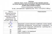
Today, all issues related to the placement of government orders are regulated by the Law on the Contract System -...
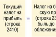
Accounting Regulations Accounting for calculations of income tax of organizations PBU 18/02 (as amended by Orders of the Ministry of Finance of the Russian Federation...

A trainee salesperson is usually called those salespeople who are not yet ready to work completely independently. Process...

Educational institution "Gomel State Medical University" Department of Neurology and Neurosurgery...

One of the most controversial and controversial methods for the early development of children was developed in the 80s by a sociologist...

Contents Dietary supplement based on an extract obtained from the fly beetle (or...

Ekaterina MirimanovaSystem minus 60. RevolutionSystem minus 60 with Ekaterina Mirimanova“System minus 60....
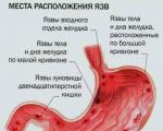
Heaviness and bloating are the causes of both ordinary overeating and more serious digestive problems...

The second blood group, Rh-negative, appeared many years ago, when a person ceased to be...

PSYCHOLOGICAL ASPECTS OF ANOREXIA PHENOMENON (EXPERIMENTAL STUDY) T. V. Tarasova, E. V. Arsentieva...

Contents Since the skin in this area is thin, it is more prone to the appearance of various types of spots....

vseslav Sat, 10/17/2015 - 20:50 Vasileostrovskaya station is one of the oldest stations...

P. S. Pallas (1741 - 1811) - naturalist and traveler-encyclopedist, who glorified his name with major contributions...
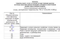
Today, all issues related to the placement of government orders are regulated by the Law on Contract...