Philosophical vision of the problem
The meaning of life is related to the question “What to live for”, and not to the question of how to maintain life. A person's attitude is...
Dogs often suffer from severe nervous system disorders that are difficult to treat, even in humans. Brain pathologies rarely occur without serious consequences that deprive the animal of the opportunity to enjoy a happy life.
Dogs, like people, get sick with a terrible disease - meningitis, which can cause the death of a beloved animal or incurable nervous disorders.
Meningitis is an inflammatory process in the membranes of the brain and spinal cord. In dogs, it is a complication of infectious diseases caused by pathogenic microorganisms, viruses, fungi, protozoa, and sometimes snake or insect venoms. The disease develops when the pathogen penetrates into the cranial cavity or the spinal canal from nearby foci of inflammation during purulent otitis, sinusitis or gingivitis, with an open skull injury or through the hematogenous route - with blood flow from the affected organs.
With meningitis, the pathogen multiplies in the pia mater and arachnoid mater with the formation of fluid between them. As a result of irritation of the meninges, neurological disorders occur, accompanied by depression, severe headaches and loss of coordination. If untreated, the infection penetrates the blood and spreads throughout the body, forming multiple foci of inflammation. A complication may be meningococcal sepsis, in which there is blockage of blood vessels, numerous hemorrhages, a sharp drop in blood pressure and the rapid death of the pet.
Kinds
Meningitis in dogs develops for the following reasons:
Meningitis in dogs is characterized by a clear clinical picture of severe neurological disorders:
If you suspect meningitis, you must immediately take the animal to a veterinary clinic; the outcome of the disease directly depends on the stage of neglect of the pathology and the timeliness of the owner contacting a specialist for appropriate treatment.
In a veterinary clinic, a comprehensive examination is carried out using an electroencephalogram, cerebrospinal fluid puncture, radiographic, ultrasound and laboratory tests of the animal’s blood, urine and feces. Magnetic resonance imaging and computed tomography are considered effective methods for diagnosing meningitis in dogs.
Treatment of meningitis in dogs is aimed at eliminating the causative agent of the disease and alleviating the symptoms of the pathological process.
Symptomatic therapy involves the use of sedative, cardiovascular, analgesic, detoxification, antiepileptic, vitamin and immunomodulatory drugs.
In advanced cases, the disease cannot be completely cured; the dog is prescribed a special diet and hormonal medications for the rest of its life.
Prevention of meningitis is annual vaccination and deworming of the pet, timely treatment of infectious and non-communicable diseases of the animal and regular examinations of the dog by a veterinarian with urine and blood tests.
Meningitis is a very serious pathology; in advanced stages, treatment does not always help, and the pet dies in the arms of the owner. Carefully monitor changes in the behavior of your four-legged friend; timely therapeutic measures begin provide a real chance for a complete cure for your dog.
Inflammatory diseases of the nervous system in dogs
Inflammatory diseases of the nervous system of dogs cover a fairly large group of diseases - meningoencephalitis/meningomyeloencephalitis of various etiologies (causality).
Meninitis is inflammation of the meningeal membranes of the central nervous system, myelitis is inflammation of the spinal cord, encephalitis is inflammation of the parenchyma (tissue itself) of the brain. Meningitis is characterized by inflammation involving the subarachnoid space, that is, inflammation of non-neuronal (containing nerve cells) tissue.
As a rule, one cannot talk about specific meningitis or encephalitis, because both processes occur simultaneously, because inside the skull, the tissues are anatomically located very close to each other, so it is more legitimate to use the term meningoencephalitis.
Meningoencephalitis, regardless of the cause, is not a widespread disease, but in general it makes up a significant percentage of the total number of neurological diseases.
Inflammatory diseases of the central nervous system (meningoencephalomyelitis) according to etiology are divided into infectious and non-infectious.
When infected by fungi or rickettsia, signs of damage to both those and other formations may be observed (diffuse symptoms).
TO non-infectious include steroid-dependent meningitis, granulomatous meningoencephalitis, and several breed-specific meningoencephalitis.
It is assumed that the pathology is based on immunological disorders, since almost all animals respond to treatment with immunosuppressive doses of glucocorticoids.
Granulomatous meningoencephalitis(GME) is a non-purulent inflammatory disease in which limited (focal) or diffuse (multifocal) damage to the central nervous system occurs.
There are 3 forms: limited GME involving the brain stem; disseminated GME, which damages the cerebrum, lower brainstem, cerebellum and cervical spinal cord; Visual GME is characterized by damage to the eyes and optic nerves.
The cause of GME is unknown, but the disease is believed to be immune in nature. Treatment includes administration of glucocorticoids; the prognosis is uncertain, especially in the long term. With rapid progression of the disease, it is always unfavorable.
Beagle pain syndrome - a severe form of steroid-dependent meningitis with polyarthritis causing pain in the cervical spine.
It is assumed that the disease is caused by immune disorders, since steroid therapy leads to complete remission.
Bernese Mountain Dog meningitis- this breed is susceptible to necrotizing vasculitis and polyarteritis (aseptic meningitis). The cause of the disease has not been established, but clinical manifestations disappear in almost all animals when treated with steroids.
Pug meningoencephalitis– a disease of young and middle-aged dogs (9 months – 4 years), characterized, as a rule, by a rapid course and a poor prognosis.
At the onset of the disease, convulsions and a picture of diffuse damage to the central nervous system occur. Characteristic features include circling, ataxia (shaky, uncoordinated gait), “head resting” against the wall, blindness, and pain in the cervical spine.
Therapy with steroids and anticonvulsants does not give good results; animals usually die within a few weeks after the onset of symptoms.
Clinical manifestations of inflammatory diseases of the central nervous system can be varied, depending on which area is affected and how severely; disorders can be limited (focal), diffuse (spread), and quickly develop from limited to diffuse.
Classic signs of meningitis are pain (usually in the neck) and fever. Animals resist being taken on a leash, they exhibit hyperesthesia (increased sensitivity to touch and influence) and rigidity (tone) of the neck muscles. In severe cases, lateral positioning, opisthotonus and hyperextension of the front legs occur.
The nature of the clinical manifestations of encephalitis is due to damage to the brain parenchyma. Violations are usually asymmetrical. The severity of symptoms can increase gradually - impaired consciousness (depression) up to stupor, coma; changes in behavior; visual impairment (while maintaining the normal reaction of the pupils to light - so-called central blindness); impaired coordination of movements and voluntary motor functions; convulsions.
In the presence of encephalomyelitis, sensory ataxia (impaired gait and body position in space, impaired posing reactions), motor dysfunction and dysfunction of the cranial nerves are detected.
A diagnostic method for identifying meningoencephalitis and its cause is cerebrospinal fluid (CSF) analysis. requires general anesthesia, and is associated with a certain risk (both anesthetic and surgical, since a puncture of the occipital cistern is performed). Non-invasive methods (however, also performed under general anesthesia) are CT (computed tomography) and MRI (magnetic resonance imaging), however, on the basis of these studies it is not always possible to accurately make this diagnosis, because changes may be specific to pathologies of different causes (for example, autoimmune, fungal, bacterial cannot be verified).
Therapy depends on the cause - in most cases, the use of steroids in immunosuppressive doses, antibiotics and symptomatic therapy (anticonvulsants, infusion) is indicated. The prognosis depends on the cause, except in cases of steroid-dependent encephalitis, unfortunately the prognosis is guarded to poor.
Encephalitis is an inflammation of the brain. Encephalitis in dogs is divided into meningoencephalitis (inflammation of the brain and its membranes), encephalomyelitis (inflammation of the brain and spinal cord) and meningoencephalomyelitis (inflammation of the brain, spinal cord and membranes).
Encephalitis, depending on the cause, is divided into infectious and immune-mediated.
Bacterial encephalitis can be primary (listeriosis) or secondary (with the spread of infection due to sepsis, otitis media, or as a result of skull injuries).
Viral lesions of the nervous system appear with canine distemper, rabies, parvovirus, and herpesvirus. Such encephalitis occurs with symptoms of the underlying disease and often develops after the first signs of the disease appear.
Fungal encephalitis is caused by Aspergillus, Blastomycetes and Histoplasma. This type of damage to the nervous system is most rarely encountered in the practice of a veterinary neurologist.
The most common autoimmune encephalitis in dogs is granulomatous meningoencephalomyelitis, necrotizing meningoencephalitis, and steroid-dependent meningitis. These diseases are common in dwarf dogs and young toy breeds; they are less common in large dogs and mixed breeds.
With encephalitis in dogs, fever, convulsions develop (often with the development of status epilepticus), behavior changes (lethargy, depression, etc.), damage to the vestibular apparatus is possible (in this case, head tilt, walking in a circle, uncoordinated movements are observed), damage to the cranial - brain nerves (changes in the size of the pupils, paralysis of the facial muscles, drooping eyelids, the appearance of drooling and impaired swallowing, blindness), pain when rotating the neck and/or palpating the spinal column.
To determine the cause of inflammation in the brain, a neurologist:
1. Determines the localization of the pathological process during a neurological examination, during which he evaluates the reaction of the cranial nerves (if they are disturbed, the doctor concludes that the process is localized in the brain) and conducts staged reactions and measures tendon reflexes: if they are absent, weakened or strengthened you can determine the location of the lesion in the spinal column;
2. Conducts a general clinical blood test to detect an increase in leukocytes in case of a bacterial infection or a decrease in lymphocytes in the case of a viral nature of the disease;
3. Conducts a biochemical blood test to differentiate encephalitis from encephalopathies (non-inflammatory diseases of the brain);
4. Conducts x-ray examination to detect foreign objects and gross violations of the integrity of the skull/spinal column;
5. Conducts analysis of cerebrospinal fluid to differentiate infectious encephalitis from immune-mediated encephalitis and determine the causative agent of the disease and select therapy;
6. Conducts magnetic resonance therapy (MRI) to confirm the diagnosis of encephalitis;
Fig 1. MRI scan of a dog with necrotizing meningoencephalomyelitis
7. Conducts electroencephalography (EEG):
To determine the localization of the pathological focus in the brain before MRI;
 Rice. 2. Taking an EEG for a dog with epileptic seizures before an MRI
After diagnosis, to determine the effectiveness of the therapeutic solution;
 Rice. 3. Electroencephalogram of a dog admitted with status epilepticus
8. Conducts PCR/ELISA diagnostics and cultures to identify the pathogen in infectious encephalitis.
Depending on the cause of the encephalitis, treatment will vary.
If the cause of the disease is bacterial, a course of antibiotics is prescribed. They are selected based on the results of cerebrospinal fluid culture and titration (determining the sensitivity of bacteria to antibacterial drugs). Until the results are obtained, broad-spectrum drugs are prescribed, with preference given to cephalosporins and fluoroquinolones.
If encephalitis is caused by a fungus, then antifungal drugs are prescribed, and if the inflammation of the brain is viral, symptomatic therapy and treatment of the underlying disease are used.
When an animal is admitted in a state of status epilepticus, it is immediately placed in an inpatient unit and anticonvulsant therapy is started, intensive measures are taken to relieve seizures and control cerebral edema.
For immune-mediated encephalitis, the mainstay of treatment is corticosteroids. If they do not show sufficient effectiveness, immunosuppressants are additionally prescribed.
There is no universal treatment regimen for encephalitis - each animal requires adjustment of prescriptions depending on the results of examinations.
Therefore, the neurologist prescribes follow-up examinations and additional examinations to determine the timing of therapy. For immune-mediated meningoencephalitis, treatment will often be lifelong and require constant monitoring of the animal by the attending physician. For steroid-dependent meningitis, the prognosis is good and the dog can stop taking the drugs after long-term corticosteroid therapy. Therefore, it is very important to make an accurate diagnosis in time and decide on treatment tactics.
To monitor the course of the disease, repeated EEGs and MRIs, blood tests are sometimes necessary to assess the functioning of the body as a whole and monitor the side effects of drugs. Less often, a repeat examination of the cerebrospinal fluid is required. During therapy with anticonvulsants, the level of drugs in the blood is periodically monitored and the dose of the antiepileptic drug is adjusted.
The prognosis depends on the cause of encephalitis and the severity of the lesion.
Favorable prognosis in animals with infectious encephalitis and limited small lesions that respond well to therapy with antibiotics/antifungals, etc.
For animals with immune-mediated encephalitis, the prognosis is most often guarded. The disease may be treatable and the dog will feel well for a long time, but a sudden rapid progression of the disease with an extremely negative prognosis, even death, cannot be ruled out.
For autoimmune encephalitis, the prognosis depends on the time of initiation of treatment. That is, the sooner the correct diagnosis is made, the greater the likelihood of a favorable outcome of the disease. Therefore, if you notice signs of encephalitis in an animal, immediately consult a neurologist.
Necrotizing meningoencephalitis (NME) is an inflammatory disease of the central nervous system (CNS) that occurs more commonly in young and typically miniature breed dogs. Necrotizing meningoencephalitis has been described in many small dog breeds, including the pug, maltese, and chihuahua.
Without supportive aggressive immunosuppressive treatment, necrotizing meningoencephalitis is quite rapidly fatal and often fatal, even despite adequate therapy. Similar to granulomatous menigoencephalitis, it is difficult to diagnose and only histological analysis of the affected tissue can definitively make a diagnosis.
Clinical picture of necrotizing meningoencephalitis in the Yorkshire terrier and possibilities of supportive treatment
NME is characterized by multiple, often asymmetric, necrotizing, nonexudative meningoencephalitis and is mainly localized in the cortex and subcortical white matter of the brain. Necrotizing menigoencephalitis is identified as a separate nosology. The most commonly affected animals are: Yorkshire Terrier, French Bulldog, Spitz, Miniature Pinscher, Pekingese. Unlike granulomatous meningoencephalitis, when the accumulation of cells in the affected areas prevails, in necrotizing meningoencephalitis the processes of alteration (destruction) prevail. These changes can be detected using research methods such as MRI and CT. Intense inflammation and softening of areas are detected, usually limited to the subcortical white matter of the brain and most often in the region of the brain stem and cerebellum.
For maintenance treatment, immunosuppressive therapy is used. Treatment is basically the same.
Meningoencephalitis- inflammation of the brain and its membranes. The disease occurs with profound dysfunction of the cerebral cortex and subcortical centers.
Dogs of small and dwarf breeds, such as Chihuahuas, toy terriers, Yorkshire terriers, and Spitz, are most predisposed to this disease. Representatives of large breeds can also get sick, but this disease is observed in them much less often.
There are mainly 2 forms of the disease:
As a rule, cerebrospinal fluid analysis helps us determine what type of disease we are faced with. But in most cases, in our practice we are faced with a non-infectious type of meningoencephalitis in dogs.
The clinical picture of the disease directly depends on which part of the brain is affected, as well as on the degree of its damage. As a rule, common manifestations include ataxia, convulsions, changes in consciousness, pain, paresis, paralysis, as well as various vestibular disorders associated with damage to the cerebellum: manipulative movements, head tilt, nystagmus (involuntary movements of the pupils in any direction), etc.
 |  |
|
| Normal brain, sagittal plane, Fig. 1 | Normal brain, axial plane, Fig. 2 |
 |  |
|
| Brain, meningoencephalitis, lesion marked in red, Fig. 3 | Brain, meningoencephalitis, lesion marked in red, Fig. 4 |
Diagnostic procedures usually begin with a neurological examination, the approximate location of the lesions, as well as their extent. It is also advisable to take blood tests, which will allow you to assess the general condition of the animal. Next, a more complex diagnosis is carried out, which includes MRI (the method of choice) or CT of the brain (if MRI is not possible).
An MRI study, aimed in particular at visualizing the organs of the central and peripheral nervous system, will help us determine the location and extent of lesions with an accuracy of several millimeters.
Well, in order to differentiate idiopathic meningoencephalitis from infectious meningoencephalitis, a cerebrospinal fluid analysis is taken.
After the diagnostic measures have been carried out, we will be able to accurately determine the type of disease, its location and extent, in order to prescribe an individual treatment for your animal.
The article has been prepared
veterinary neurologist "MEDVET"
© 2015 SEC "MEDVET"

The meaning of life is related to the question “What to live for”, and not to the question of how to maintain life. A person's attitude is...
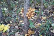
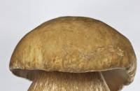
A mushroom is a living organism that forms a separate kingdom of the same name. For a long time they were classified as part of the plant kingdom. But in...
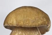
For lovers of “quiet” hunting, mushroom season begins in early summer and lasts until late autumn. And rarely do they...
Aleshnikova, V.I. Use of professional consultants. - M.: Infra-M, 1999. - 240 p. 2. Beich, E....

Orange juice. The symbolic meaning of orange juice in dream books is pleasure and temptation. Quite often we...

We most often overcome all kinds of difficulties encountered along the path of life. Of course, for this we make...

This year your patron Neptune will be in your constellation and this is a good sign, because you will...

1993 who? 1993 is the year of which animal? — According to the Chinese horoscope, 1873, 1933, 1993 belonged to the years of the Black...

Wave-particle duality of light means that light simultaneously has the properties of continuous...

The role of biology is enormous in our world. Although it is not one of the priority subjects, most schoolchildren and...

Amines are organic derivatives of ammonia containing an NH 2 amino group and an organic radical. In general...

How to answer questions in Part BThe second part of the social studies work consists of 7 tasks with a short answer....

Form TORG-15 is drawn up in the case when during transportation, movement between and within the warehouse, when...

Nutritionists say that for good health and a slim figure, you must include snacks in your...

A mushroom is a living organism that forms a separate kingdom of the same name. For a long time they were classified as a kingdom...
For lovers of “quiet” hunting, mushroom season begins in early summer and lasts until late autumn. And rarely do they...