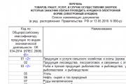
Procedure for filling out an income tax return
An example of a correct income tax return in 2017, download a new current form for free in Excel. What...
23-11-2012, 14:24
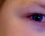
Eye damage due to syphilis . Parenchymal syphilitic keratitis can be congenital and acquired. The disease is rare and more often develops with congenital syphilis.
Parenchymal keratitis occurs in children and adults, but is especially common between the ages of six and twenty years. There are known cases of typical parenchymal keratitis occurring in early childhood and adulthood. The syphilitic etiology of the disease is confirmed by serological reactions. Positive Wasserman reaction observed in 80-100% of cases.
Proof that parenchymal keratitis is a consequence of congenital syphilis is a number of characteristic symptoms:
Pathogenesis this disease is quite complex. The main link in the pathogenesis of syphilitic inflammation is vasculitis, and there are no vessels in the cornea. It is now generally accepted that parenchymal keratitis in the fetus and newborn is caused by spirochetes that penetrate the cornea and the period of intrauterine development, when there were vessels in it. Another pathogenesis of late congenital stromal keratitis, which develops in the absence of blood vessels, is an allergic reaction of the cornea in an organism affected by syphilis.
At the end of the period of intrauterine development, when the vessels are reduced, sensitization of the corneal tissue to the decay products of spirochetes occurs. As a result, in the first two decades of life, when congenital syphilis is activated, when the concentration of spirochete breakdown products in the blood is increased, any provoking factor (trauma, colds) leads to the development of an anaphylactic reaction in the cornea. There is other evidence that syphilitic keratitis is caused by a special form of filterable spirochetes.
Factors favoring the development of parenchymal keratitis include general diseases, endocrine disorders and eye injuries. Acquired syphilis is rarely the cause of parenchymal keratitis.
Deep punctate keratitis, pustular deep Fuchs keratitis and gumma cornea are very rare.
Inflammatory process begins with the appearance of subtle pinpoint lesions in the peripheral part of the cornea, often in the upper sector. Subjective symptoms and pericorneal vascular injection are mild. The number of infiltrates gradually increases, they can occupy the entire cornea. On external examination, the cornea appears diffusely cloudy, resembling frosted glass. Bnomncroscopy shows that the infiltrates are deep and have unequal shapes (dots, spots, stripes). Located in different layers, they overlap each other, resulting in the impression of diffuse turbidity. The surface layers, as a rule, are not damaged, and epithelial defects are not formed. An optical section of the cornea can be doubled in thickness. The clinical picture is very diverse. During the most commonly brewed typical form, three periods are distinguished: infiltration, vascularization and resorption.
In the first period(progressive period or infiltration period), which lasts 3-4 weeks, moderate pericorneal injection and mild irritation phenomena (lacrimation, photophobia, minor pain) are observed. A grayish-white infiltrate appears in the corneal stroma. When examining it through a magnifying glass, it can be noted that it consists of individual point infiltrates located in the middle and deep layers of the cornea. Together they, located one above the other, create the impression of diffuse infiltration. Infiltration most often begins from the upper part of the cornea and slowly spreads down into its transparent part. At the same time, the corneal epithelium is also involved in the process; it becomes dull and uneven. Near the limbus, the opacities resolve, but the number of vessels leading to new foci in the center increases. By the end of this period, the entire cornea is penetrated by a dense network of deep vessels. In this case, superficial neovascularization may also occur. Sometimes “deep” newly formed vessels appear.
The second period is the period of vascularization, which lasts on average 6-8 weeks. Pain, photophobia, lacrimation intensify, the intensity of clouding of the cornea increases, sometimes it all becomes cloudy and dull, like frosted glass. Deep vessels in the form of panicles or brushes grow into the thickness of the cornea from the upper limbus from the sclera and episclera. The number of newly formed vessels can be very large, so that the entire vascularized cornea resembles a ripe cherry. In other cases, there are few vessels, sometimes they may be completely absent. In this case, superficial neovascularization may also occur.
In parenchymal keratitis, infiltration and newly formed vessels are usually located at a certain level between the layers of the corneal stroma, without passing from one layer to another.
Course of parenchymal keratitis often (up to 50%) complicated by inflammation of the iris, cyclitis is often observed. Symptoms of iridocyclitis are as follows: pericorneal injection of blood vessels increases, the pattern of the iris becomes blurred, the pupil contracts, and precipitates appear that are difficult to see behind the shadow of corneal infiltration. The progression of the disease lasts 2-3 months, then the third stage begins - the period of regression, or the period of resorption of opacities, the duration of which is 1-2 years. During this period, the phenomena of irritation decrease, the infiltrates resolve, and the resorption occurs in the same order as the infiltrates developed, i.e., the upper part of the cornea clears first, and later its center. The newly formed vessels gradually become hollow. The resorption process is slow. It takes 4-6 months, and in severe cases - a year or more, until the cornea clears up.
In addition to the typical picture of diffuse parenchymal keratitis, atypical forms (central, ring and avascular keratitis) are very rarely observed.
Parenchymal keratitis is characterized by cyclical course and damage to the second eye. The disease of the second eye rarely begins at the same time; usually the second eye becomes ill when the process in the first eye reaches its maximum development.
In recent years, avascular keratitis with a tendency to relapse has become more common. The disease can recur after various periods.
Complications of parenchymal keratitis include iridocyclitis, sometimes with hypertension, and anterior chorioretinitis.
In patients with parenchymal syphilitic keratitis, an active inflammatory process is detected in the optic nerve and retina, which can adversely affect vision.
Forecast with parenchymal keratitis, it is serious, since the infiltrate does not always completely resolve. As a result of the disease, cicatricial opacities of the cornea can form, leading to decreased vision and even blindness.
In most cases, the outcome is parenchymal keratitis vision is restored. In approximately 25% of patients, vision is restored completely, in 50% it remains at least 0.5, in 15% it is not less than 0.1, and only 10% of patients have vision below 0.1, because the corneal stroma during this process is not destroyed, and the infiltrate resolves.
But still, after suffering parenchymal keratitis, traces of deserted and separately semi-deserted vessels, foci of atrophy in the iris and choroid will remain in the corneal stroma for the rest of your life.
If parenchymal keratitis is detected in a child, consultation with a venereologist is necessary not only for the child, but also for members of his family.
Parenchymal keratitis in acquired syphilis . The disease develops extremely rarely and is unilateral with mild symptoms. Corneal vascularization and iritis are usually absent. The recovery process may subside without leaving any traces. Differential diagnosis is carried out with diffuse tuberculous keratitis.
Gummy keratitis - This is a focal form of inflammation, rarely observed in acquired syphilis. Gumma is always located in deep layers. The process is complicated by iritis or iridocyclitis. When the lesion disintegrates, a corneal ulcer may form. This form of keratitis must be differentiated from deep focal tuberculous keratitis.
Treatment carried out jointly by a venereologist and an ophthalmologist, since the main disease and cause of keratitis is syphilis.
Syphilitic treatment does not prevent the development of parenchymal keratitis in the second eye, but significantly reduces the frequency of relapses. Patients are prescribed penicillin, bicillin, povarsenol, miarsenol, bitoquinol, osarsol, iodine preparations according to available regimens, desensitizing and vitamin preparations.
Local treatment is aimed at resolving infiltrates in the cornea, preventing iridocyclitis and random corneal erosions. To prevent the development of iridocyclitis, instillation of mydriatics is prescribed once a day or every other day under the control of pupil dilation. If iritis occurs, the number of instillations is increased to 4-6 times a day (atropine sulfate solution). If adhesions have formed and the pupil has not dilated, electrophoresis with atropine is used. Corticosteroids (dexazone, dexamethasone) in the form of subconjunctival injections and installations provide a good therapeutic effect. Due to the fact that treatment is carried out over a long period of time (1-2 years), it is necessary to alternate drugs within the same group of drugs and periodically discontinue them. The administration of mydriatics must also be stopped for several days. If the pupil has narrowed, it is dilated again. This procedure is called gymnastics of the iris. It prevents the fusion of the immobilized wide pupil with the lens. During the regression period, drops and ointments are prescribed to improve trophism and prevent the formation of corneal erosions. In order to resolve corneal opacities, it is advisable to use phonophoresis of lidase and aloe. If, two years after active treatment, there remains opacification in the cornea that reduces vision by less than 0.1, then keratoplasty can be performed.
Tuberculous keratitis . Tuberculous keratitis is divided into two groups: hematogenous keratitis, which arises from bacterial metastases into the corneal tissue, and allergic-tuberculous keratitis, in which the corneal process is only a paraspecific allergic reaction of the sensitizing tissue.
Hematogenous tuberculous keratitis always arises secondary to bacterial metastasis of the anterior vascular tract. The mechanism of involvement of the cornea in the process may be different. Play an important role precipitates, around which corneal infiltrates form. In addition, the process can directly pass into the cornea from the ciliary body and iris. In the clinic (there are three forms of metastatic tuberculous keratitis: deep limited, deep diffuse keratitis, sclerosing keratitis.
Deep limited keratitis . With this form of keratitis, limited, deep-lying infiltrates are located in the transparent stroma of the cornea or among diffuse opacification. It is characteristic that these infiltrates, accompanied by moderate vascularization, are localized in the most posterior layers of the cornea, directly at Descemet's membrane. Often, along with deep corneal infiltrates, superficial ones are also observed. Irritation of the iris and ciliary body is often noted (constriction of the pupil, deposition of precipitates on the posterior surface of the cornea). Vascularization is insignificant. Newly formed vessels grow in the form of a horn towards the source of inflammation and have an unusual appearance for deep vessels - they branch. The course of the disease is long, and relapses may occur. Healing of focal keratitis is accompanied by the formation of a cataract.
Diffuse tuberculous keratitis . The clinical picture of diffuse tuberculous keratitis is varied. Most often, with this form of keratitis, among diffuse opacities, large yellowish-gray infiltrates are found, lying in the middle and deep layers of the cornea. Infiltrates are located in isolation, without a tendency to merge. The process almost never occupies the entire cornea. The deep vessels located at the posterior surface of the cornea do not have | rectilinear, twist and branch. Along with deep vessels, there are almost always superficial ones. One eye is affected. The course of the disease is long, with periodic exacerbations. The inflammation ends with the formation of a rough vascularized cataract, the elimination of which requires surgical treatment.
Focal and diffuse hematogenous tuberculous keratitis almost always complicated by iridocyclitis. Metastatic keratitis, both focal and diffuse, parenchymal, is accompanied by neovascularization of the cornea due to the ingrowth into it of deeply located branches of the anterior ciliary vessels, which are embedded in the tissue in the form of a narrow capillary loop. In contrast to the superficial wide-loop vascularization characteristic of allergic tuberculous keratitis, these vessels are visible only in the corneal tissue itself, but not in the conjunctiva. They are distinguished by dichotomous branching and are located in different levels of the stroma, the transition from one layer to another. This is due to caseous tissue necrosis in the infiltration zone, as a result of which the cornea loses its density and properties of the horn with the ability to resist this type of neovascularization. When differentiating from parenchymal syphilitic keratitis, it is taken into account that in the latter case the vessels do not actively branch and are located in one layer of the cornea.
The outcome of metastatic keratitis, especially often recurrent, remains persistent vascularized stromal opacification in different, but mainly middle and deep layers of the cornea.
Sclerosing tuberculous keratitis develops simultaneously with inflammation of the sclera. The sclera near the limbus and the limbus itself swell. From this area towards the center of the cornea, the infiltrate slowly spreads, localized in the deep layers and shaped like a rounded triangle. The base of the triangle at the limbus is intensely turbid, towards the apex the infiltrate gradually becomes more transparent. Vascularization is weak, the epithelium above the infiltrate is swollen in the form of small blisters, but no ulceration is observed. Often, new infiltrates appear in neighboring areas of the limbus, moving toward the center in the form of tongues. Course of the disease long-term, with variable exacerbation and subsidence of the process. Gradually, the phenomena of irritation subside, the infiltrate slowly resolves, but its base is mixed with connective tissue, leaving behind intense turbidity. Damage to the vascular tract either precedes tuberculous keratitis or occurs as the process progresses. Sclerosing keratitis is always accompanied by iritis or iridocyclitis, and is often complicated by secondary glaucoma. Sclerosing keratitis can occur not only with tuberculosis, but also with syphilis, rheumatism and gout.
Etiological diagnosis tuberculous keratitis causes significant difficulties. A characteristic clinical picture should be noted: the presence of more or less active tuberculous changes in the lungs or bronchopulmonary lymph nodes, a characteristic temperature curve, a positive Mantoux test. All this makes it possible to establish the tuberculous etiology of keratitis. Only etiological stenosis can help reduce the duration of treatment and prevent relapses of the disease. The faster the inflammatory process in the eye stops, the fewer complications will arise during the course of the disease and the greater the hope for preserving vision.
Tuberculous-allergic keratitis - a frequently occurring form of tuberculous lesions of the cornea in children and adults. Most patients are children and adolescents.
Allergic phlycticulous keratitis is a tuberculous-allergic eye disease, similar to phlyctic conjunctivitis.
Small infiltrates appear in the superficial layers of the cornea - conflicts. Phlyctena is a nodule consisting mostly of lymphocytes, but often in its center there are epithelial cells, and sometimes giant ones.
These nodules are very similar to a tuberculous tubercle. However, Mycobacterium tuberculosis is never found in them and there is no curdled disintegration. Nodules develop in any part of the cornea (usually at the limbus) and have the appearance of grayish translucent elevations. Their number and size are different: from barely noticeable single (miliary) to large single (solitary) conflicts. The larger the conflicts, the fewer there are. A bundle of superficial dilated vessels usually approaches the conflicts.
Conflicts always appear accompanied by severe eye irritation, such as basal injection around the entire cornea or only according to the position of conflicts, profuse lacrimation, severe photophobia. Patients hide their faces from the light, their eyelids are convulsively compressed (blepharospasm), their skin is macerated. Very painful cracks often form in the corners of the eyelids, which causes children to squeeze their eyelids even more tightly. Spasm of the eyelids impedes blood and lymph circulation in them, the eyelids swell and become cyanotic. The facial skin of scrofulous patients is pasty. Bloody crusts are often visible on the nasal mucosa.
Phlyctenes can be resorbed without leaving a trace, but they can ulcerate, then after their healing a thin cloudiness remains in the form of a cloud and a spot.
In conflicts it is often observed iris irritation. If pathogenic microbes enter the defect area, a purulent ulcer may develop, which tends to go deeper into the corneal tissue and sometimes reach Descemet's membrane. Under the influence of intraocular pressure, which increases with sharp compression of the eyelids, Descemet's membrane breaks through, and perforation of the ulcer occurs. When the anterior chamber is emptied, especially with rapid loss of moisture, the iris may fall out and become pinched into the corneal defect. After perforation, scarring begins, resulting in the formation of a corneal cataract, fused with the iris.
Especially often, marginal (limbal) conflicts take this course in weakened children.
Wandering phlyctena, tufted keratitis . The phlyctena, located at the limbus, begins to slowly spread along the surface of the cornea towards the center, followed by a bundle of blood vessels.
Its edge, facing the limb, is cleaned. The edge facing the center of the cornea remains infiltrated and raised above the rest of the surface. In the development of such a conflict, after healing, cloudiness remains.
Allergic tuberculosis of the cornea can develop according to the so-called scrofulous pannus. In this case, the cornea in a certain area of greater or lesser extent is diffusely cloudy and penetrated by superficial vessels. In appearance, scrofulous pannus is similar to trachomatous pannus, but trachomatous pannus is necessarily localized in the upper part of the cornea, and scrofulous pannus occurs in various parts of the limbus.
The main difference between scrofulous pannus is absence of conjunctival changes typical for trachoma. Scrofulous pannus is warmed up with frequent relapses of phlyctenular keratitis.
Scrofulous-allergic eye diseases are observed mainly in children and young people suffering from tuberculosis of the cervical, submandibular, bronchial lymph nodes or pulmonary tuberculosis in the active period.
The basis of the disease is severe tissue allergy, and any damage to the conjunctiva and cornea (trauma, conjunctivitis) can serve as a resolving factor (Arthus phenomenon) contributing to the development of a specific inflammatory focus.
Much less often, conflicts can appear as a result of a reaction of an allergic cornea or conjunctiva to a nonspecific, i.e., non-tuberculous, irritation.
Finally, conflicts can sometimes develop as allergic keratitis in people who do not suffer from tuberculosis.
Prognosis for scrofulous disease is always placed carefully: firstly, due to the tendency to relapse over many years; secondly, because of the cloudiness that remains after conflict. If the corneal phlyctena is located opposite the pupil, then the remaining cloudiness can lead to a persistent decrease in visual acuity.
This disease is chronic, relapsing. Upon reaching puberty, relapses in most cases stop. However, people who suffered from this disease in childhood may develop conflicts at a later age. Relapses occur in the spring months when the reactivity and degree of sensitization of the body are increased, which is apparently due to a lack of solar radiation and dietary habits in winter and spring. For the increase in allergic diseases in the spring, a lack of vitamins in food, especially A and C, is known to be important.
Along with these reasons, the increased incidence of phlyctenulosis in early spring plays a role deterioration of general hygienic conditions in winter(insufficient exposure to air and lack of sunlight).
Treatment. The choice of the general treatment regimen for tuberculosis is carried out by a phthisiatrician. It determines a rational scheme for combining first- and second-line drugs, the duration of the course of treatment, the timing of the second course, taking into account the immune status of the patients, diet and the need for climatotherapy.
Goal of local treatment- suppression of the inflammatory process in the eye, prevention of the formation of posterior synechiae, improvement of metabolism in the corneal tissue. In the form of instillations, a 3% solution of tubazide, a 5% solution of saluzide, streptomycin-calcium chloride complex (50,000 units in 1 ml of distilled water), hydrocortisone or dexamethasone are prescribed. Mydriatics are used to prevent or treat iritis and iridocyclitis. The frequency of instillations is determined depending on the stage of the inflammatory process. At night, 5-10% PASK ointment or vitamin ointments, 20% Actovegin gel are placed behind the eyelid. Dexazone is administered under the conjunctiva, alternating it with a 5% solution of saluzide, every other day or with another frequency during different periods of treatment. In the scarring stage, the doses of anti-inflammatory drugs are reduced, physiotherapy is carried out, vitamin preparations and enzymes (trypsin, fibrolysin) are used to resolve adhesions.
In the treatment of tuberculous-allergic keratitis, they are of great importance desensitization therapy, diet with limited consumption of carbohydrates and table salt, climate therapy.
Leprosy keratitis . Damage to the cornea in patients with leprosy was observed very often in previous years - in 72.6% of cases. Currently, there is a decrease in the frequency of leprosy keratitis and a more benign course. The cornea is affected in all types of leprosy, most often in the lepromatous form. In lepromatous, tuberculous and borderline forms of leprosy, keratitis is specific; in subdifferentiated forms, it is nonspecific, as it develops as a result of lagophthalmos. Specific keratitis is usually bilateral. The appearance of inflammatory infiltration in the cornea is preceded by a change in the pain and tactile sensitivity of the cornea, which is determined primarily in its peripheral parts. In the central part of the cornea, normal sensitivity remains much longer. Hypo- and anesthesia of the cornea are caused by dystrophic changes in the trigeminal nerve. During biomicroscopy, predominantly at the limbus in the upper outer segments, distinct thickenings of the corneal nerves in the form of shiny nodules. These limited thickenings of the corneal nerves are characteristic of leprosy eye lesions. A typological study reveals perineural infiltration in them.
Specific keratitis can be diffuse and nodular. A more severe course is observed with nodular keratitis. With diffuse inflammation of the cornea, sclerosing or diffuse vascular inflammation develops, and with limited inflammation, punctate or subspinal keratitis develops.
With sclerosing keratitis near focal infiltration of the sclera at the limbus, clouding of the deep layers of the cornea. In the area of turbidity, focal hypo- or anesthesia and a few newly formed vessels are noted. Foci of deep corneal infiltration never ulcerate. The course of the disease is areactive, chronic, with periodic exacerbations, accompanied by the appearance of new foci of opacification in the deep layers of the cornea.
For diffuse vascular keratitis the process usually begins in the upper third of the cornea and gradually spreads to most of it. In the deep layers of the cornea, diffuse inflammatory infiltration and a significant number of newly formed vessels are observed. Leprous pannus of the cornea differs from trachomatous pannus in the deep location of newly formed vessels. The corneal infiltrate in diffuse vascular keratitis never ulcerates. The sensitivity of the cornea is reduced or completely absent. The course of the disease is areactive, chronic, with periodic exacerbations.
For punctate leprosy keratitis Usually, in the upper third of the cornea, pinpoint infiltrates are found, located mainly in the middle layers, corresponding to the location of the thickened nerves of the cornea. The development of newly formed vessels is not observed. Histological studies indicate that pinpoint infiltrates of the cornea are miliary lepromas. The course of the disease is areactive, chronic, relapsing.
Subsular leprosy keratitis - the most severe, acute form of specific keratitis. It is observed during the development of leprosy reactions, i.e. during exacerbation of the disease. Typically, in the area of the upper limbus, lepromas of dense consistency appear, fused with the bulbar conjunctiva. The inflammatory process progresses, spreading to most of the corneal stroma, iris and ciliary body tissue. Leukomas remain in place of corneal lepromas that have healed. In severe cases, the inflammatory process spreads to all the membranes of the eyeball, resulting in its atrophy. The disease progresses with periodic exacerbations.
In the undifferentiated type of leprosy, damage to the linden and trigeminal nerves leads to the development lagophthalmos, anesthesia and impaired trophism of the cornea. Infiltrates are located in the superficial layers of the cornea. The epithelium covering them is often torn off, and corneal erosion is formed. Keratitis of this type is non-reactive, has a chronic form, and periodically worsens. Due to disturbances in the trophism of the cornea, dystrophic keratitis such as band-shaped, circular, and bullous can also be observed.
Thus, keratitis, which is the most common clinical form of ocular leprosy, occurs predominantly in an unresponsive, chronic manner, with periodic exacerbations. The types of leprosy keratitis described above are not strictly isolated clinical entities, since depending on the development trend of the leprosy process transitions from one form of keratitis to another are possible.
A clinical feature of specific keratitis in patients with leprosy is their frequent combination with damage to the iris and ciliary body. Exacerbations of leprosy keratitis, as a rule, coincide with exacerbations of the general leprosy process. The specific etiology of keratitis is confirmed by the detection of Mycobacterium leprosy in the cornea during bacterioscopic and histological studies.
Brucellosis keratitis . There are superficial and deep keratitis. Superficial (coin-shaped) keratitis is characterized by the appearance of yellowish infiltrates located over the entire surface of the cornea. With timely treatment, infiltrates can completely resolve or undergo decay and ulceration due to secondary infection. Deep brucellosis keratitis is often unilateral, has a recurrent course with the localization of the main focus in the center, influx of Descemet's membrane folds, and precipitates. Initially, the process is avascular, then slight vascularization occurs. Changes in the cornea in brucellosis do not have any specific pattern, and the diagnosis is established using serological tests.
Rheumatic lesion of the cornea. Sclerosing keratitis - a rare but serious eye disease, one of the causes of which may be rheumatic disease. The process is predominantly bilateral, occurs more often in elderly and old people, begins with limited swelling of the sclera or with annular scleritis, in which inflammatory mifiltration of the sclera in a ring surrounds the cornea. The infiltrate forms a gelatinous shaft up to 1 cm wide, starting with a steep rise, slightly retreating from the limbus to the arches of the conjunctival sac. The conjunctiva covering the shaft is pale or slightly hyperemic, the eye is moderately irritated. Massive infiltration of predominantly deep corneal layers often spreads from the shaft into the cornea in separate wide “tongues”. The initially grayish areas of infiltration eventually turn white and become similar to sclera. This impression is reinforced by the piercing opacity and isolated blood vessels. Sometimes infiltrations and subsequent opacities surround all or most of the periphery of the cornea, but the center remains transparent and appears in the opacification ring like a microcorneal contact lens, so this pattern is called a “contact lens” cornea. Patients are worried pain in the eyes, moderate photophobia, lacrimation, blurred vision due to reactive edema of the epithelium of the center of the cornea. A similar, but more severe lesion of the sclera, complicated by iridocyclitis, retinal detachment, and sometimes secondary glaucoma, was previously described under the name “progressive scleroperikeratitis”. In cases of scleral swelling in a limited area (or areas) in the cornea, one, sometimes 2 - 3 deep limbal infiltrates with individual vessels, less often - without them, appear. Turning into persistent opacities, their “tongues” make the contour of the cornea scalloped.
No matter how rheumatic sclerosing keratitis begins, it is usually accompanied by varying degrees of anterior uveitis or panuveitis, occurs cyclically, with long-term remissions and exacerbations, often associated with colds, tonsillitis, etc.
Viral keratitis . Herpetic keratitis ranks first in the frequency of diseases of the cornea and occurs in two types: herpes simplex and herpes zoster (shingles).
Herpes simplex characterized by a varied clinical picture with the formation of a group of small transparent epithelial vesicles in combination with subepithelial tender infilphates. The rash of blisters is accompanied by severe symptoms of irritation: photophobia, lacrimation, blepharospasm, pericorneal injection and especially severe pain.
The blisters usually burst quickly, and in their place superficial, very sluggishly healing ulcers with a soft gray bottom are formed, sometimes enlarging due to new rashes along their edges. Keratitis goes away without a trace or leaves behind a gentle cloud-like cloudiness.
Type of herpes - dendritic keratitis, accompanied by significant symptoms of irritation. In this case, infiltrates in the cornea are located in the form of tree-like branches. On the surface of the infiltrates, thin grooves are visible, limited by gray, slightly raised edges. There are often small button-like extensions at the ends of the branches. This form of herpetic keratitis, more persistent in its course, requires long-term treatment and will leave behind intense opacities.
The prognosis for dendritic keratitis should be made with caution. Even in the most favorable case, the long course of the disease must be taken into account.
Sometimes dendritic keratitis can develop into another form of herpetic keratitis. In this case, corneal infiltration is diffuse character with various outlines; the epithelium is swollen in places in the form of separate blisters, and in places it is desquamated. The blisters quickly burst, ulcers form, which, in severe cases, merge together and form one long-lasting ulcer with an undermined progressive edge directed towards the center of the cornea. In the future, against the background of scar opacification, fresh areas of infiltration may appear. At the same time, branched infiltrates may appear and bubbles may appear along the edge of the infiltrate or ulcer. This form of corneal herpes is the most severe, lasting several months, during which exacerbations are followed by remissions. In some cases, the corneal process is accompanied by phenomena of irritation of the iris and ciliary body.
For all forms of herpes corneal sensitivity is sharply reduced, and sometimes completely absent. At the same time, there is a decrease in sensitivity in the unaffected eye.
Etiology. Herpetic lesions of the cornea are caused by the herpes virus. There are many strains of this virus. The herpes virus is neurotropic. The virus, having entered the body, remains in it, forming intranuclear pegs in the epithelium, surrounded by a transparent membrane. Corneal herpes appears simultaneously with herpes of the lips and nose, but is often limited to the cornea only. Herpes usually occurs during a febrile state, which is accompanied by a wide variety of acute and chronic infections, such as influenza, seasonal catarrh of the upper respiratory tract, pneumonia and other infectious diseases. In addition, it can develop due to diseases of the paranasal cavities and teeth, or minor trauma to the cornea. The share of all acute and chronic infections in the occurrence of corneal herpes varies depending on the time of year, area, and outbreak of a particular infection.
Herpetic lesions of the cornea occur most often with influenza, both in the acute period and in the recovery period. They become more frequent during an influenza epidemic.
Herpetic keratitis usually recurs. In case of relapse, infiltrates are localized in the healed lesion.
Herpes zoster (shingles) observed when the first branch of the trigeminal nerve is affected.
The cornea is affected only if the process involves nasociliary nerve. The eruption of vesicles is preceded by neurological pain in the area of the branching of the first branch. Then, usually simultaneously with the rash of blisters on the eyelid and skin of the forehead, blisters appear on the cornea. In this case, the blisters are large, and the infiltrates involve deeper layers of the cornea than with herpes simplex. After healing, more intense opacities remain. Sensitivity of the cornea is always absent. The iris and ciliary body are often affected.
Discoid keratitis is a herpetic lesion of the deep layers of the cornea. Sometimes it develops from dendritic keratitis, and sometimes it occurs after minor damage to the cornea. In these cases, the epithelium is quickly restored, and a gray, disc-shaped infiltrate appears in the central part of the cornea, delimited from the transparent part of the cornea by a sharply outlined gray line. Sometimes discoid keratitis is one of the forms of the degenerative process. In the center of the disk there is usually an intense gray spot. The infiltrate covers the entire layer of the cornea's own tissue up to Descemet's membrane. The epithelium over it is swollen in the form of tiny bubbles, sometimes folds of Descemet's membrane appear. Iridocyclitis with precipitates on the posterior surface of the cornea is often observed, mainly corresponding to the infiltrate. The phenomena of irritation are usually mild and observed only at the beginning of the disease; later they are absent. With this disease, there is an almost complete absence of tactile sensitivity of the cornea. Over time, deep vessels appear in the cornea. The infiltrate never disintegrates, and no epithelial defect is observed.
The course of the disease is long, often several months. In this case, persistent intense clouding almost always remains, sharply reducing visual acuity.
Treatment of herpetic keratitis complex and long-lasting. It is aimed at suppressing the vital activity of the virus, improving trophic processes in the cornea, accelerating the epithelization of defects, and increasing local and general immunity.
Antiviral treatment includes chemotherapy, nonspecific and specific immunotherapy. At different stages of the disease, appropriate combinations of drugs are used. At the beginning of the disease, frequent instillations of kerecid, deoxyribonuclease are made daily, ointments with tebrofen, florenal, bonafton, oxolin are applied, and Zovirax is prescribed 3-4 times a day. The medications are changed every 5-10 days. Acyclovir is taken orally for 10 days. If the eye disease is combined with herpetic inflammation of another location, then the duration of the course of treatment is increased to 1-2 months. In case of severe complications, intravenous infusions of acyclovir are administered every 8 hours for 3-5 days. This is a highly effective drug, but has a narrow spectrum of action, so it is used against herpes simplex and herpes zoster viruses.
Used simultaneously with chemotherapy agents of nonspecific antiviral action I (interferon in stones and subconjunctivally), as well as drugs that stimulate the production of endogenous interferon (interferonogens), preventing the multiplication of the virus in the cell, for example poludan (course dose - 2000 units), pyrogenal in antipyrogenic doses, proligiosan (3-5 intramuscular injections) . In case of decreased immunity, chronic and relapsing course of the disease, immunostimulants are prescribed: levomisol, thymalin. For specific immunotherapy, human immunoglobulin and antiherpetic vaccine are used.
For cleaning ulcerated surfaces for necrotic masses, a cryoprobe or laser coagulation is used. For long-term non-healing herpetic keratitis, a cornea transplant is performed for therapeutic purposes.
In cases where bacterial flora is involved, sulfonamide drugs, antibiotics, and non-steroidal anti-inflammatory drugs in the form of drops and ointments are additionally prescribed. For the prevention and treatment of concomitant iritis and iridocyclitis, mydriatics are used
In addition to the main antiviral treatment, prescribe vitamins and drugs that improve corneal trophism, and if necessary, use antiallergic drugs.
Corticosteroid drugs They are used with great caution only in the regression stage of deep stromal keratitis, under constant monitoring of the condition of the cornea and intraocular pressure, since they can complicate the course of herpetic keratitis, and in the interictal period they contribute to the occurrence of relapses due to a pronounced immunosuppressive effect.
After the end of anti-inflammatory and symptomatic treatment of keratitis, when the eye has completely calmed down, restorative treatment is required - corneal transplantation for optical purposes.
Keratitis with chickenpox, measles, rubella e. The varicella zoster virus belongs to the group of herpetic viruses, it is an analogue of the causative agent of herpes zoster. Against the background of a sharp increase in body temperature, a macular vesicular rash appears, in particular on the face and eyelids. This is accompanied by photophobia, lacrimation, and conjunctival hyperemia, on which blisters may also appear. The discharge from the conjunctival cavity is mucous, subsequently containing elements of pus.
Emerging keratitis often has a superficial, point-like character. But there may be a creeping ulcer with hypopyon, and perforation often occurs. The prognosis is poor and is done during the period of pustule eruption. Treatment boils down to the administration of globulin by injection, smearing the elements of the rash with brilliant green, washing the eyes with tea infusion, instilling interferon, a 20% solution of albucid, and placing 1% erythromycin or tetracycline ointment behind the eyelids at night.
Keratitis with measles is caused by a pathogen related to paramyxoviruses, which are transmitted by airborne droplets through the lymphoid tissue of the nasopharyngeal ring. The clinical picture of conjunctivitis, sometimes with severe photophobia, blepharospasm and swelling of the eyelids, is complemented by epithelial keratitis with the presence of corneal erosions. When the body's defenses are weakened, a secondary infection may occur. Against the background of proper treatment (β-globulin in injections and drops, interferon, vitamin and desensitizing drugs), the general and local processes are completed and favorably. Otherwise, deep keratitis, corneal ulceration, and iridocyclitis may develop, resulting in severe clouding of the cornea with decreased vision.
For rubella simultaneously with general clinical manifestations, catarrhal conjunctivitis and superficial keratitis occur, requiring only symptomatic treatment and interferon.
For malaria keratitis does not have specific features, it occurs in the form of herpetic keratitis, since high temperature promotes the activation of the herpes simplex virus. There are superficial keratitis, interstitial (infiltration of nerve endings along the buttocks - “tree branches”) and deep. The infiltrate can be purulent or in the form of hypopyon.
Corneal damage in epidemic hemorrhagic conjunctivitis caused by an RNA virus (enterovirus). Characterized by high contagiousness (100% in the absence of preventive measures). The onset is acute: severe pain in the eyes, inability to look at the light. Hemorrhages in the tissue of the conjunctiva and under the conjunctiva, individual follicles are typical.
Keratitis in epidemic hemorrhagic conjunctivitis - superficial epithelial localization, small infiltrates. After a few days it disappears without a trace. The sensitivity of the cornea decreases.
With adenoviral conjunctivitis, the onset is less acute, there is no such acute pain, pain in the eyes. Marked injection and swelling in the area of the transitional fold are noted. A large number of small gray follicles. Serous-mucous discharge.
Keratitis occurs 2-3 weeks after the onset of the disease, infiltrates appear at the limbus, spread to the center, and are localized in the superficial layers of the stroma under the epithelium. Infiltrates are round, coin-shaped, in quantities from 2 to 100, and take a long time to resolve - from one month to a year.
In rare cases, adenoviral conjunctivitis begins with damage to the cornea.
Laboratory diagnostics includes methods such as:
Comprehensive treatment:
Drugs.
Synonyms: keridid, detsdrid, herplex, emanil, iduridine - 0.1% aqueous solution. Drops are more effective for superficial keratitis, but ineffective for deep herpes. Resistance to this drug easily develops, so use should not exceed 10 days.
In addition, it delays the regeneration of the epithelium and has a toxic effect on the epithelium, especially in combination with dexazone. Unstable, quickly destroyed at room temperature. Drops are used once a day for herpes, herpes zoster, and chickenpox.
You can introduce ready-made interferon or stimulators of its formation into the body: interferon in drops (150-200 units 5 - 6 times a day); interferon and 0.1 mg of methyluracil (diluted in 1 ml of interferon).
Baralkan - drops that improve reparative processes
Corticosteroids are used for superficial keratitis, herpetic keratouveitis with deep localization of infiltrate.
Microdex (0.001% drops 2-4 times a day) is prescribed for dendritic keratitis and postherpetic keratitis.
For pain, periorbital novocaine blockades and analgesics are indicated.
Physiotherapeutic methods:
Surgery
Indications:
A therapeutic deep layer-by-layer corneal transplant is performed, and a soft contact lens impregnated with IMU is inserted.
You will learn what parenchymal keratitis is. It can also be called syphilitic or interstitial in another way. Let's find out the symptoms and course of this disease. We will also touch on the etiology and treatment.
Parenchymal keratitis
As a rule, the disease itself manifests itself at the stage of late syphilis.
In general, treatment of this disease is usually very difficult for the body to tolerate. Therefore, it is very important to identify the symptoms of the disease as early as possible.
Causes of the disease:
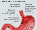
Syphilitic keratitis can begin either in the center or at the periphery of the cornea. If it first affects the center of the cornea, then small gray, blurry spots appear in this place, located in the middle and deep layers of the cornea.

The surface is smooth, but without shine, dull.
The number of spots gradually increases. They are increasingly spreading towards the periphery. But in the center they are always denser than in other places. So here they often merge.
Since the cornea is opaque in the spaces between the spots and shows a subtle diffuse cloudiness, in severe cases the entire cornea can become uniformly gray, resembling milk glass.
When corneal opacification reaches a more or less significant degree, vascularization begins. Moreover, the vessels penetrate the cornea from various places on its edge.
These vessels are deep!
They branch in the form of a brush in the deep layers of the cornea. They are often very dimly visible and appear dirty red or gray-red due to the fact that they are covered with cloudy layers of the cornea.
With this form, a very cloudy ring often forms, which in the further course becomes more and more contracted towards the center of the cornea. Or the central, most cloudy part of the cornea is sharply separated in the form of a white disk from its less cloudy peripheral parts.
Much more often, parenchymal keratitis begins at the periphery of the cornea. In addition to a slight ciliary injection, only very slight turbidity and dullness are initially noticed in this place.
The cloudiness is deep! When viewed with the naked eye, it appears uniformly gray. Under a magnifying glass, it breaks up into separate specks or vague parallel stripes.
Soon similar opacities also appear in other places on the edge of the cornea and move concentrically from all sides to the center of the cornea. Subsequently, after the appearance of peripheral opacities, an injection appears at the corresponding places of the limbus and the growth of blood vessels from the corneal edge begins.
Since the new vascular formation comes from the marginal looped network in the limbus, it soon ends. So the limbus extends only slightly onto the cornea and appears red and thickened.
The latter is soon reversed. But on the other hand, the deep vessels located under the limbus begin to grow, moving further into the cornea, following the clouding in front of them.
It seems as if they are chasing a cloud in front of them. As with the first form, the characteristic branching and dull tone of these vessels indicate their deep position.
When interstitial keratitis reaches its highest point, the cornea is often so cloudy that the iris can barely be seen through it.

At the same time, it completely loses its shine and looks as if it had been greased. Through a magnifying glass, numerous tiny elevations of the epithelium are visible. They give the surface of the cornea a gently shagreen appearance.
Vision deteriorates so much that only the ability to count fingers near the eyes remains, or even only the movements of the hand are distinguished. Then gradual regression begins and, moreover, from the periphery, where the cornea clears up first.
At the same time, the number of vessels gradually decreases.
The central part of the cornea remains cloudy for the longest time. But it, too, eventually clears up so much that only a slight cloudiness remains, which has little effect on vision.
This turbidity, as well as individual, very small, deep vessels visible only through a magnifying glass, are sure signs of previous parenchymal keratitis.
Not all cases proceed as described!
Often there are milder ones in which the changes do not go so far. Therefore, they end in a shorter time. The matter may be limited, for example, to the appearance of several spots, which gradually disappear, without being accompanied throughout the entire time by particularly noticeable inflammatory phenomena.
If the clouding begins on the periphery of the cornea, it is often limited to only the area where it began. If it moves from here a certain distance towards the center, then only one sector becomes cloudy, and not the entire cornea.
It also happens, on the contrary, severe cases in which thick opacities remain forever. Inflammatory infiltration can lead to softening of the cornea. The latter then succumbs to intraocular pressure and develops keratectasia. In such cases, the cornea also remains very cloudy forever.
But even without obvious ectasia, the convexity of the cornea sometimes changes in such a way that it turns out incorrect astigmatism, which, together with clouding, weakens vision.
The most unfavorable cases are those in which the cornea flattens and a thick tendon-white cloudiness remains, since then vision is completely or almost completely lost.
In some cases, the lower part of the cornea becomes cloudier, as if inflammatory products are located here, obeying the law of gravity. Such turbidity is limited at the top by a convex line or forms a triangle. Its base is located at the lower edge of the cornea, and the apex faces upward.
The cloudiness bears the greatest resemblance to those that remain after iridocyclitis, if the effusion has been adjacent to the posterior surface of the cornea at the bottom of the chamber for a long time.
Both the density and prevalence of infiltration and vascularization are expressed to varying degrees. In some cases, the cornea is so richly vascularized that it resembles red cloth.
In other cases, it is almost devoid of vessels and looks more like white milk glass.
There are numerous transitional cases when vessels develop only from several places on the edge of the cornea so that only one sector of it is red, or only single vascular brushes are found. Therefore, according to the nature of the vessels, one can distinguish vascular And avascular form.
However, the question has not yet been resolved whether the avascular form exists in the strict sense of the word!
In any case, this can be established only after the complete end of syphilitic keratitis. And the cornea must maintain a sufficient degree of transparency so that not a single vascular bundle escapes observation.
The vessels are located deep.
However, in older cases, some superficial vessels are also quite often found. All vessels are directed to the center of the cornea, but do not reach it. So what remains here is an avascular roundish spot the size of a millet grain or slightly larger.
If there are many vessels, then parts of the cornea have a red appearance and rise above the level of the avascular center, colored gray or even yellowish-gray.
Fuchs even observed two cases in which a perforation occurred in the center of the cornea.
The anterior chamber is often deep, even if the corneal convexity is unchanged. In most of these cases, the issue is a backward displacement of the iris due to an increase in the amount of chamber moisture. This can be facilitated by both irritation of the uveal membrane and altered filtration conditions.
The uveal membrane is more or less involved in the inflammatory process in all keratitis. But with interstitial keratitis, its participation is especially noticeable.
In mild cases, this is manifested by hyperemia of the iris. In most cases, a clear effusion appears in the form of precipitates; (which are rarely absent), posterior synechiae and even seclusio or occlusio pupillae.
In particularly severe cases it develops plastic iridocyclitis resulting in atrophy of the eyeball.
Intraocular pressure is mostly reduced. An increase in pressure is rarely observed and only in later stages or after inflammation has passed, usually if keratectasia has formed. However, it is independent of ectasia.
Finally, in many cases of parenchymal keratitis we find choroiditis. This inflammation is localized in the very anterior parts of the choroid, which is covered with numerous small, mostly black spots.
Usually, choroiditis is detected only after the inflammation of the cornea has ended, since at the height of the disease it is impossible to perform an ophthalmoscopic examination.
Due to the fact that the same peripheral choroiditis is sometimes found in the other, still healthy eye, the latter is not always a companion or consequence of parenchymal keratitis. And often it is only one of the signs of the underlying disease, equivalent to other “stigmas”.
A rare complication of interstitial keratitis is diffuse scleritis around the cornea. It can subsequently give rise to scleral ectasia.
If we take into account how different individual cases are in terms of opacification and vascularization, it is clear that the picture of syphilitic keratitis is very varied. Therefore, its recognition is often difficult for a novice doctor.
However, in most cases, a diagnosis can be made confidently based on the symptoms common to all cases:
Parenchymal keratitis is always protracted. Inflammatory phenomena increase over 1-2 months until the disease reaches its highest point.
After this, the inflammatory phenomena soon disappear, and the clearing of the cornea occurs at first in rapid steps. Then it slows down again. The central part of the cornea remains cloudy for a particularly long time. It generally takes from six months to 1 year or even more until the disease is completely over.
Syphilitic keratitis usually affects both eyes. Moreover, more often not simultaneously, but one after another. Sometimes there is even a gap of several years between the disease in both eyes.
Relapses occur, although not often.
Thus, the prognosis for parenchymal keratitis is unfavorable with regard to the duration of the disease, since the disease can drag on for many months. And along with all the phenomena associated with it, so do the years.
Regarding the outcome, the prognosis is generally favorable, since in most cases satisfactory vision is still restored. With these chances of restoring vision, it is necessary to maintain the courage of patients who are prone to completely lose hope due to the slow progression of the disease.
On the other hand, complete restoration of good conditions and normal function can only be counted on in very rare cases.
is a disease of young people. It develops between 6 and 20 years of age. And only in rare cases earlier or later (sometimes even after 30 years).
Women get it more often than men!
However, in most cases this is not necessary, since hereditary syphilis can be confidently diagnosed based on a number of symptoms. We'll talk about them below.
It is often useful to find out during a survey how many children have died in the family (the mortality rate for children of syphilitic parents is on average 50%). Have you had a premature birth? Especially the birth of dead or macerated fruits, etc.
Symptoms of hereditary syphilis, which are often found in patients with parenchymal keratitis, are as follows:

It is important to examine all these symptoms, because each of them separately is not evidence for lues hereditaria. On the other hand, one cannot expect all of the listed changes to be clearly expressed at the same time in one person.
If any case still remains in doubt, then you should also resort to a blood test, which is positive in most cases lues hereditaria. The more thoroughly the research is carried out, the more one comes to the conclusion that in the vast majority of cases parenchymal keratitis is explained by hereditary syphilis.
In very rare cases, this keratitis is also observed with acquired syphilis. We should also not forget that the latter can be acquired in childhood. For example, through a wet nurse.
Isolated cases develop due to scrofula or tuberculosis. In some cases, it is not possible to find a clear cause of eye disease at all.
The typical course and involvement of both eyes in parenchymal keratitis has long suggested a constitutional cause.
So Mackenzie gave an excellent description of this disease called corneitis scrofulosa, simultaneously indicating a number of accompanying symptoms that he considered signs of scrofulosis.
Hutchinson is credited with adding to the range of these symptoms and at the same time proving that they do not relate to scrofula, but to hereditary syphilis.
This view found recognition for a long time. Many initially admitted it only in relation to a limited number of cases and distinguished two forms of parenchymal keratitis - keratitis scrofulosa And keratitis syphilitica.
But the more accurately the symptoms of hereditary syphilis were studied, the more confidently they came to the conclusion that the latter underlies most cases of interstitial keratitis, no matter what form the keratitis occurs.
This disease belongs to one of the later manifestations of hereditary syphilis and therefore is quite rightly considered as one of the most important and most common symptoms of lues hereditaria tarda.
Treatment of parenchymal keratitis usually begins with iodine preparations. Next, injections of bioquinol are prescribed. Doctors may give you a penicillin solution for a couple of weeks. As a rule, treatment is carried out by a urologist and an ophthalmologist.
Parenchymal keratitis occurs when a disease such as syphilis affects the eyes. That is why this type of keratitis is often called syphilitic, which is associated with the proliferation of pale bacteria. This disease most often appears between the ages of 5 and 20 years, less often at a younger age - from 1 to 5 years. It is important to understand that the disease can manifest itself even after several generations (two or three).
Interstitial (parenchymal) keratitis is the most common complication of congenital syphilis. The most common etiology of the disease is associated with the proliferation of bacteria on the cornea - spirochetes.
The development of this disease today causes a lot of controversy among ophthalmologists. Some believe that bacteria are constantly present in the patient’s body, but are inactive. Others think that spirochetes are present in the body only until the birth of a child (that is, during intrauterine development), and after birth these bacteria die. Traces of the decay of spirochetes contaminate the cells of the visual organs of newborns, as a result of which the inflammatory process is activated.
Despite many controversies and discussions, scientists agree that the following cases are the provoking factors of parenchymal keratitis:

To identify parenchymal (syphilitic) keratitis, you need to pay attention to the following symptoms:
As the disease develops, complete clouding of the cornea is observed, most often it acquires a white tint. Often the disease is accompanied by the formation of new vessels in the visual organ.
With syphilitic (interstitial) keratitis, the development of such ophthalmological diseases as.
If the above symptoms of the disease are present, patients undergo tests: for example, for Lyme disease, Epstein-Barr virus, and serological testing.
Treatment of syphilitic keratitis is carried out by combating the main disease (the patient is given antisyphilitic therapy). Antibiotics and other medications are prescribed (which must be taken at a strictly defined time).
Treatment procedures are performed at a venereology clinic and carried out jointly with an ophthalmologist.
According to the results of observations, timely and high-quality treatment leads to positive results, and the risk of relapse is minimized.
Keratitis of tuberculous etiology is divided into tuberculous-allergic, as a local manifestation in conditions of sensitization of the body, and true hematogenous tuberculous keratitis, caused by the influence of tuberculous mycobacteria.
Tuberculous-allergic keratitis are the most common form of corneal tuberculosis. This disease has several names: phlyctenulous, scrofulous, eczematous keratitis. The disease is more common in childhood, but can also occur in adults, usually against the background of inactive primary tuberculosis of the lungs and peripheral lymph nodes.
Grayish translucent rounded lesions appear on the cornea, resembling a bubble (phlyctena) in appearance, hence the name of the disease, proposed by Hippocrates. Despite the fact that it has been proven that phlyctenular keratitis is not a vesicle, but a nodule consisting of lymphocytes and epithelioid cells, the term “phlyctenular keratitis” has become firmly established in the clinical practice of ophthalmologists.
The number, size and location of lesions may vary. Small flick-tenes (miliary), smaller than a millet grain, are, as a rule, multiple. Single (solitary) conflicts can reach 3-4 mm in diameter. Phlyctens are always located in the superficial layers of the cornea, but can also affect deep layers. Following the appearance of conflicts, superficial vessels are introduced into the cornea, which in the form of bundles stretch towards the lesion. The appearance of conflicts in the cornea is accompanied by severe photophobia, which reaches such a high degree that the child’s eyelids are convulsively compressed. Blepharospasm and excessive lacrimation lead to maceration of the skin of the eyelids and their swelling. The nose and lips also swell. Cracks may appear in the corners of the mouth. The picture is so typical that the diagnosis of phlyctenulous keratitis can be made at a distance. The disease is prone to relapse. In some cases, disintegration of the phlyctena with destruction of the stroma is observed, up to the appearance of descemetocele or even perforation of the cornea.
Along with the typical form of phlyctenular keratitis (Figure 8.11, see inset), there are other varieties: fascicular keratitis, phlyctenular pannus.
Diagnosis Tuberculous-allergic keratitis is diagnosed based on clinical signs of the disease and general examination data (tuberculin tests, X-ray examination, blood test). In 97% of young children, tuberculin tests are positive. X-ray examination reveals fresh forms of tuberculosis, damage to the paratracheal glands, and, less commonly, infiltrative pneumonia in 82% of cases.
Treatment treatment of phlyctenulous keratitis should be complex, including general and local effects. General treatment is carried out in contact with a phthisiatrician. Corticosteroids are used locally in drops and subconjunctivally, mydriatics, magnetophoresis with an anti-inflammatory mixture, and helium-neon laser irradiation.
Hematogenous tuberculous keratitis
In their development, a large role is given to the vascular tract, which is affected primarily. The process can pass to the cornea directly from the ciliary body through the moisture of the anterior chamber. The lesion may spread to the cornea and sclera.
The most common three forms are: deep diffuse keratitis; deep corneal infiltration; sclerosing keratitis.
With deep diffuse keratitis (Figure 8.12, see inset), the cornea becomes cloudy in the deep and middle layers, and yellowish-gray large non-confluent foci stand out against the background of general clouding. Vascularization of the cornea is superficial and deep, moderate. As a rule, one eye is affected. Remissions alternate with periods of exacerbation, which significantly delays the course. The outcome is unfavorable.
Deep corneal infiltrate is characterized by a deeply located inflammatory focus with slight deep vascularization. With a favorable course, the infiltrates undergo resorption, sometimes necrotization with ulceration of the cornea can occur.
Sclerosing keratitis develops in the presence of deep scleritis. Infiltration of deep layers occurs first at the limb in a limited area, then the process spreads towards the center. Infiltrated areas are tongue-shaped or crescent-shaped. The epithelium over the affected area is swollen, but ulceration never occurs. Vascularization is absent or weakly expressed. The greatest intensity of opacification is observed at the limbus. The disease lasts a long time, the iris and ciliary body are involved in the process, remissions are replaced by new exacerbations. The prognosis is unfavorable, since the infiltrated corneal tissue is replaced by a scar.
A reliable criterion for tuberculous metastatic keratitis is a focal reaction in the affected eye to subcutaneous injection of tuberculin (Mantoux reaction). A focal reaction can be expressed in a rash of phlyctenae, increased pericorneal injection and vascularization, and an increase in exudation.
Treatment tuberculous metastatic keratitis is carried out together with a phthisiatrician. Instillations of a 3% tubazide solution, a 5% saluzide solution, and subconjunctival injections of a 5% saluzide solution are performed. Additionally, corticosteroids and mydriatics are used locally. Magnetic therapy and helium-neon laser irradiation are useful.
syphilitic keratitis
Syphilitic parenchymal or interstitial keratitis is a late manifestation of congenital syphilis, sometimes occurring after two or three generations. The disease usually occurs in childhood and adolescence (6-20 years), extremely rarely in middle-aged and elderly people. Syphilitic etiology is confirmed by serological reactions in almost 80-100% of patients. Parenchymal keratitis in 60-70% is accompanied by other signs of congenital syphilis: Hutchinson's teeth, saddle nose and others.
Clinic parenchymal keratitis is not of the same type, its forms are diverse, which is generally characteristic of syphilis, but the most typical features can be identified. The disease is characterized by cyclicality, bilateral lesions, frequent involvement of the vascular tract in the process, absence of relapses, and a relatively favorable outcome. There are three periods during parenchymal keratitis: the stage of infiltration, vascularization and resorption. In the first period, a diffuse infiltration of a grayish-white color appears in the corneal stroma at the limbus, consisting of individual dots, dashes, and strokes. The surface above the infiltrate is rough due to the spread of edema to the epithelium. Gradually, the infiltration becomes more intense, spreads throughout the entire cornea, and thickens by one and a half times. This period takes 3-4 weeks; at the 5th week, deep vessels begin to grow into the cornea. The limbus becomes swollen, as if moving onto the cornea. The entire cornea resembles frosted glass with a rough surface (Figure 8.13, see inset). During this period, 90% of patients show signs of iridocyclitis. The period of vascularization lasts 6-8 weeks. Gradually, a period of resorption or regression begins, which lasts 1-2 years. Eye irritation is reduced. Resorption of infiltration begins from the limbus and gradually moves towards the center in the same sequence in which it spread. The thickness of the cornea returns to normal, the folds of Descemet's membrane straighten, and precipitates disappear. In severe cases, complete clearing of the cornea does not occur. The vessels gradually become empty.
Diagnosis of syphilitic keratitis is relatively simple. The set of typical clinical signs, positive serological reactions, and family history allow an early diagnosis to be made. It should be differentiated from tuberculous keratitis.
Treatment should be aimed at eliminating the underlying cause, which has a beneficial effect on the outcome of the general disease and the local process. General treatment is carried out jointly with a dermatovenerologist. Corticosteroids, mydriatics, magnetic therapy, and helium-neon laser irradiation are used locally.
But therapy aimed at treating and inhibiting the reproduction of the pale spirochete is quite difficult to tolerate by the body. Therefore, it is necessary to distinguish the symptoms of syphilitic keratitis so that it is possible to begin treatment as early as possible and complete it most successfully.
Syphilitic keratitis is a congenital disease. It manifests itself in the 2-3 generation of infected carriers. Infection with syphilis during fetal development causes not only the occurrence of keratitis. This course of the disease is characterized by a number of specific symptoms caused by the influence of the pale spirochete on the developing organism:

The cause of parenchymal keratitis is the proliferation of pale spirochetes on the cornea. At the moment, doctors cannot accurately determine the pathogenesis of the disease.
Some believe that spirochetes are in a latent state in the body of such patients, causing exacerbations only during periods of decreased immunity. Another group of scientists suggests that spirochetes live in the organs of vision only during intrauterine development, and die after birth, increasing the sensitization of the cornea.
The following can be called provoking factors for the occurrence of syphilitic keratitis:
The pathology is characterized by cyclical exacerbations. As a rule, both eyes are affected. Corneal ulceration is not observed, and the choroid is often involved in the inflammation process. In the process of exacerbation of pathology, three main periods are distinguished:

The diagnosis is made by an ophthalmologist. Based on blood tests and microscopic examination of the cornea, he can make a diagnosis.
The course of treatment for parenchymal keratitis is compiled individually for each patient, based on the results obtained.
To learn more about eye diseases and their treatment, use the convenient site search or ask a specialist a question.

An example of a correct income tax return in 2017, download a new current form for free in Excel. What...

P. S. Pallas (1741 - 1811) - naturalist and traveler-encyclopedist, who glorified his name with major contributions to...
Today, all issues related to the placement of government orders are regulated by the Law on the Contract System -...
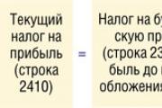
Accounting Regulations Accounting for calculations of income tax of organizations PBU 18/02 (as amended by Orders of the Ministry of Finance of the Russian Federation...

A trainee salesperson is usually called those salespeople who are not yet ready to work completely independently. Process...

Educational institution "Gomel State Medical University" Department of Neurology and Neurosurgery...

One of the most controversial and controversial methods for the early development of children was developed in the 80s by a sociologist...

Contents Dietary supplement based on an extract obtained from the fly beetle (or...

Ekaterina MirimanovaSystem minus 60. RevolutionSystem minus 60 with Ekaterina Mirimanova“System minus 60....
Heaviness and bloating are the causes of both ordinary overeating and more serious digestive problems...

The second blood group, Rh-negative, appeared many years ago, when a person ceased to be...

PSYCHOLOGICAL ASPECTS OF ANOREXIA PHENOMENON (EXPERIMENTAL STUDY) T. V. Tarasova, E. V. Arsentieva...
Contents Since the skin in this area is thin, it is more prone to the appearance of various types of spots....

vseslav Sat, 10/17/2015 - 20:50 Vasileostrovskaya station is one of the oldest stations...

P. S. Pallas (1741 - 1811) - naturalist and traveler-encyclopedist, who glorified his name with major contributions...
Today, all issues related to the placement of government orders are regulated by the Law on Contract...