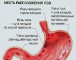
Procedure for filling out an income tax return
An example of a correct income tax return in 2017, download a new current form for free in Excel. What...
Streptococcus pneumoniae are gram-positive diplococci, usually lanceolate or arranged in the form of chains, having a polysaccharide capsule, which allows them to be easily “typed” with specific antisera. Pneumococci are immobile and do not form spores; facultative anaerobes. When cultivated on artificial nutrient media, they lose the capsule and pass from the S- to the R-form. They grow well on blood and serum media. Highly virulent for white mice (sepsis). Based on the capsular antigen, pneumococci are divided into 85 serovars.
Role in human pathology is a natural inhabitant of the human upper respiratory tract. Streptococcus pneumoniae can cause pneumonia, usually lobar, sinusitis and otitis media, and meningitis, which is most often secondary. May also cause osteomyelitis, septic arthritis, endocarditis, peritonitis, cellulitis and brain abscesses. Streptococcus pneumoniae is a leading invasive infection in both young and old people.
Bacteriological A sputum sample collected before the start of antibiotic therapy, obtained by deep coughing and meeting the following criteria is considered suitable for analysis; sputum examination should be performed no later than 2 hours after its collection.
Bacterioscopic- sputum smear and gram stain.
The main pathogenicity factors pneumococcus is considered capsule and substance C.
The pneumococcal capsule is the main virulence factor. It protects bacteria from the microbicidal potential of phagocytes and the action of opsonins. Non-capsulated strains of pneumococcus are practically avirulent and are rarely found. The majority of the pool of antipneumococcal ATs consists of ATs to Ag capsules.
Scarlet fever- an acute infectious disease characterized by sore throat, general intoxication, and the appearance of red kidney rashes on the neck and chest.
The causative agent of scarlet fever is S. pyogenes.
Pathogenesis and clinic. The entrance gate of infection is the mucous membrane of the pharynx. The source of infection is patients, convalescents and bacteria carriers. Infection occurs by airborne droplets. Children aged 1 to 5 years are mainly affected; there are two periods of infection: the first is toxic, accompanied by the appearance of a rash; the second is characterized by allergic reactions. Not only the erythrogenic toxin but also the microbe itself plays a role in the pathogenesis of scarlet fever.
Immunity. After an illness, it is durable, predominantly antitoxic. Recurrent diseases are extremely rare.
Microbiological diagnostics. The diagnosis is usually made based on the clinical picture. In doubtful cases use:
1) Dick's reaction; can be positive. As a result of the disease, antitoxic immunity is developed and the reaction becomes negative. The use of pure toxin, purified from the protein fraction, in the Dick reaction gives consistently clear results and indicates the specificity of this reaction for scarlet fever
2) the phenomenon of rash extinction (intradermal injection of 0.1 ml of convalescent antitoxic serum);
3) isolation of hemolytic streptococcus from the throat;
4) detection of specific precipitins in the urine of patients.
Prevention and treatment. The main preventive measures come down to timely identification of patients, their hospitalization, and quarantine measures. Contact weakened children are administered 1.5-3 ml of gamma globulin. Despite numerous attempts, it was not possible to obtain an effective vaccine against scarlet fever.
Dick's reaction: intradermal test (a type of allergy skin test in which the allergen is injected intradermally) with purified streptococcal toxin, used in the diagnosis of scarlet fever and in determining susceptibility to it.
13. Meningococci, classification, serological groups, characteristics of biological properties, pathogenicity factors, pathogenesis of infection. Laboratory diagnosis of various clinical forms of meningococcal infection, bacterial carriage, specific prevention.
The causative agent of meningococcal infection belongs to the family Neisseriaceae, genus Neisseria, species - N. meningitidis. Meningococci are gram-negative diplococci in the form of coffee beans, often inside leukocytes - incomplete phagocytosis. They do not have spores or flagella and form a capsule. They are strict aerobes. They grow on media with the addition of serum at a temperature of 37⁰, forming S forms of colonies (small, delicate, colorless). Growth is stimulated by increased CO2 concentrations and high humidity. They ferment glucose, maltose, and are oxidase positive.
They have capsular polysaccharide antigens, according to which meningococci are divided into serogroups, typical protein antigens, a common protein species-specific antigen and lipopolysaccharide antigens - 8 serotypes.
Pathogenicity factors:
1. Pili and outer membrane proteins – adhesion and colonization factors
2. Hyaluronidase and neuraminidase are invasion factors
3. Capsule – protection from phagocytosis
4. LPS – has toxic, pyrogenic, necrotic and lethal effects.
Pathogenesis Infection occurs by airborne droplets. The entrance gate is the nasopharynx, from here meningococci penetrate into the lymphatic vessels and blood. Meningococci cause the following clinical forms of infection: nasopharyngitis, meningococcemia, epidemic cerebrospinal meningitis, meningococcal endocarditis. After an illness, a persistent long-term immunity against all serogroups of meningococci.
For laboratory diagnostics meningococcal infection is used:
Bacteriological method, in which a pure culture is isolated from pathological material (blood, cerebrospinal fluid, exudate, mucus from the pharynx and nasopharynx).
Serological– antibodies are detected in RPGA and ELISA.
Actually immune– antigen is detected by ELISA
Microscopic- a smear prepared from cerebrospinal fluid is stained with Gram stain. The presence of a large number of leukocytes in the smear and the presence of gram-negative diplococci in them allows us to confirm the diagnosis of meningococcal meningitis.
As specific prevention Vaccines containing polysaccharide antigens of various serogroups have been proposed, but they only form group-specific immunity.
Treatment– antibiotics (b-lactams, penicillins, cephalosporins.)
14. Causative agents of wound anaerobic infection: clostridia and bacteroides. Their classification, characteristics of biological properties, pathogenicity factors. Pathogenesis of wound infection, laboratory diagnostic methods, specific prevention and therapy.
Clostridia. A wound infection caused by bacteria of the genus Clostridium (C.perfringens, C.septicum, C.novyi, C.histolyticum, etc.) is called gas gangrene.
Morphology and cultivation
Gram-positive, spore-forming, rod-shaped bacteria that form a capsule in the affected tissues.
Obligate anaerobes.
Pathogenicity factors
They produce exotoxins specific to each species, affecting the central nervous system.
They form aggressive enzymes - collagenase, proteases, hyaluronidase, deoxyribonuclease, hemolysins.
Resistance and ecology
The causative agents of gas gangrene are normal inhabitants of the intestines. humans and animals, spores with feces enter the environment, where they remain viable for decades.
Pathogen spores are resistant to boiling and disinfectants
Epidemiology
Gas gangrene is widespread, especially often recorded in cases of mass wounds and injuries (wars, disasters) and untimely surgical treatment of wounds.
Pathogenesis and clinical picture
Gas gangrene develops as a result of pathogen spores entering the wound, especially if there is necrotic tissue in the wound and a decrease in the body’s resistance. Vegetative forms of clostridia produce toxins and enzymes that damage body tissues.
The incubation period is 1-3 days. The clinical picture is varied and boils down to swelling, gas formation in the wound, suppuration with intoxication. The course of the infection is complicated by secondary infection (staphylococci, Proteus, Escherichia coli, bacteroides, etc.)
Immunity
Past infection does not create immunity. The leading role belongs to antitoxic immunity.
Microbiological diagnostics
They examine pieces of affected tissue, wound discharge, and blood. Microscopy is performed.
Bacteriological studies are carried out under anaerobic conditions; toxin identification using RN in mice with diagnostic anti-gangrenosis antitoxic sera.
Treatment and prevention
Treatment includes surgical excision of all dead tissue, broad-spectrum antibiotics, and administration of an anti-gangrenous antitoxic medicinal serum.
Prevention - proper surgical treatment of wounds, compliance with asepsis and antisepsis during operations; For active immunization, toxoids against gas gangrene in the composition of sextaanatoxin are used; such vaccination is carried out for special indications (military personnel, diggers, etc.)
Non-spore-forming anaerobes ( non-clostridial) are gram negative ( bacteroides, fusobacteria, veillonella, etc.) and gram-positive (actinomycetes, peptococci, peptostreptococci, etc.), rod-shaped and coccoid bacteria with various biological properties. They constitute a large group of obligate anaerobes. Consisting of various taxonomic groups.
They are cultivated under strict anaerobic conditions and die quickly in the presence of oxygen.
They differ in polymorphism.
Various biochemical activities.
Antigenic properties have not been sufficiently studied.
Pathogenicity factors: capsule, aggression enzymes, cell wall LPS in gram-negative bacteria.
Antibiotic sensitivity individual for each species. Some representatives are sensitive to metronidazole (Trichopol), clindamycin and some other broad-spectrum drugs.
Non-spore-forming anaerobes are representatives of the normal human microflora. The mucous membranes of the oral cavity, colon and genital organs of women are especially abundantly contaminated with them.
Diseases most often have an endogenous origin and occur against the background of a decrease in the body’s resistance.
pathogenesis Non-spore-forming anaerobes cause a variety of clinical diseases: purulent-inflammatory diseases of the maxillofacial area, affecting the genitourinary system, musculoskeletal system, liver, causing appendicitis, peritonitis, sepsis.
Usually these are mixed infections caused by associations of anaerobes and aerobes.
Immunity poorly studied.
diagnostics Microscopic and bacteriological studies are carried out, gas-liquid chromatography, ELISA, etc. are used.
Treatment often surgical in combination with antibacterial therapy (metranidazole, clindamycin, cephalosporins, chloramphenicol, erythromycin).
Specific prevention absent.
MINISTRY OF HEALTH OF THE RUSSIAN FEDERATION KAZAN STATE MEDICAL UNIVERSITY DEPARTMENT OF MICROBIOLOGY
PNEUMOCOCCUS
KAZAN 1999
UDC 576.851.21(07)
Published by decision of the Central Coordination and Methodological Council of the Kazan State Medical University.
Compiled by:
(Head of the Department of Microbiology, Doctor of Medical Sciences, Professor O.K. Pozdeev, Assistant of the Department of Microbiology, Candidate of Medical Sciences, E.R. Fedorova.
Reviewers:
Head of the Department of Epidemiology, Kazan State Medical University, Doctor of Medical Sciences, Associate Professor M.Sh. Shafeev, Head of the Department of Epidemiology, Kazan State Medical Academy, Doctor of Medical Sciences, Professor V.E. Grigoriev.
Pneumococci /O.K. Pozdeev, E.R. Fedorov - Kazan: KSMU, 1999. - 14 s.
Kazan State Medical University, 1999
Pneumococcus (Streptococcus pneumoniae) was first isolated by Pasteur (1881) while working on an anti-rabies vaccine and was initially considered the causative agent of rabies. The etiological role of the microorganism in the development of pneumonia in humans was proven by Frenkel and Weichselbaum (1884). The bacteria colonize human and animal organisms and belong to the group of so-called “oral” streptococci. They are the main causative agents of pneumonia, and can also cause pleurisy, meningitis, creeping corneal ulcers, purulent inflammation of the middle ear, septic conditions and other diseases. In the IX edition of Bergey's guide to bacteria (1994), pneumococci are included in group 17, “Gram-positive cocci.”
Epidemiology of lesions. Pneumococcus is one of the main causative agents of bacterial pneumonia recorded outside hospitals (2-4 cases per 1000 people); Every year, at least 500,000 cases of pneumococcal pneumonia are observed in the world (the real value is much higher). Children and the elderly are most susceptible to infection. The reservoir of infection is patients and carriers (20-50% of preschool children and 20-25% of adults); the main transmission route is contact; during outbreaks it is also airborne. The peak incidence occurs in the cold season. In the vast majority of cases, clinical forms of infection develop when the body’s resistance is impaired (including due to cold stress), as well as against the background of other pathologies (sickle cell anemia, Hodgkin’s disease, HIV infection, myeloma, diabetes mellitus, conditions after splenectomy, etc.) or with alcoholism. Options 1, 2 and 3 play the greatest epidemiological significance in pathology in adults; for children - 1, 2, 3 and 14 options. The virulence of serovars in descending order is variants 3, 1, 2, 5, 7 and 8. White mice (if infected, they die from septicemia within 24 hours), calves, lambs, piglets, dogs and monkeys are sensitive to pneumococci.
Morphology. Pneumococci are immobile, they do not form spores, and have a slightly elongated shape, reminiscent of the contours of a candle flame. In smears of clinical material, they are arranged in pairs, each of which is surrounded by a thick capsule. In smears from culture media, they may be located in short chains and be more rounded. On simple media they form a thin capsule; its development is stimulated by the introduction of blood, serum or ascitic fluid. Capsule formation is most pronounced in type III bacteria. When arranged in chains, the capsule may be common.
Cultural properties. Pneumococci are aerobes or facultative anaerobes; When cultivating, capnophilic conditions (5-10% CO2) are preferred. They are chemoorganogrophs and grow well on blood or serum media supplemented with 0.1% glucose. They can grow in the temperature range of 28-42 °C, optimum - 37 °C. Optimum pH -7.6-7.8. On dense media they form delicate, translucent, clearly defined colonies with a diameter of about 1 mm. Sometimes they can be flat with a central depression; like other streptococci, colonies never fuse
between themselves. On blood agar they form small translucent greenish-gray colonies. The center of the colonies is darker, the periphery is lighter. Under the colony and along its periphery, a zone of a-hemolysis is visible in the form of a greenish discolored zone (which is due to the transition of hemoglobin to methemoglobin). Colonies of type III pneumococcus often have a mucous consistency and are up to 2 mm in size. They are a bit cloudy and can merge with each other. They form S- and R-forms of colonies. When transitioning from the S- to the R-form, they lose the ability to synthesize the capsule. On liquid media with serum and 0.2% glucose they give uniform turbidity and a small flocculent sediment. With prolonged cultivation, the sediment increases.
Sustainability. Pneumococci belong to the group of unstable microorganisms. They persist in dry sputum for up to two months. Able to be stored for a long time at low temperatures; at a temperature of 60 °C they die within 3-5 minutes. A 3% solution of carbolic acid kills them in 1-2 minutes. Optochin (at a concentration of 1:100,000) and bile have a detrimental effect on pneumococci, which is used to identify bacteria.
Pneumococci differ from other microorganisms in a number of properties (Table 1).
Table 1 Biochemical properties of pneumococci
|
Test substrate |
Result |
Test substrate |
Result |
|
Growth at 100°C |
|||
|
Raffinose |
|||
|
Wednesday with 6.5% Nad |
|||
|
a-hemolysis |
|||
|
B-hemolysis |
Trehalose |
||
|
Phosphatase |
|||
|
Hippurate |
β-galactosidase |
||
|
Glycerol |
|||
|
Designations: “+” - 90% or more strains are positive; (+) - 80-89% of strains are positive; d - 21-79% of strains are positive; (-) - 11-20% of strains are positive; “- - 90% or more of the strains are negative. |
|||
Antigenic structure. Several types of antigens have been found in pneumococci: polysaccharide, 0-somatic antigen, located in the cell wall; polysaccharide capsular K-antigens and M-protein. The polysaccharide somatic antigen is similar to the C-substance of other streptococci. The relationship determines the similarity in the chemical structure of ribitteichoic acids associated with choline phosphate. Capsule antigens also have a polysaccharide nature, consisting of monosaccharides repeated in various combinations: D-glucose, D-galactose and L-rhamnose. Based on the structure of capsular antigens, pneumococci are divided into 84 serovars. It should be remembered that capsular antigens cross-react with antisera to antigens of group A and B streptococci, as well as with sera to Klebsiella and Escherichia antigens. During the transition from S to R form, capsular antigens are lost. For serotyping of pneumococci, group sera are produced, designated by Latin letters (A, B, C, D, etc.) and serovariant sera, designated by Roman numerals. Agglutinating serum III is not included in serum mixtures. It is released separately and cannot be bred. In humans, pneumococci of serovariants I, II and III are most often isolated. They are the most virulent for humans, so the agglutination test is initially performed using antisera to these variants. If the result is negative, the agglutination reaction is performed with a mixture of sera A, B, C, etc. (until a positive result is obtained), and then with separate antisera. For faster identification of serovars, the Neufeld reaction (immune capsule swelling) is used. The method is based on the ability of pneumococcal capsules to increase in volume in the presence of homologous antiserum, which is recorded by light optical microscopy. Capsular polysaccharides have sensitizing properties, manifested in the development of a delayed-type hypersensitivity reaction, detected using skin tests.
Pathogenicity factors. The main factor is the capsule, which protects bacteria from the microbicidal potential of phagocytes and diverts them from the action of opsonins. Non-encapsulated strains are practically avirulent and are rare. The majority of the pool of antipneumococcal ATs consists of ATs to Ag capsules. An important function of the capsule and M-protein is also to ensure adhesion to the mucosa. Substance C is essential, as it specifically interacts with C-reactive protein. The consequence of such a reaction is the activation of the complementary cascade and the release of mediators of the acute phase of inflammation; their accumulation in the lung tissue stimulates the migration of polymorphonuclear phagocytes. The formation of powerful inflammatory infiltrates is accompanied by a disruption of the homeostasis of the lung tissue and its hepatization. Pneumococci produce endotoxin, a- and beta-hemolysins (pneumolysins), and leukocidin. α-pneumolysin is a thermolabile protein capable of neutralizing O-streptolysin,
erythrogenic substance, neuraminidase. Pneumococci also synthesize a number of enzymes that contribute to the pathogenesis of lesions - muramidase, hyaluronidase (promotes the spread of microorganisms in tissues), peptidase (breaks down IgA).
Pathogenesis of lesions. The biotope of pneumococci is the upper respiratory tract. The pathogenesis of most pneumonias involves aspiration of saliva containing S. pneumoniae and penetration of bacteria into the lower airways. Violation of protective drainage mechanisms - cough impulse and mucociliary clearance - is essential. In adults, lobar lung lesions are more often observed; in children and the elderly, peribronchial or focal lesions predominate. The formation of powerful inflammatory infiltrates is accompanied by a disruption of the homeostasis of the lung tissue and its hepatization. Infections with the most virulent serovar 3 may be accompanied by the formation of cavities in the lung parenchyma. From the primary focus, the pathogen can penetrate the pleural cavity and pericardium or disseminate hematogenously and cause meningitis, endocarditis and joint lesions
Clinical manifestations. Classic pneumococcal pneumonia begins suddenly; They note a rise in body temperature, a productive cough and chest pain. In weakened persons and the elderly, the disease develops slowly, with slight fever, impaired consciousness and signs of pulmonary heart failure. Streptococcal meningitis is registered in all age groups; they are characterized by a violent onset with a rise in body temperature, stiffness of the neck muscles, headache, nausea and vomiting. Lesions of the vessels of the meninges are often accompanied by loss of consciousness; among children and the elderly, mortality can reach 80%. Quite often, hematogenous pneumococcal lesions, as well as sinusitis, mastoiditis, otitis media, endocarditis and peritonitis, are noted in people with immunodeficiencies (for example, HIV-infected) or patients with splenectomy. After an illness, unstable immunity develops, which is type-specific and is caused by the appearance of antibodies against the typical capsular polysaccharide.
Treatment. The basis of treatment for pneumococcal infection is antibiotics - penicillin, tetracycline, chloramphenicol, vancomycin, rifampicin, ceftriaxone.
Prevention. Nonspecific prevention of pneumococcal infections is aimed at identifying patients and carriers with their subsequent treatment. For specific prevention of infection, children over two years of age, adults at risk, as well as healthy individuals during a disease outbreak are vaccinated with the polyvalent polysaccharide vaccine "Pneumovex 23". The drug contains 23 capsular polysaccharide antigens of pneumococci (1, 2, 3, 4, 5, 6B 7F, 8, 9N, 9V, 10A, 11A, 12F, 14, 15B, 17F, 18C, 1-9F, 19A, 20, 22F , 23F, 33F). Antigens
pneumococci were obtained from 90% of the strains isolated from the blood of patients with invasive pneumococcal infection in the USA and corresponding to the strains found in Russia. Immunization is carried out twice with an interval of 5-8 years.
After vaccination, artificial, active, type-specific immunity is created.
Laboratory diagnostics. The “gold standard” is pathogen isolation. It should be remembered that the material must be examined quickly, because bacteria are prone to rapid autolysis due to the activity of intracellular enzymes. The material for the study is sputum, pleural effusion and other exudates, cerebrospinal fluid, blood, mucus from the nose and pharynx, discharge from eye ulcerations, discharge from the ear, urine, pieces of organs (in case of death of the patient). A signal response to pneumococcal infection can be issued when neutrophils and gram-positive lanceolate diplococci (at least 10 per field of view) are detected in sputum smears. Otherwise, they resort to isolating the pathogen.
The first stage of the study. Pathological material is subjected to preliminary bacterioscopy (except blood). The sputum is placed in a sterile Petri dish, washed, a purulent mucous lump is captured with a loop, ground on a glass slide, dried and stained with a Gram stain. The smear reveals gram-positive lancet-shaped or oval-shaped cocci surrounded by a capsule (capsule formation is observed only in pneumococci isolated from sick and infected animals). Detection of pneumococcal capsules can be carried out using the Burri-Gins method. Inoculation of pathological material is carried out on 5-10% blood or serum agar and on enrichment medium (8-10% whey broth). If sepsis of a pneumococcal nature is suspected, 5-10 ml of the patient's blood is inoculated into 45-90 ml of whey broth. The cerebrospinal fluid, if it is clear, is centrifuged and a few drops from the sediment are inoculated onto nutrient media. Semi-solid whey agar is used as an enrichment medium. The crops are incubated at 37 °C for 24 hours. The best method for isolating a pure culture of pneumococci is to infect white mice with pathological material. Sputum, washed in a Petri dish with sterile saline, is ground in a sterile mortar with a sterile pestle or broken glass while adding saline in a ratio of 1:2-1:5. The suspension is settled, the supernatant liquid in an amount of 0.5-1 ml is administered intraperitoneally to white mice. If pneumococci are present in the material, mice die within 72 hours. Typical pneumococci are found in smears from organs and blood. Organs and blood are also cultured on whey broth and on Petri dishes with blood or serum agar.
Second stage of the study. The growth pattern on nutrient media is studied. On blood agar, colonies of pneumococci are small, round, with smooth
edges, tender, surrounded by a zone of greening of the medium (which is very reminiscent of the growth of viridans streptococci). On serum agar, the colonies are delicate, translucent and transparent. During bacterioscopy of Gram-stained smears. Gram-positive diplococci without capsules are detected. After bacterioscopy, colonies suspected of pneumococci are subcultured onto slanted serum or blood agar or into whey broth. When microscopying smears from the enrichment medium, along with various microflora, one can detect gram-positive cocci, arranged in pairs or short chains. The material from the enrichment medium is transferred to solid nutrient media. The crops are incubated at 37°C for 24 hours.
The third stage of the study. On slants of blood agar, pneumococci form a delicate, thin, translucent coating. In whey broth, pneumococci cause turbidity and a slight sediment. In smears from solid culture media, pneumococci can have different appearances. Along with diplococci of elongated shape with pointed outer ends, reminiscent of a candle flame, there are cells of regular oval and round shape. In broth culture, pneumococci are often arranged in chains. Based on the morphological and cultural properties of pneumococci, it is difficult to distinguish from viridans streptococci, therefore a set of special tests has been proposed for their differentiation:
Solubility in bile (deoxycholate test);
Ability to decompose inulin;
Sensitivity to optochin;
Agglutination reaction with specific antipneumococcal sera;
The ability to decompose glucose, maltose, sucrose, lactose, mannitol, sorbitol and salicin.
The most accessible methods for differentiating pneumococci from other streptococci are a test with optochin (inhibits their growth); They are distinguished from viridans streptococci by their ability to ferment inulin, as well as sensitivity to bile.
Deoxycholate test. After preliminary bacterioscopy, 10 drops of isolated pure culture (preferably broth) are added to a test tube with 5 drops of sterile bovine bile. The control is a culture added to a test tube with 5 drops of saline solution. After 30-60 minutes of incubation at 37 °C, complete lysis of the culture is observed in the form of clearing in the test tube with bile; in the control tube the mixture remains cloudy. It should be remembered that avirulent pneumococcal cultures are resistant to bile.
Bile resistance can also be tested by culture in 10% bile broth. The test material is added to the medium, and the broth becomes cloudy. After 24 hours of incubation at 37 °C, the presence of pneumococci will be indicated by clearing of the broth as a result of lysis of bacteria.
You can also use discs soaked in a 20% bile solution. The discs are placed on the grown culture in a dish and incubated for 1-2 hours at 37 °C. In the presence of pneumococci, colonies are lysed around the disc at a distance of 1-2 mm.
Test for inulin. The pneumococcal culture is inoculated on a medium with inulin. To do this, add 200 ml of sterile distilled water, 18 ml of litmus tincture and 3 g of inulin to 100 ml of bovine serum heated at 56 °C for 30 minutes, and sterilize with running steam for 30 minutes. The crops are incubated at 37 °C for 24 hours. Pneumococcus decomposes inulin, causing the medium to turn red. Viridans streptococcus does not cause redness of the environment.
Test with optochin. The test pneumococcal culture is inoculated on whey broth with optochin at a dilution of 1:100,000 or 1:200,000. Pneumococcus does not grow on such a medium. You can also determine sensitivity to optochin by plating on 10% blood agar containing optochin at a dilution of 1:50,000. The control is to inoculate the culture on blood agar. Pneumococci do not grow on the medium with optochin; growth of pneumococci is observed on the control medium. You can use disks soaked in 6 μg of optochin, which are applied to the surface of the medium after inoculation. In pneumococci, a growth inhibition zone of at least 18 mm in diameter forms around the disc.
Virulence test. A daily culture of pneumococcus grown in whey broth is diluted with 1% sterile peptone water (pH - 7.6) or slightly alkaline broth up to 1:10. The diluted culture is administered intraperitoneally to white mice weighing 16-20 g in a volume of 0.5 ml and observed for 72 hours. The organs of a dead mouse are inoculated onto nutrient media and the fingerprint smears are examined microscopically. Highly virulent cultures include pneumococci, which cause the death of mice after the introduction of a culture at a dilution of 1:10. Avirulent cultures do not cause death in mice.
Serotyping of pneumococci. The 18-hour culture is tested in the Sabin microagglutination reaction. 4 drops of pneumococcal culture are applied to a glass slide. To 1 drop add a drop of antipneumococcal serum type 1, to the 2nd - type II serum, to the 3rd - serum - 111, to the 4th - a drop of normal serum. The mixtures on glass are mixed with a loop and examined under a magnifying glass or microscope at low magnification. In a positive case, agglutination is observed in one of the first three drops. The type of pneumococcus is determined by an agglutination reaction with specific agglutinating sera of the first three fixed types. Cultures that are not agglutinated by these types of sera are classified as X-group. The reaction is set up as follows. Pour 0.5 ml of 18-hour broth culture into test tubes. Then an equal volume of serum is added, diluted with saline in a ratio of 1:5. The controls are 2 test tubes, one of which contains the test culture mixed with
normal rabbit serum, and the other - only the test culture. The contents of the tubes are thoroughly shaken and placed in a thermostat at 37 °C for 2 hours, after which a preliminary calculation of the reaction is carried out. Final results are noted after additional storage at room temperature for 20 hours. Agglutination is assessed as four pluses if the contents of the tubes are completely cleared and the agglutination culture is a dense film that does not break when shaken; three pluses if, when the contents of the tube are completely cleared, the agglutinating culture easily breaks into parts; two pluses - if clearing does not occur, particles of the agglutinated culture are clearly visible to the naked eye in the turbid contents of the test tube; with agglutination for one plus, a fine-grained mixture of glued pneumococci is found in the test tube. In case of a negative reaction visible to the eye, agglutination is not observed;
The contents of the test tubes after shaking are uniformly cloudy.
Typing of X-group pneumococci is carried out using group
sera containing a mixture of typical agglutinating sera taken
in equal volumes. Prepare the following group sera by
mixing equal volumes of undiluted standard diagnostic
serums (Lund, I960):
A -1, II, IV, V, XVIII serovars;
B - VI, VIII, XIX serovars;
C - VII, XX. XXIV, XXXI, XL serovars;
D - IX, XI, XVI, XXXVI. XXXVII serovars;
E - X, XXI. XXXIII, XXXIX serovars;
F - XII. XVII. XXII, XXXVII, XXXII, XLI serovars;
G - XIII, XXV. XXIX, XXXIV, XXXV, XXXVIII, XLII, XLVII serovars;
J - XLIII. XLIV, XLV, XLVI serovars.
Type III agglutinating serum is used per se (without mixing with other standard sera) due to the difficulty of obtaining it in a sufficiently high titer. Typing is carried out in two stages: first with the help of group sera, and then with individual sera of the group with which a positive reaction was obtained. Serotyping of pneumococci is used primarily for epidemiological studies of the results of specific serotherapy and seroprophylaxis.
Microagglutination of pneumococci using the Sabin method can be obtained by mixing anti-pneumococcal sera with exudate from the abdominal cavity of a mouse contaminated with the sputum of a patient. Already four hours after infection, a pure culture of pneumococci is detected in the exudate, giving a positive Sabin agglutination.
Accelerated methods for detection and typing of pneumococci. 1. Neufeld's method or the phenomenon of swelling of the pneumococcal capsule. One lump of freshly secreted sputum of the patient is applied to three
coverslips, to each of them add a drop of undiluted specific antipneumococcal serum (types 1, II, III) and a drop of Loeffler's blue. The drops are thoroughly mixed and covered with a glass slide with a well smeared around the edges with Vaseline. After two minutes, the hanging drops are examined under a microscope with an immersion system. In a positive case, a sharp increase in pneumococcal capsules is visible. If the result is negative, the capsules are hardly treasured. The swelling reaction is specific and does not give a positive result with other capsular bacteria. I do not use it for examining sputum from patients treated with sulfonamides and antibiotics, because in this case, non-capsular pneumococci can be isolated.
2. Precipitation method. 5-10 ml of sputum is boiled in a water bath until a dense clot is obtained. The clot is ground and a small amount of saline is added and boiled again for several minutes to extract the specific polysaccharide from pneumococci. The suspension is centrifuged, and a ring precipitation reaction is performed with the resulting clear liquid and specific standard sera in precipitation tubes. The appearance of a ring at the interface between the liquids indicates a positive result.
3. Determination of pneumococcal capsules according to Burri. A drop of the test material and a drop of ink are applied to the end of the slide. The mixture is mixed and a smear is made, dried in air and, without fixing, examined under a microscope. The background of the preparation is dark smoky; microbial bodies and their capsules are not stained. The preparation prepared according to Burri can be fixed with Nikiforov’s mixture, rinsed with water, and stained with Ziel fuchsin diluted 1:3 for 3-5 minutes. Against the dark background of the smear, unpainted capsules stand out, inside of which there are bacteria of a bright crimson color (Hins method).
Pneumococci (synonym: Pneumococcus Talamon - Frankel, Streptococcus lanceolatus Pasteur, Micrococcus pneumoniae, Diplococcus pneumoniae Frankel, Streptococcus pneumoniae) are lanceolate diplococci isolated in human pneumonia. Discovered in 1881 by L. Pasteur and independently by G. M. Sternberg in the USA. The etiological relationship of pneumococci to human pneumonia was established by Frenkel and Weichselbaum (A. Frankel, A. Weichselbaum) in 1884.
Pneumococci isolated from the human or animal body are oval or lanceolate cocci, arranged in pairs; stained positively by Gram, with a value of about 1 micron. Each pair is surrounded by a thick capsule, revealed by eosin staining [T. J. Mackie, J. E. McCartney]. Pneumococci usually grow on artificial nutrient media in the form of chains. The chain of pneumococcus is usually shorter than that of streptococcus pyogenes. In culture, pneumococci are less lanceolate and more round, nonmotile and do not form spores. The capsule of pneumococcus is clearly visible on preparations from animal and human exudates, when growing on nutrient media to which blood, blood serum or ascitic fluid is added, but is poorly visible when growing on ordinary nutrient media. Pneumococci are aerobes or facultative anaerobes, easily stained with ordinary aniline dyes and Gram positive, although they become Gram negative in old cultures.
The growth of pneumococci on ordinary nutrient media is poor, but is significantly improved by adding glucose (0.1%), blood, serum or ascitic fluid to the nutrient medium. Pneumococci grow well in an atmosphere containing 5-10% carbon dioxide. The optimal growth temperature is 37°, maximum 42°, minimum 25°. Pneumococci are sensitive to changes in the pH of the nutrient medium; the optimal pH is 7.8, the acidity limit is 6.5, and the alkalinity limit is 8.3. On nutrient agar, pneumococci grow, forming small colonies 1 mm in diameter, tender, translucent, resembling dewdrops, not merging with each other. Colonies of pneumococci on special nutrient media, for example on blood agar (5%), are small, moist, transparent, with well-demarcated edges, exhibit α-hemolysis, appear surrounded by a greenish discolored zone, similar to that observed when viridans streptococcus (Streptococcus) grows on blood agar viridans).
When growing on colored nutrient media, pneumococci ferment carbohydrates with the formation of acid, but without the formation of gas. Fermentation of inulin is an important distinguishing feature of pneumococci (viridans streptococcus does not have the ability to decompose inulin). Pneumococci exhibit the ability to undergo rapid autolysis under the influence of bile salts. Bile or bile salts dissolve pneumococcus, which also distinguishes it from streptococcus.
Pneumococci are more sensitive than many other microorganisms to the bactericidal action of quinine and some of its derivatives. For example, optochin (ethylhydrocuprein) kills pneumococci at a concentration of 1:500,000, and streptococci at a concentration of 1:5000.
Pneumococci quickly lose virulence when stored in ordinary nutrient media, but can survive for months in the cold in the vacuum-dried spleen of a white mouse that died from pneumococcal septicemia. The most sensitive to pneumococci are white mice and rabbits, guinea pigs are less sensitive, and cats, dogs, chickens and pigeons are highly resistant. Pneumococcus does not produce a true toxin, but produces hemolysins that are active against red blood cells of sheep, guinea pigs and humans, as well as hyaluronidase, lencocidin and a necrotizing substance. The virulence of pneumococcus does not depend on these toxic formations, but on the presence of a specific soluble substance inherent in the pneumococcus of the corresponding type.
Pneumococci contain several antigens. Deep in the microbial cell there is a nucleoprotein component associated with species specificity. Closer to the surface is a species-specific polysaccharide (C-antigen) - a somatic antigen that is immunologically identical in all pneumococci. A type-specific protein (M-antigen), similar to the M-antigen of hemolytic streptococci, is also located close to the surface of the microbial cell. The capsule located more superficially consists entirely or partially of a polysaccharide specific to each type of pneumococcus and is closely related to the virulence of the living microbe. This antigen - a polysaccharide hapten or specific soluble substance (SSS) - is type-specific and serves to differentiate the immunological types of pneumococci.
Each type has an individual antigenic structure and virulence. Pneumococci isolated during pneumonia are divided on the basis of immunological reactions into types I, I, III and IV. Type IV pneumococci are immunologically heterogeneous. This type includes all pneumococci that do not belong to the first three types. Typing of pneumococci was important in its time due to the fact that the effect of serotherapy for pneumonia with specific serum was directly dependent on the type of pathogen.
Microbiological diagnosis of pneumococci consists of microscopic examination and isolation of pneumococci on artificial nutrient media. The type of pneumococcus is determined by: the capsule swelling reaction, the microagglutination reaction on glass (Sabin method) and the macroscopic agglutination reaction. If for some reason the white mouse cannot be used, then sputum or other pathological material is inoculated into blood broth with glucose, which is then used as an antigen in the same immunological reactions.
Pneumococci are found on the mucous membranes of the mouth and upper respiratory tract more often than in the human environment. Pneumococci are transmitted by airborne droplets. Type IV pneumococci are much more common than types I, II and III. Recently, the incidence of pneumococci in pneumonia has sharply decreased, while at the same time the incidence of staphylococci has increased significantly. With a significant decrease in the inoculation rate of pneumococci and, consequently, with a decrease in their importance as etiological agents, Escherichia coli, enterococci, proteus and other microorganisms began to be isolated in larger quantities.
Acquired immunity to pneumococcus is obviously associated with a capsular antigen, upon immunization with which a clear correlation of resistance is established with an antibody response to this antigen. See also Bacteria.
Biochemical properties mostly typical for the genus Salmonella Distinctive features are: the absence of gas formation during fermentation of S. Typhi, the inability of S. Paratyphi A to produce hydrogen sulfide and decarboxylate lysine.
Epidemiology.Typhoid fever and paratyphoid fever are anthroponoses, i.e. cause disease only in humans. The source of infection is the patient or the bacteria carrier, who release the pathogen into the external environment with feces, urine, and saliva. The causative agents of these infections, like other salmonellae, are stable in the external environment and persist in soil and water. S. Typhi can become uncultivable. A favorable environment for their reproduction is food products (milk, sour cream, cottage cheese, minced meat, jelly). The pathogen is transmitted by water, which currently plays a significant role, as well as by nutritional and household contact routes. The infecting dose is approximately 1000 cells. The natural susceptibility of people to these infections is high.
Pathogenesis and clinical picture. Once in the small intestine, typhoid and paratyphoid pathogens invade the mucous membrane when
with the help of effector proteins TTSS-1, forming the primary focus of infection in Peyer's patches. It should be noted that in the submucosa the osmotic pressure is lower compared to the intestinal lumen. This promotes intensive synthesis of Vi-antigen, which increases the antiphagocytic activity of the pathogen and suppresses the release of proinflammatory tissue mediators by submucosal cells. The consequence of this is the lack of development of inflammatory diarrhea in the initial stages of infection and the intensive proliferation of microbes in macrophages, leading to inflammation of Peyer's patches and the development of lymphadenitis, resulting in a violation of the barrier function of the mesenteric lymph nodes and the penetration of salmonella into the blood, resulting in the development of bacteremia. This coincides with the end of the incubation period, which lasts 10-14 days. During bacteremia, which accompanies the entire febrile period, pathogens of typhoid and paratyphoid fever spread throughout the body through the bloodstream, settling in the reticuloendothelial elements of parenchymal organs: liver, spleen, lungs, as well as in the bone marrow, where they multiply in macrophages. From the Kupffer cells of the liver, salmonella enter the gallbladder through the bile ducts, into which they diffuse, into the gallbladder, where they also multiply. Accumulating in the gallbladder, salmonella cause inflammation and reinfect the small intestine with a flow of bile. The repeated introduction of Salmonella into Peyer's patches leads to the development of hyperergic inflammation in them according to the Arthus phenomenon, their necrosis and ulceration, which can lead to intestinal bleeding and perforation of the intestinal wall. The ability of typhoid and paratyphoid pathogens to persist and multiply in phagocytic cells when the latter are functionally insufficient leads to the formation of bacterial carriage. Salmonella can also persist for a long time in the gallbladder, excreted in feces for a long time, and contaminate the environment. By the end of the 2nd week of the disease, the pathogen begins to be excreted from the body in urine, sweat, and breast milk. Diarrhea begins at the end of the 2nd or beginning of the 3rd week of the disease, from which time the pathogens are cultured from the feces.
Table of contents of the topic "Streptococci. Hemolytic streptococci. Pneumococcus. Non-hemolytic streptococci.":First Pneumococcus isolated by Pasteur (1881) while working on an anti-rabies vaccine and initially considered it the causative agent of rabies. Etiological role pneumococcus in the development of pneumonia in humans was proven by K. Frenkel and A. Weichselbaum (1884).
Pneumococcus bacteria do not contain group Ag and are serologically heterogeneous - 84 serovars are distinguished based on the Ag of capsular polysaccharides. Strains are known that colonize human and animal organisms.
Pneumococcus- one of the main causative agents of community-acquired bacterial pneumonia (2-4 cases per 1000 people). At least 500,000 cases are reported worldwide every year pneumococcal pneumonia, and children and the elderly are most susceptible to infection.
Reservoir of pneumococcal infection- patients and carriers (20-50% of preschool children and 20-25% of adults), main transmission route of pneumococcus- contact, and during outbreaks also airborne. The peak incidence occurs in the cold season.
In the vast majority of cases clinical forms of pneumococcal infection develop when the body's resistance is impaired (including due to cold stress), as well as against the background of concomitant pathology (sickle cell anemia, Hodgken's disease, HIV infection, myeloma, diabetes mellitus, conditions after splenectomy) or alcoholism.
Pneumococci are represented by oval or lanceolate cocci with a diameter of about 1 micron. In smears from clinical material pneumococci arranged in pairs, each pair surrounded by a thick capsule (Fig. 12-10).
Formation of capsules by pneumococci stimulates the introduction of blood, serum or ascitic fluid into the medium. On agar pneumococci form delicate translucent, clearly defined colonies with a diameter of about 1 mm; sometimes they can be flat with a depression in the center. Like other streptococci, colonies never merge with each other. On the KA, the colony is surrounded by a zone of a-hemolysis in the form of a greenish discolored zone.

An example of a correct income tax return in 2017, download a new current form for free in Excel. What...

P. S. Pallas (1741 - 1811) - naturalist and traveler-encyclopedist, who glorified his name with major contributions to...
Today, all issues related to the placement of government orders are regulated by the Law on the Contract System -...
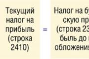
Accounting Regulations Accounting for calculations of income tax of organizations PBU 18/02 (as amended by Orders of the Ministry of Finance of the Russian Federation...
A trainee salesperson is usually called those salespeople who are not yet ready to work completely independently. Process...
Educational institution "Gomel State Medical University" Department of Neurology and Neurosurgery...

One of the most controversial and controversial methods for the early development of children was developed in the 80s by a sociologist...

Contents Dietary supplement based on an extract obtained from the fly beetle (or...

Ekaterina MirimanovaSystem minus 60. RevolutionSystem minus 60 with Ekaterina Mirimanova“System minus 60....
Heaviness and bloating are the causes of both ordinary overeating and more serious digestive problems...

The second blood group, Rh-negative, appeared many years ago, when a person ceased to be...

PSYCHOLOGICAL ASPECTS OF ANOREXIA PHENOMENON (EXPERIMENTAL STUDY) T. V. Tarasova, E. V. Arsentieva...

Contents Since the skin in this area is thin, it is more prone to the appearance of various types of spots....

vseslav Sat, 10/17/2015 - 20:50 Vasileostrovskaya station is one of the oldest stations...

P. S. Pallas (1741 - 1811) - naturalist and traveler-encyclopedist, who glorified his name with major contributions...
Today, all issues related to the placement of government orders are regulated by the Law on Contract...