The most complete interpretation of sleep orange
Orange juice. The symbolic meaning of orange juice in dream books is pleasure and temptation. Quite often we...
Brain cancer occurs in one in a hundred cases of tumor formation. The danger lies in the often sparse neurological symptoms, which at an early stage are mistaken for banal fatigue. As cancer cells develop, the clinical picture increases, but it may be too late for effective treatment. Therefore, it is important to seek qualified medical help if you detect one sign or a combination of several that persist for more than two weeks. The sooner you can recognize the symptoms and signs of brain cancer, the higher the likelihood of a full recovery. A favorable result is shown by treatment when the tumor size is less than 2 cm.
There is no single clinical picture for a brain affected by cancer cells. Symptoms vary depending on the size of the tumor and location. General cerebral and focal signs are distinguished. The first group includes headache, vomiting, dizziness. This clinical picture is typical for cancer due to increased intracranial pressure (hypertension).
The first signs of brain cancer include severe pain, which is sharp, spreading, increasing in nature, and occurs at night or in the morning. It is not relieved by analgesics and goes away on its own within a few hours. Symptoms worsen during coughing, defecation and other physical exertion on the abdominal muscles, or when changing body position.
Advice. If a severe headache persists for more than two weeks and is not relieved by painkillers, you should consult a doctor immediately!
In the first stages of tumor development, vomiting often occurs. It is not associated with dysfunction of the gastrointestinal tract or food intake, so there is no feeling of nausea. Vomiting is gushing, reflexive in nature, and sometimes occurs on an empty stomach. Dizziness is one of the signs of early stage brain cancer, characterized by dysfunction of the vestibular apparatus or pituitary gland. The patient is confident that he is moving, turning, remaining in place. Dizziness occurs regardless of body position and lasts for a long time.
Common focal symptoms of early stage brain cancer include:
First-degree cancer often manifests itself with the following neurological symptoms:
The affected area of the brain forms focal symptoms, which become more active as cancer cells develop. A tumor of the frontal lobe is characterized by epileptic seizures, often mental disorders (unmotivated actions, inadequate humor). In this case, symptoms are observed:
The patient smells an odor of unknown origin or aromas that are unusual for a specific object. Sometimes speech is partially lost. Signs of brain cancer in adults of the temporal lobe are olfactory and taste disturbances. The patient experiences auditory hallucinations in the form of endless monotonous knocking and ringing. Symptoms include epileptic seizures and impaired articulation (sensory aphasia). The latter implies that the patient hears sounds, but is deprived of the ability to analyze the speech of the people around him, and perceives his native language as a foreign language.
Extremely rare cancers of the occipital lobe. They affect the optic nerve and, depending on the degree of development, cause symptoms:
Parietal lobe cancer characterized by damage to the limbs. There is unsteadiness of gait, instability in the Romberg position, and impaired pain and tactile sensitivity of the side opposite to the tumor location. A quarter of neoplasms are meningiomas. This type of brain cancer is common in women and is characterized by the following symptoms:
Cerebellar cancer in adults it manifests itself as a lack of coordination, movements become unclear and sweeping. Muscle hypotonia and pendulum syndrome, when the eyes dart from side to side, are often noted.
Cranial nerve tumors more common in women. Distinctive feature neuronyms is early-stage hearing loss, which may be the only symptom. Sometimes paralysis of half the face develops on the side where the cancerous tumor is located, along with a dull aching pain. Women often experience diplopia and inactivity of the masticatory muscles. If cancer is detected at an early stage, the probability of five-year survival depends on how old the patient is, the location and size, type of tumor, and its spread to other organs.
In the treatment of brain cancer, it is important to recognize it in time and begin to act as early as possible. It often happens that symptoms do not appear for quite a long time. For example, such a common symptom of cancer in the form of headaches in the first and second stages appears only in half of the cases.
It is impossible to say exactly how many people live with stage 4 brain cancer. Even with a clear clinical picture, results of computed tomography, MRI, histology and other data, doctors may make a mistake in predicting the patient’s life expectancy.
There are five stages of brain cancer in total, but the fourth is considered final. At the same time, there is no hope for a complete recovery; only in one out of five cases can the patient’s life be prolonged. A terminal cancer tumor implies death. Patients over 65 years old can live another 2-3 years. The chances are higher for people aged 20-45, who have a greater potential to fight cancer. How much time is allocated to the patient depends on him.


Advice. Do not underestimate the role of psychological attitude. Cancer puts moral pressure on the patient, which inhibits the body’s ability to fight the disease. Support is extremely important for the patient.
The most dangerous type of brain cancer is glioblastoma, which forms stellate cells. Patients with this stage 4 tumor live for a maximum of a year. The reason is the progressive metastasis of cancer cells to healthy ones, which sharply reduces the chances of removing the tumor without serious consequences. Glioblastoma is immediately classified as stage 4; such cancer is initially considered inoperable.
Among the most serious and dangerous diseases is brain cancer, the causes of which are not fully understood by scientists. This disease is quite difficult to treat. The danger of the disease lies in its asymptomatic course, which entails the tumor growing into the last stage. Quite often, patients consult a doctor with symptoms that indicate other diseases. It is very important at this moment to conduct a competent diagnosis, which will allow timely detection of cancer.
Brain oncology is not a common occurrence, but it carries a huge danger. Formations are not always deadly. Tumors are divided into two types:
Tumors that develop from the membrane of the brain are called. A neoplasm formed directly in the brain tissue is called. Malignant formations affect the sheath of the cranial nerves, and therefore have a name.

To protect yourself and your loved ones, it is important to know what causes brain cancer. To the question: why cancer occurs, scientists cannot give a comprehensive answer, but today a list of the most common factors that cause this disease has been compiled. The main causes of the formation of cancerous tumors are:
Numerous studies have shown that women and people with fair skin most often suffer from this disease. Most often, brain cancer occurs in adult patients. In children, this disease is much less common. The tumor is localized in the lining of the brain, affecting the pineal gland and pituitary gland. In children, the disease has a negative impact on the cerebellum and organ trunk. Brain cancer can also form in the presence of cancerous tumors in other organs of the body. Metastases penetrate the circulatory system, causing cancer cells to easily pass through the skull into the brain tissue.
To determine therapeutic and surgical options, it is necessary to know how brain cancer develops. Brain cancer has the following stages of development:

Early identification of symptoms will help to recognize the disease in its first stages.
For timely detection of the disease, it is necessary to know how brain cancer manifests itself in the primary stages. The first thing you need to know is that signs of the disease can be single and appear almost imperceptibly. Regardless of the location of cancer, they can be focal or cerebral. The second type appears precisely in the first stages:

Focal symptoms depend on the location of the tumor and manifest themselves as:
These symptoms may be a sign of other dangerous diseases. Therefore, if at least one of the symptoms appears, you should consult a doctor who will prescribe a series of diagnostic procedures.
It is important to know how brain cancer manifests itself in the later stages in order to take measures to improve the patient's condition. In later stages, the following symptoms appear:
This symptomatology makes a person incapacitated and dependent. Therefore, you should not delay your visit to the doctor.
Doctors have found that 16% of cancer patients are children. These tumors are called medullablastomas. This species rarely metastasizes, and symptoms manifest themselves in the pressure of the tumor on certain areas of the brain. The main sign of tumor development in children is considered to be an increase in head circumference, and in infants the fontanelle swells. In older children, there is a strong increase in intracranial pressure, resulting in divergence of the sutures of the skull. Basically, all the symptoms of brain cancer occur the same in adults and children. The problem is that a small child cannot explain his feelings. You can guess the problem by observing his behavior. As a rule, children become nervous, often cry, and constantly pull their hands to their head. Doctors can determine the disease by the child's fundus. During the examination, swelling and hemorrhages in the retinal area are visible. These symptoms are conditional and an accurate diagnosis can only be made after a series of diagnostic procedures.

It is simply unrealistic to understand that you have brain cancer without a certain examination. If any symptom appears, it is necessary to undergo an examination, which will be prescribed by a doctor after collecting an anamnesis. Diagnosis of brain cancer includes:
In this article we will look at the symptoms and signs of brain cancer. What kind of disease is this?
Brain cancer is a rare disease and at the same time little studied. It is often fatal. At the same time, as doctors say, a characteristic feature of cancer patients is almost always the extreme neglect of the disease, when the chances of a cure are much less than they could be. Let's find out what are the first signs of early-stage brain cancer in adult patients.
This is an extremely dangerous disease that is difficult to treat and can lead to the death of the patient. The greatest threat is posed by the asymptomatic course of the disease. Basically, the fourth stage is characterized by pronounced symptoms, but at this stage the disease is difficult to treat, and the prognosis for such people is disappointing.
The symptoms of brain cancer in women are not particularly different from those in men.
At the same time, the symptoms with which a patient may consult a doctor can easily be confused with signs of other diseases. For example, headaches along with vomiting and dizziness in combination with blurred vision are often observed with migraines and hypertensive crisis. In addition, headaches can be caused by osteochondrosis. In this regard, therapy depends on the level of qualifications of the doctor to whom the patient turns for diagnosis. It is extremely important that the specialist is able to detect dangerous symptoms in time and conduct the necessary examination, which can help identify oncological processes.
Tumors in medicine are classified according to the tissues in which they grow. Thus, a tumor that develops from the lining of the brain is called meningioma. A tumor that arises in brain tissue is a ganglioma or astrocytoma, and their common name will be neuroepithelial neoplasms. Neuroma is a malignant tumor affecting the nerve sheath of the skull.

Gliomas make up about eighty percent of malignant neoplasms; meningiomas are also classified as common tumors; doctors note them in thirty-five percent of brain oncology cases. Now let's find out what are the main reasons for the appearance of this dangerous disease.
Let's look at the signs of brain cancer below.
It must be said that the causes of brain tumors have not yet been fully studied. As practice shows, in ten percent of cases, cancer is provoked by hereditary gene diseases. Secondary neoplasms arise due to the spread of metastases against the background of cancer of other organs. Today, doctors identify several causes of brain cancer.
Secondary tumors are a consequence of other oncological processes that occur in the body: metastases penetrate into the skull through the circulatory system and contribute to the appearance of a malignant neoplasm. Such tumors often occur against the background of breast cancer and other oncological diseases.
In brain oncology, symptoms are of two types: focal and cerebral. General cerebral symptoms are typical for all cases of cancer, while focal symptoms directly depend on the location of the tumor. Focal symptoms can be very diverse, its type and severity depend on the area of the brain that is affected by the disease, as well as on the functions for which it is responsible: be it memory, counting, written speech, and so on. Among the focal symptoms of the brain, the following signs are distinguished:

All tumors are characterized by common symptoms (brain cancer is no exception), which are associated with increased intracranial pressure, and, in addition, the mechanical effect of the tumor on different centers of the brain. So, the following symptoms are observed:
Signs of early-stage brain cancer often go undetected for a long time.
Many people want to know how brain cancer manifests itself. The first signs are not limited to this.
Now let's look at the symptoms that occur in later stages:

We now know what signs of cancer a person may develop.
Types of diagnosing brain cancer include the following procedures:

After diagnosis, the doctor draws up an individual treatment plan.
Signs of brain cancer in men and women depend on the stage of the disease.
Due to the almost asymptomatic course of the disease, it is difficult to accurately determine its stage. This is especially difficult to do due to the fact that the disease passes from one stage to another quickly and unexpectedly. This is especially true for cancers in the brain stem. The stage of the disease is accurately determined only after a post-mortem autopsy; therefore, the slightest signs of pathology should be treated carefully from the very first days. Unfortunately, at the last stage, cancer is not amenable to surgical therapy, and, in addition, responds extremely poorly to medications and other types of treatment. There are four stages in total:
Symptoms and signs of brain cancer in adults can be determined by a qualified physician.

As part of predicting the development of the disease and assessing the health status of patients with brain cancer, the concept of “five-year survival” is used. Patients who have been diagnosed with this disease are assessed, regardless of the course of therapy used. Some patients live longer than 5 years after successful treatment, while others are forced to undergo regular therapeutic procedures. The average survival rate for people with tumors located in the brain is thirty-five percent. As for malignant tumors, of which the majority are gliomas, in this case the survival rate is only five percent.
We looked at the symptoms and signs of brain cancer.
Brain tumors are composed of cancer cells that exhibit abnormal growth in the brain. They can be benign (meaning they do not spread elsewhere or invade surrounding tissue) or malignant (cancerous). Brain cancers are also divided into primary and secondary.
Primary brain tumors.
Primary tumors begin in the brain, while secondary tumors spread from the brain to other organs, such as the breasts or lungs. (In this article, the term “brain tumor” refers primarily to the primary malignant tumor unless otherwise noted.)Primary benign brain tumors account for half of all brain tumors. Their cells appear relatively normal, grow slowly and do not spread (metastasize) to other parts of the body or invade brain tissue. However, benign tumors can also be a serious problem, even life-threatening, if they are in a vital area of the brain where they put pressure on sensitive nerve tissue, or if they increase pressure on the brain.
Although some benign brain tumors can pose health risks, including the risk of disability and death, most can usually be treated successfully with treatments such as surgery.
Primary malignant brain tumors originate in the brain itself. Although they often spread cancer cells to other parts of the central nervous system (brain or spinal cord), they rarely spread to other parts of the body.
Brain tumors are generally named and classified according to the following criteria:
The type of brain cells they come from;
- the place where cancer develops.
The biological diversity of these tumors, however, makes classification difficult.
Secondary malignant (metastatic) brain tumors. Secondary, metastatic, brain tumors occur when cancer cells spread to the brain from a primary cancer in other parts of the body. Secondary brain tumors occur approximately three times more often than primary ones.
Single metastases from brain cancer can occur, but are less common than multiple tumors. Most often, cancers that have spread to the brain and cause secondary brain tumors arise in the lungs, breasts, kidneys, or from skin melanoma.
All metastatic brain tumors are malignant.
- Primary brain tumors are gliomas. About 80% of primary malignant brain tumors are known as gliomas. It is not a specific type of cancer, but the term is used to describe tumors that arise in glial cells (neuroglia or glia - these cells surround nerve cells and play a supporting role; glial cells, other than microglia, have common functions and partly a common origin, they constitute a specific microenvironment for neurons, providing conditions for the transmission of nerve impulses). Glial cells are the building blocks of connective or supporting tissue cells in the central nervous system (CNS).
Gliomas are divided into four classes, which reflect the degree of malignancy. Classes (grades) I and II are considered low-grade, and classes III and IV are considered high-grade. Classes I and II are the slowest growing and the least malignant. Class III are considered malignant tumors and grow at a moderate rate. Class IV malignancy – tumors such as glioblastoma are the fastest growing and most malignant primary brain tumors. Gliomas can develop from several types of glial cells.

- Astrocytomas. Astrocytomas of primary brain tumors are derived from astrocytes, also glial cells. Astrocytomas account for about 60% of all malignant primary brain tumors.
- Oligodendrogliomas develop from oligodendrocytes - glial cells that form protective coverings around nerve cells. Oligodendrogliomas are classified as low-grade (grade II) or anaplastic (grade III). Oligodendrogliomas are rare. In most cases they occur in mixed gliomas. Oligodendrogliomas usually occur in young and middle-aged people.
- Ependymomas are derived from ependymal cells in the lower part of the brain and the central canal of the spinal cord. They are one of the most common types of brain tumors in children. They can also occur in adults between 40 and 50 years of age. Ependymomas are divided into four categories (classes): myxopapillary ependymomas (class I), subependymomas (class I), ependymomas (class II), and anaplastic aependymomas (classes III and IV).
Mixed gliomas contain a mixture of malignant gliomas. About half of these tumors contain cancerous oligodendrocytes and astrocytes. Gliomas may also contain cancer cells other than glial cells derived from brain cells.
- Non-gliomas. Malignant types of brain tumors - non-gliomas - include:
- Medulloblastomas. They are always found in the cerebellum, which lies towards the back of the brain. These fast-growing, high-grade tumors account for about 15-20% of pediatric and 20% of adult brain tumors.
- Pituitary adenomas. Pituitary tumors (also called "pituitary adenomas") account for about 10% of primary and often benign brain tumors that grow slowly in the pituitary gland. They are more common in women than men.
- CNS Lymphomas.
The CNS can affect people with both a healthy immune system and those with immunodeficiency due to other diseases (organ transplant recipients infected with HIV, etc.). CNS lymphomas most often arise in the cerebral hemispheres, but can also develop in the cerebrospinal fluid, eyes, and spinal cord.
Benign types of non-gliomas of the brain include:
- Meningiomas. These are usually benign tumors that develop in the membranes covering the brain and spinal cord (meninges). Meningiomas account for about 25% of all primary brain tumors and are most common in women 60–70 years of age. Meningiomas are classified as: benign meningiomas (grade I), atypical meningiomas (grade II), and anaplastic meningiomas (grade III).
- Genetics. Only 5-10% of primary brain tumors are associated with hereditary, genetic disorders.
For example, neurofibromatosis is associated with 15% of cases of pilocytic astrocytoma, the most common type of childhood glioma.
Many different cancer-causing genes (oncogenes) are involved in the growth of brain tumors. Receptors stimulate cell growth. Epidermal growth factor receptor plays an important role in the full-blown brain tumor glioblastoma. Knowing the molecular origin of a brain tumor can determine the course of treatment for both standard chemotherapy and “targeted therapy” with biological drugs.
Most genetic abnormalities that cause brain tumors are not inherited but result from environmental or other factors that affect the genetic material (DNA) in cells. Researchers are studying various environmental factors (viruses, hormones, chemicals, radiation, etc.) that can cause genetic disorders that lead to brain tumors. They also work to identify specific genes that are affected by these environmental triggers (ie, irritants, catalysts).
Primary malignant brain tumors account for about 2% of all cancers. However, tumors of the brain and spinal cord are, after leukemia, the second most common form of cancer in children.
- Floor. Brain tumors are slightly more common in men than in women. Some types (such as meningiomas) are more common in women.
- Age. Most brain tumors in adults occur between the ages of 65 and 79 years. Brain tumors usually occur in children under 8 years of age.
- Race. The risk of primary brain tumors is higher in whites than in other races.
- Environmental and occupational risk factors. Exposure to ionizing radiation, usually from radiation therapy, is the only environmental risk factor that is associated with brain tumors. People who receive radiation therapy to the head during treatment for any cancer have an increased risk of developing brain tumors 10 to 15 years later.
Nuclear workers are also at increased risk.
Research into metals, chemicals and other substances, including vinyl chloride, petroleum products, lead, arsenic, mercury, pesticides, etc. continues.
- Medical conditions. People with impaired immune systems have an increased risk of developing CNS lymphoma. Organ transplants, HIV infections and chemotherapy are medical factors that can weaken the immune system.
Malignant primary brain tumors are classified according to the grade(s) of malignancy. Grade I is the least cancerous, grades III and IV are the most dangerous. Classifying tumors can help predict how quickly they will grow and how they will spread.
Tumor cells of classes I and II are clearly defined and appear almost normal under the microscope. Some primary low-grade brain tumors are curable only with surgery, and some are curable with surgery and radiation therapy. Low-grade tumors tend to have better survival outcomes. However, this is not always the case. For example, some low-grade II gliomas have a very high risk of progression.
Higher grade tumor cells (III and IV) are adrenal and more diffuse in nature, indicating more aggressive behavior (high grade brain tumors usually require surgery, radiation therapy, chemotherapy, etc.). In tumors that contain a mixture of different classes of cells, the tumors differentiate depending on the highest grade of cells in the mixture.
Brain tumors produce a variety of symptoms. They often imitate other neurological disorders, which is also dangerous (it is not always possible to diagnose immediately). The problem occurs if the tumor directly damages nerves in the brain or central nervous system, or if its growth puts pressure on the brain. Symptoms may be mild and gradually get worse, or they may occur very quickly.
Main symptoms: headache; gastrointestinal symptoms including nausea and vomiting; seizures, etc.

Tumors can be localized and affect areas of the brain. In such cases, they can cause partial seizures, where the person does not lose consciousness, but may experience confusion, twitching, tingling, or confusion about mental and emotional events. Generalized seizures, which can lead to loss of consciousness, are less common because they are caused by damage to nerve cells in diffuse areas of the brain.
Mental changes as symptoms of brain tumors may include:
Memory loss;
- impaired concentration;
- problems with reasoning;
- changes in personality and behavior;
- increase in sleep duration.
- gradual loss of movement or sensation in the arms or legs;
- instability and balance problems;
- unexpected visual disturbances (especially if associated with headache), including loss of vision (usually peripheral) in one or both eyes, double vision;
- hearing loss with or without dizziness;
- difficulty speaking.
Brain tumors can cause seizures, mental changes, and emotional mood changes. The tumor can also impair muscle function, hearing, vision, speech, and other neurological activities. Many children who survive brain tumors are at risk for long-term neurological complications. Children under 7 years of age (especially those under 3 years of age) are at greatest risk for full cognitive development. These problems can arise as a result of the tumor and its treatment (cranial radiation therapy, chemotherapy, etc.).

A neurological examination is usually performed when a patient complains of symptoms suggestive of a brain tumor. The examination includes testing of eye movement, hearing, sensation, muscle strength, smell, balance and coordination. The doctor also checks the patient's mental state and memory.
Advanced imaging techniques have greatly improved the diagnosis of brain tumors:
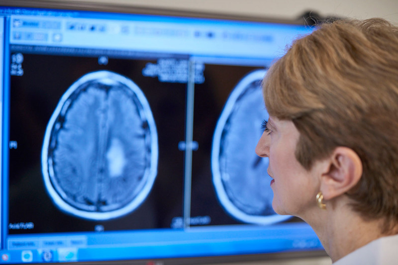
- Magnetic resonance imaging (MRI). MRI of the brain provides images from multiple angles that can help doctors build clear, three-dimensional images of tumors near bones, brain stem tumors, and low-grade tumors. MRI also shows tumor size during surgery to accurately map the brain and response to therapy. MRI creates a detailed image of the complex structures of the brain, allowing doctors to more accurately identify tumors or aneurysms.

- Computed tomography (CT) helps determine the location of the tumor and can sometimes help determine its type. It can also help detect swelling, bleeding and related symptoms. In addition, CT is used to evaluate the effectiveness of treatment and monitor tumor recurrence. A CT or MRI should usually be performed before a lumbar procedure to ensure that the procedure can be performed safely.

- Positron emission tomography (PET) provides insight into brain activity by tracking sugars that have been tagged with radioactive tracers, sometimes distinguishing between recurrent tumor cells and dead cells or scar tissue caused by radiation therapy. PET is not routinely used for diagnosis, but may complement MRI to determine tumor extent once diagnosis is made. PET data can also help improve the accuracy of new radiosurgery techniques. PET scans are often done in conjunction with CT scans.
- Single photon emission computed tomography (SPECT) helps distinguish tumor cells from destroyed tissues after treatment. It can be used after CT or MRI to help differentiate between low-grade and high-grade cancer.
- Magnetoencephalography (MEG) scans measurements of magnetic fields created by nerve cells producing electrical current. Used to evaluate the functioning of different parts of the brain. This procedure is not widely available.
- MRI angiography assesses blood flow. MR angiography is usually limited to planning surgical removal of tumors suspected of containing blood reserves.
- Spinal tap (lumbar puncture) used to obtain a sample of cerebrospinal fluid, which is examined for the presence of tumor cells. The cerebrospinal fluid may also be tested for the presence of certain tumor markers (substances that indicate the presence of a tumor). However, most primary brain tumors are not detected by tumor markers.

- Biopsy. This is a surgical procedure in which a small sample of tissue is taken from suspected tumors and examined under a microscope for malignancy. Biopsy results also provide information about the type of cancer cells. A biopsy may be performed either as part of surgery to remove the tumor or as a separate diagnostic procedure.
In some cases, such as brainstem glioma, a standard biopsy may be too dangerous because removing any healthy tissue from the area may affect vital functions. In these cases, surgeons may use alternative methods such as stereotactic biopsy. This is a computer-guided type of biopsy that uses images from an MRI or CT scan to provide precise information about the location of the tumor.
- Standard treatment.
The standard approach to treating brain tumors is to shrink the tumor as much as possible through surgery, radiation therapy, or chemotherapy. Such approaches are used individually or, more often, in combination with each other.
The intensity, combinations and sequence of treatments depend on the type of brain tumor (there are more than 100 types), its size and location, as well as the patient's age, health and medical history. Unlike other types of cancer, there is no system of organization for brain tumors.
Some very slow-growing cancers that originate in the brain or optic nerve pathways may require patients to be watched closely and not treated until the tumor shows signs of growth.
- TTF therapy. TTF therapy literally translates as “tumor treating fields”. The fundamental principle of the method is the effect of an electric field on cancer cells, leading to their apoptosis. A low intensity electric field is used to disrupt the rapid division of cancer cells. The system for treating adults with glioblastoma so that it does not recur or progress despite chemotherapy and radiation is a new device using electrodes placed on the patient's scalp at the site of the tumor, where an alternating electric field is delivered that affects only the area where the tumor is located. By selecting the frequency of the alternating electric field, it is possible to influence only a certain type of malignant cells without causing harm to healthy tissues.
- Radiation therapy. Radiation therapy, also called radiation therapy, plays a central role in the treatment of most brain tumors.
Radiation is usually received externally, from a source outside the body that directs beams of radiation. Even when all tumors have been surgically removed, microscopic cancer cells often remain in the surrounding tissue. The goals of radiation are to reduce the size of the residual tumor or stop its development. If the entire tumor cannot be removed, postoperative radiation therapy is recommended. Even some benign gliomas may require radiation, as they can become life-threatening if their growth is not controlled.
Radiation therapy can also be used instead of surgery for tumors that are difficult to reach and those tumors that have properties that are particularly responsive to radiation therapy.
Combining chemotherapy with radiation therapy is useful for some patients with high-grade tumors.
Conventional radiotherapy uses external beams aimed directly at the tumor, which is generally recommended for large or penetrating tumors. Conventional radiation therapy begins approximately one week after surgery and continues on an outpatient basis 5 days a week for 6 weeks. Older people tend to have a more limited response to external beam radiation therapy than younger people.
Three-dimensional conformal radiation therapy uses computer-generated images to scan tumors. Radiation beams are then used to match the three-dimensional shape of the tumor.
Researchers are studying drugs that can be used together with radiation to increase the effectiveness of treatment: radioprotectors, radiosensitizers, etc.
- Stereotactic radiosurgery (stereotactic radiation therapy, or stereotaxis), an alternative to conventional radiotherapy, allows radiation to be precisely targeted directly to small tumors, while avoiding healthy brain tissue. The destruction is so precise that it acts almost like a surgical knife. The advantages of stereotactic radiosurgery: it allows high-dose beams to be precisely targeted to target gliomas, with minimal damage to surrounding tissue. Stereotactic radiosurgery can help reach small tumors deep in the brain that were previously considered inoperable.
- Chemotherapy. Chemotherapy uses drugs to kill or change cancer cells. Chemotherapy is not an effective treatment for early low-grade brain tumors, mainly because standard drugs have difficulty getting into the brain as the brain protects itself across the blood-brain barrier. Additionally, not all types of brain tumors respond to chemotherapy. It is usually given after brain tumor surgery or radiation therapy.
- Interstitial chemotherapy It uses disc-shaped polymer plates (called Gliadel plates) impregnated with Carmustine, a standard chemotherapy drug for brain cancer. Plate implants are removed directly into the cavity following surgical tumor surgery.
- Intrathecal chemotherapy provides administration of chemotherapy drugs directly into the cerebrospinal fluid.
- Intra-arterial chemotherapy delivers high-dose chemotherapy into brain arteries using tiny catheters.
- Chemotherapy drugs and treatment regimens.
Many different drugs and drug combinations are used for chemotherapy. The standard ones are: Temozolamide (Temodar), Carmustine (Biknu), PVC (Procarbazine, Lomustine and Vincristine).
Platinum-based drugs: Cisplatin (Platinol) and Carboplatin (Paraplatin) are standard cancer drugs that are sometimes used to treat glioma, medulloblastoma, and other types of brain tumors.
Researchers are studying drugs used to treat other types of cancer that may have benefits in treating brain tumors. These drugs include: Tamoxifen (Nolvadex) and Paclitaxel (Taxol), which are used to treat breast cancer, Topotecan (Hicamtin), which is used to treat ovarian and lung cancer, Vorinostat (Zolinza), which is approved for the treatment cutaneous T-cell lymphoma, Irinotecan (Camptostar) is another anticancer drug being studied in combination treatment.
- Biological drugs (targeted therapy). Traditional chemotherapy drugs can be effective against cancer cells, but because they do not differentiate between healthy and cancer cells, their high generalized toxicity can cause serious side effects. Meanwhile, targeted biological therapies work at the molecular level, blocking certain mechanisms associated with cancer growth and cell division. Because they selectively target cancer cells, these biologics may cause less serious side effects. They also hold the promise of creating more personalized cancer treatment options based on a patient's genotype.
Bevacizumab (Avastin) is a biologic drug that blocks the growth of blood vessels that feed tumors (a process called angiogenesis). Approved for the treatment of glioblastoma in patients whose brain cancer continues to progress after prior treatment with chemotherapy and radiation.
Targeted treatments undergoing clinical trials include: vaccines; tyrosine inhibitors, which block proteins involved in the growth of tumor cells; tyrosine kinase inhibitors and other new drugs.
Patients may also take part in clinical trials that study new treatments for brain tumors.
Surgery is usually the mainstay of treatment for most brain tumors. In some cases, however (brain stem gliomas and other tumors located deep inside the brain), performing surgery can be dangerous. The goal of most brain tumor surgeries is to remove or shrink the tumor as much as possible. By reducing the size of the tumor, other types of therapy, particularly radiation, may be more effective.
- Craniotomy. The standard surgical procedure is called craniotomy. The neurosurgeon removes part of the skull bone to expose the area of the brain above the tumor. The tumor site is then removed.

There are various surgical methods to destroy and remove the tumor. They include:
Laser microsurgery, which produces heat that concentratedly vaporizes tumor cells;
- Ultrasonic aspiration, which uses ultrasound to break glioma tumors into small pieces, which are then sucked out.
A relatively benign class of glioma can only be treated with surgery. Most malignant tumors require additional treatment, including repeated surgeries.
Imaging techniques such as CT and MRI are used in conjunction with surgery.
The neurosurgeon's skill in removing the tumor is critical to the patient's survival. An experienced surgeon can work with many high-risk patients.
- Bypass surgery(shunts - flexible pipes) . Sometimes a brain tumor can create blockages in blood vessels and cerebrospinal fluid will accumulate excessively in the skull, causing increased intracranial pressure. In these cases, the surgeon may implant a ventriculoperitoneal shunt (VP) to drain the fluid.
The most serious concern from brain surgery is the preservation of brain function. Surgeons must be conservative in their approach to limit the removal of tissue that could lead to loss of function. Sometimes bleeding, blood clots and other complications occur. Postoperative complications include: swelling in the brain, which is usually treated with corticosteroid medications. Measures are taken to reduce the risk of blood clots during the postoperative period.
- Peritumoral edema and hydrocephalus. Some tumors, especially medulloblastomas, interfere with the flow of cerebrospinal fluid and cause hydrocephalus (fluid accumulation in the skull), which in turn causes fluid to accumulate in the ventricles (cavities) of the brain. Symptoms of peritumoral edema include: nausea and vomiting, severe headaches, lethargy, difficulty staying awake, seizures, blurred vision, irritability and fatigue. The ventricles of the brain are hollow chambers filled with cerebrospinal fluid (CSF), which supports brain tissue.

Corticosteroids (steroids) - such as Dexamethasone (Decadron) are used to treat peritumoral edema. Side effects include: high blood pressure, mood swings, increased risk of infection, increased appetite, facial swelling, fluid retention. A shunt procedure may be performed to drain the fluid (shunts allow fluid to be redirected and drained).
- Seizures. Seizures occur in common cases of brain tumors in younger, high-risk patients. Anticonvulsants, such as carbamazepine or phenobarbital, can treat seizures and are useful in preventing relapse. These drugs are not useful in preventing the first seizures, but they should not be used routinely to treat patients with a newly diagnosed brain tumor. Anticonvulsants should only be used for patients who have had a seizure.
Medicines including Paclitaxel, Irinotecan, Interferon and Retinoic acid may interact with chemotherapy used to treat brain cancer. However, patients should be sure to discuss all of these interactions with their doctors.
- Depression. Antidepressants may help treat the emotional side effects associated with brain tumors. Support groups can also be used successfully for patients and their families.
Recent advances in surgical and radiation therapy have significantly extended the average survival time for patients with brain tumors. These advanced treatments can often help reduce the size and progression of malignant gliomas.
Survival for people with brain tumors depends on many different variables:
Tumor type (eg, astrocytoma, oligodendroglioma, or ependymoma);
- location and size of the tumor (these factors influence whether the tumor can be removed surgically);
- degree of tumor differentiation;
- age of the patient;
- the patient’s ability to function and move;
- how far the tumor has spread.
Patients with some types of tumors have relatively good survival rates. The five-year survival rates for patients with ependymoma and oligodendroglioma, respectively, are 86% and 82% for people aged 20-44 years and 69% and 48% for patients aged 55 to 64 years.
Glioblastoma of the brain has a worse prognosis for 5-year survival: only 14% for people aged 20-44 years and 1% for patients aged 55-64 years. Survival rates are highest in younger patients and decrease as patients age.
Intracranial neoplasms, including both tumor lesions of cerebral tissues and nerves, membranes, vessels, and endocrine structures of the brain. They manifest themselves as focal symptoms, depending on the topic of the lesion, and general cerebral symptoms. The diagnostic algorithm includes examination by a neurologist and ophthalmologist, echo-EG, EEG, CT and MRI of the brain, MR angiography, etc. The most optimal is surgical treatment, supplemented with chemotherapy and radiotherapy if indicated. If this is not possible, palliative treatment is carried out.

Brain tumors account for up to 6% of all neoplasms in the human body. Their frequency of occurrence ranges from 10 to 15 cases per 100 thousand people. Traditionally, cerebral tumors include all intracranial neoplasms - tumors of cerebral tissue and membranes, formations of cranial nerves, vascular tumors, neoplasms of lymphatic tissue and glandular structures (pituitary gland and pineal gland). In this regard, brain tumors are divided into intracerebral and extracerebral. The latter include neoplasms of the cerebral membranes and their choroid plexuses.
Brain tumors can develop at any age and can even be congenital. However, among children the incidence is lower, not exceeding 2.4 cases per 100 thousand children. Cerebral neoplasms can be primary, initially originating in the brain tissue, and secondary, metastatic, caused by the spread of tumor cells due to hemato- or lymphogenous dissemination. Secondary tumor lesions occur 5-10 times more often than primary tumors. Among the latter, the proportion of malignant tumors is at least 60%.
A distinctive feature of cerebral structures is their location in a limited intracranial space. For this reason, any volumetric formation of intracranial localization to one degree or another leads to compression of brain tissue and increased intracranial pressure. Thus, even brain tumors that are benign in nature, when they reach a certain size, have a malignant course and can lead to death. Taking this into account, the problem of early diagnosis and adequate timing of surgical treatment of cerebral tumors becomes particularly relevant for specialists in the field of neurology and neurosurgery.
The occurrence of cerebral neoplasms, as well as tumor processes of other localizations, is associated with exposure to radiation, various toxic substances, and significant environmental pollution. Children have a high incidence of congenital (embryonic) tumors, one of the causes of which may be a violation of the development of cerebral tissues in the prenatal period. Traumatic brain injury can serve as a provoking factor and activate a latent tumor process.
In some cases, brain tumors develop during radiation therapy for patients with other diseases. The risk of developing a cerebral tumor increases when undergoing immunosuppressive therapy, as well as in other groups of immunocompromised individuals (for example, with HIV infection and neuroAIDS). A predisposition to the occurrence of cerebral neoplasms is noted in certain hereditary diseases: Hippel-Lindau disease, tuberous sclerosis, phakomatoses, neurofibromatosis.
Among primary cerebral neoplasms, neuroectodermal tumors predominate, which are classified into:
Intracerebral cerebral tumors are classified according to localization into sub- and supratentorial, hemispheric, tumors of the middle structures and tumors of the base of the brain.
Suspicion of a brain mass is a clear indication for computed tomography or magnetic resonance imaging. A CT scan of the brain allows one to visualize a tumor formation, differentiate it from local edema of cerebral tissues, establish its size, identify the cystic part of the tumor (if any), calcifications, a zone of necrosis, hemorrhage into the metastasis or tissue surrounding the tumor, and the presence of a mass effect. MRI of the brain complements CT and makes it possible to more accurately determine the spread of the tumor process and assess the involvement of border tissues in it. MRI is more effective in diagnosing tumors that do not accumulate contrast (for example, some gliomas of the brain), but is inferior to CT when it is necessary to visualize bone destructive changes and calcifications, to distinguish the tumor from the area of perifocal edema.
In addition to standard MRI, MRI of brain vessels (study of vascularization of the tumor), functional MRI (mapping speech and motor areas), MR spectroscopy (analysis of metabolic abnormalities), MR thermography (monitoring thermal destruction of the tumor) can be used in the diagnosis of a brain tumor. PET of the brain makes it possible to determine the degree of malignancy of a brain tumor, identify tumor relapse, and map the main functional areas. SPECT using radiopharmaceuticals specific to cerebral tumors makes it possible to diagnose multifocal lesions, assess the malignancy and degree of vascularization of the tumor.
In some cases, stereotactic biopsy of a brain tumor is used. During surgical treatment, tumor tissue is collected intraoperatively for histological examination. Histology makes it possible to accurately verify a neoplasm and establish the level of differentiation of its cells, and therefore the degree of malignancy.
Conservative therapy for a brain tumor is carried out with the aim of reducing its pressure on cerebral tissue, reducing existing symptoms, and improving the patient’s quality of life. It may include painkillers (ketoprofen, morphine), antiemetic drugs (metoclopramide), sedatives and psychotropic drugs. To reduce brain swelling, glucocorticosteroids are prescribed. It should be understood that conservative therapy does not eliminate the root causes of the disease and can only have a temporary alleviating effect.
The most effective is surgical removal of the cerebral tumor. The surgical technique and access are determined by the location, size, type and extent of the tumor. The use of surgical microscopy allows for more radical removal of the tumor and minimizes injury to healthy tissue. For small tumors, stereotactic radiosurgery is possible. The use of the CyberKnife and Gamma Knife techniques is permissible for cerebral formations with a diameter of up to 3 cm. In case of severe hydrocephalus, shunt surgery can be performed (external ventricular drainage, ventriculoperitoneal shunting).
Radiation and chemotherapy can complement surgery or be a palliative treatment. In the postoperative period, radiation therapy is prescribed if the histology of the tumor tissue reveals signs of atypia. Chemotherapy is carried out with cytostatics selected taking into account the histological type of tumor and individual sensitivity.
Benign brain tumors of small size and localization accessible for surgical removal are prognostically favorable. However, many of them tend to recur, which may require repeated surgery, and each surgical intervention on the brain is associated with trauma to its tissues, leading to persistent neurological deficits. Tumors of a malignant nature, hard-to-reach localization, large size and metastatic nature have an unfavorable prognosis, since they cannot be radically removed. The prognosis also depends on the patient’s age and the general condition of his body. Old age and the presence of concomitant pathologies (heart failure, chronic renal failure, diabetes mellitus, etc.) complicate surgical treatment and worsen its results.
Primary prevention of cerebral tumors consists of excluding oncogenic environmental influences, early detection and radical treatment of malignant neoplasms of other organs to prevent their metastasis. Prevention of relapse includes avoiding sun exposure, head injuries, and taking biogenic stimulants.

Orange juice. The symbolic meaning of orange juice in dream books is pleasure and temptation. Quite often we...

We most often overcome all kinds of difficulties encountered along the path of life. Of course, for this we make...

This year your patron Neptune will be in your constellation and this is a good sign, because you will feel...

1993 who? 1993 is the year of which animal? — According to the Chinese horoscope, 1873, 1933, 1993 belonged to the years of the Black Water...

Wave-particle duality of light means that light simultaneously has the properties of continuous electromagnetic...

The role of biology is enormous in our world. Although it is not a priority subject, most students and parents...

Amines are organic derivatives of ammonia containing an NH 2 amino group and an organic radical. In general...

How to answer questions in Part BThe second part of the social studies work consists of 7 tasks with a short answer....

Form TORG-15 is drawn up in the case when during transportation, movement between and within the warehouse, when...

Nutritionists say that for good health and a slim figure, you must include snacks in your...

Delicious pickled carrots for the winter can be prepared in a variety of containers, it can be a wooden...

When I have a few minutes left to prepare breakfast, the simplest and fastest recipes are used....

An elegant table, a decorated Christmas tree, tangerine spirit spilled throughout all the rooms, soon the most magical holiday -...

Each of us has repeatedly faced financial difficulties and difficult periods in life when Fortune...

We most often overcome all kinds of difficulties encountered along the path of life. Of course, for this we...

This year your patron Neptune will be in your constellation and this is a good sign, because you will...