Annunciation Cathedral of the Moscow Kremlin
One of the oldest churches in the Moscow Kremlin stands on the edge of Cathedral Square on the edge of Borovitsky Hill. Many centuries...
In 1895, the German physicist W. Roentgen discovered a new, previously unknown type of electromagnetic radiation, which was named X-ray in honor of its discoverer. V. Roentgen became the author of his discovery at the age of 50, holding the post of rector of the University of Würzburg and having a reputation as one of the best experimenters of his time. One of the first to find technical application for the discovery of X-ray was the American Edison. He created a convenient demonstration apparatus and already in May 1896 organized an X-ray exhibition in New York, where visitors could examine their own hand on a luminous screen. After Edison's assistant died from severe burns he received during constant demonstrations, the inventor stopped further experiments with X-rays.
X-ray radiation began to be used in medicine due to its great penetrating ability. Initially, X-rays were used to examine bone fractures and determine the location foreign bodies in the human body. Currently, there are several methods based on X-ray radiation. But these methods have their drawbacks: radiation can cause deep damage to the skin. The ulcers that appeared often turned into cancer. In many cases, fingers or hands had to be amputated. X-ray(synonym for transillumination) is one of the main methods of x-ray examination, which consists of obtaining a planar positive image of the object under study on a translucent (fluorescent) screen. During fluoroscopy, the subject is positioned between a translucent screen and an x-ray tube. On modern X-ray transmission screens, the image appears when the X-ray tube is turned on and disappears immediately after it is turned off. Fluoroscopy makes it possible to study the function of an organ - the pulsation of the heart, the respiratory movements of the ribs, lungs, diaphragm, peristalsis of the digestive tract, etc. Fluoroscopy is used in the treatment of diseases of the stomach, gastrointestinal tract, duodenum, diseases of the liver, gallbladder and biliary tract. In this case, the medical probe and manipulators are inserted without damaging the tissue, and the actions during the operation are controlled by fluoroscopy and visible on the monitor.
X-ray - X-ray diagnostic method with registration of a still image on a photosensitive material - special. photographic film (X-ray film) or photographic paper with subsequent photo processing; With digital radiography, the image is recorded in the computer memory. It is performed on X-ray diagnostic machines - stationary, installed in specially equipped X-ray rooms, or mobile and portable - at the patient’s bedside or in the operating room. X-rays show the structural elements of various organs much more clearly than a fluorescent screen. X-rays are performed to identify and prevent various diseases; its main purpose is to help doctors of various specialties make a diagnosis correctly and quickly. An X-ray image records the condition of an organ or tissue only at the time of shooting. However, a single radiograph records only anatomical changes at a certain moment; it gives a static process; through a series of radiographs taken at certain intervals, it is possible to study the dynamics of the process, that is, functional changes. Tomography. The word tomography can be translated from Greek as "slice image". This means that the purpose of tomography is to obtain a layer-by-layer image of the internal structure of the object under study. Computer tomography is characterized by high resolution, which makes it possible to distinguish subtle changes in soft tissues. CT can detect such pathological processes, which cannot be detected by other methods. In addition, the use of CT makes it possible to reduce the dose of X-ray radiation received by patients during the diagnostic process.
Fluorography- a diagnostic method that allows one to obtain images of organs and tissues was developed at the end of the 20th century, a year after X-rays were discovered. In the photographs you can see sclerosis, fibrosis, foreign objects, neoplasms, inflammation of a developed degree, the presence of gases and infiltration in the cavities, abscesses, cysts, and so on. Most often, chest fluorography is performed to detect tuberculosis, a malignant tumor in the lungs or chest, and other pathologies.
X-ray therapy is a modern method used to treat certain joint pathologies. The main areas of treatment of orthopedic diseases using this method are: Chronic. Inflammatory processes of the joints (arthritis, polyarthritis); Degenerative (osteoarthrosis, osteochondrosis, spondylosis deformans). The purpose of radiotherapy is the inhibition of the vital activity of cells of pathologically altered tissues or their complete destruction. For non-tumor diseases, radiotherapy is aimed at suppressing the inflammatory reaction, suppressing proliferative processes, reducing pain sensitivity and secretory activity of the glands. It should be taken into account that the sex glands, hematopoietic organs, leukocytes, and malignant tumor cells are most sensitive to X-rays. The radiation dose is determined individually in each specific case.
For the discovery of X-rays, Roentgen was awarded the first Nobel Prize in Physics in 1901, and the Nobel Committee emphasized the practical importance of his discovery.
Thus, X-rays are invisible electromagnetic radiation with a wavelength of 105 - 102 nm. X-rays can penetrate some materials that are opaque to visible light. They are emitted during the deceleration of fast electrons in a substance (continuous spectrum) and during transitions of electrons from the outer electron shells of an atom to the inner ones (line spectrum). Sources of X-ray radiation are: an X-ray tube, some radioactive isotopes, accelerators and electron storage devices (synchrotron radiation). Receivers - photographic film, fluorescent screens, nuclear radiation detectors. X-rays are used in X-ray diffraction analysis, medicine, flaw detection, X-ray spectral analysis, etc.
X-rays were discovered by accident in 1895 by the famous German physicist Wilhelm Roentgen. He studied cathode rays in a low-pressure gas-discharge tube at high voltage between its electrodes. Despite the fact that the tube was in a black box, Roentgen noticed that a fluorescent screen, which happened to be nearby, glowed every time the tube was in use. The tube turned out to be a source of radiation that could penetrate paper, wood, glass and even a one and a half centimeter thick aluminum plate.
X-ray determined that the gas-discharge tube was a source of a new type of invisible radiation with great penetrating power. The scientist could not determine whether this radiation was a stream of particles or waves, and he decided to give it the name X-rays. They were later called X-rays
It is now known that X-rays are a type of electromagnetic radiation that has a shorter wavelength than ultraviolet electromagnetic waves. The wavelength of X rays ranges from 70 nm up to 10 -5 nm. The shorter the wavelength of X-rays, the greater the energy of their photons and the greater their penetrating power. X-rays with a relatively long wavelength (more than 10 nm), are called soft. Wavelength 1 - 10 nm characterizes hard X-rays. They have enormous penetrating power.
X-rays are produced when fast electrons, or cathode rays, collide with the walls or anode of a low-pressure gas discharge tube. A modern X-ray tube is a evacuated glass cylinder with a cathode and anode located in it. The potential difference between the cathode and anode (anti-cathode) reaches several hundred kilovolts. The cathode is a tungsten filament heated by electric current. This causes the cathode to emit electrons as a result of thermionic emission. The electrons are accelerated by the electric field in the X-ray tube. Since there is a very small number of gas molecules in the tube, the electrons practically do not lose their energy on the way to the anode. They reach the anode at a very high speed.
X-rays are produced whenever electrons moving at high speed are slowed down by the anode material. Most of the electrons' energy is dissipated as heat. Therefore, the anode must be artificially cooled. The anode in the X-ray tube must be made of a metal that has a high melting point, such as tungsten.
The part of the energy that is not dissipated in the form of heat is converted into the energy of electromagnetic waves (X-rays). Thus, X-rays are the result of electron bombardment of the anode substance. There are two types of X-rays: bremsstrahlung and characteristic.
Bremsstrahlung X-ray radiation occurs when electrons moving at high speed are slowed down by the electric fields of the anode atoms. The conditions for stopping individual electrons are not the same. As a result, various parts of their kinetic energy are converted into X-ray energy.
The spectrum of X-ray bremsstrahlung does not depend on the nature of the anode substance. As is known, the energy of X-ray photons determines their frequency and wavelength. Therefore, X-ray bremsstrahlung is not monochromatic. It is characterized by a variety of wavelengths that can be represented continuous (continuous) spectrum.
X-rays cannot have an energy greater than the kinetic energy of the electrons that form them. The shortest wavelength of X-ray radiation corresponds to the maximum kinetic energy of decelerating electrons. The greater the potential difference in the X-ray tube, the shorter the wavelengths of X-ray radiation can be obtained.
The characteristic X-ray radiation is not continuous, but line spectrum. This type of radiation occurs when a fast electron, reaching the anode, penetrates the inner orbitals of atoms and knocks out one of their electrons. As a result, a free space appears that can be filled by another electron descending from one of the upper atomic orbitals. This transition of an electron from a higher to a lower energy level produces x-rays of a specific discrete wavelength. Therefore, the characteristic X-ray radiation has line spectrum. The frequency of the characteristic radiation lines completely depends on the structure of the electron orbitals of the anode atoms.
The spectrum lines of the characteristic radiation of different chemical elements have the same appearance, since the structure of their internal electron orbitals is identical. But their wavelength and frequency are due to energy differences between the internal orbitals of heavy and light atoms.
The frequency of the lines in the spectrum of characteristic X-ray radiation changes in accordance with the atomic number of the metal and is determined by the Moseley equation: v 1/2 = A(Z-B), Where Z- atomic number of a chemical element, A And B- constants.
The primary interaction between X-rays and matter is characterized by three mechanisms:
1. Coherent scattering. This form of interaction occurs when the X-ray photons have less energy than the binding energy of the electrons to the atomic nucleus. In this case, the photon energy is not sufficient to release electrons from the atoms of the substance. The photon is not absorbed by the atom, but changes the direction of propagation. In this case, the wavelength of X-ray radiation remains unchanged.
2. Photoelectric effect (photoelectric effect). When an X-ray photon reaches an atom of a substance, it can knock out one of the electrons. This occurs if the photon energy exceeds the binding energy of the electron with the nucleus. In this case, the photon is absorbed and the electron is released from the atom. If a photon carries more energy than is needed to release an electron, it will transfer the remaining energy to the released electron in the form of kinetic energy. This phenomenon, called the photoelectric effect, occurs when relatively low-energy X-rays are absorbed.
An atom that loses one of its electrons becomes a positive ion. The lifetime of free electrons is very short. They are absorbed by neutral atoms, which turn into negative ions. The result of the photoelectric effect is intense ionization of the substance.
If the energy of the X-ray photon is less than the ionization energy of the atoms, then the atoms go into an excited state, but are not ionized.
3. Incoherent scattering (Compton effect). This effect was discovered by the American physicist Compton. It occurs when a substance absorbs X-rays of short wavelength. The photon energy of such X-rays is always greater than the ionization energy of the atoms of the substance. The Compton effect results from the interaction of a high-energy X-ray photon with one of the electrons in the outer shell of an atom, which has a relatively weak connection with the atomic nucleus.
A high-energy photon transfers some of its energy to the electron. The excited electron is released from the atom. The remaining energy from the original photon is emitted as an x-ray photon of longer wavelength at some angle to the direction of motion of the original photon. The secondary photon can ionize another atom, etc. These changes in the direction and wavelength of X-rays are known as the Compton effect.
As mentioned above, X-rays are capable of exciting atoms and molecules of matter. This may cause certain substances (such as zinc sulfate) to fluoresce. If a parallel beam of X-rays is directed at opaque objects, you can observe how the rays pass through the object by placing a screen covered with a fluorescent substance.
The fluorescent screen can be replaced with photographic film. X-rays have the same effect on photographic emulsion as light. Both methods are used in practical medicine.
Another important effect of X-rays is their ionizing ability. This depends on their wavelength and energy. This effect provides a method for measuring the intensity of x-rays. When X-rays pass through the ionization chamber, an electric current is generated whose magnitude is proportional to the intensity of the X-rays.
As X-rays pass through matter, their energy decreases due to absorption and scattering. The attenuation of the intensity of a parallel beam of X-rays passing through a substance is determined by Bouguer’s law: I = I0 e -μd, Where I 0- initial intensity of X-ray radiation; I- intensity of X-rays passing through the layer of matter, d- absorbent layer thickness , μ - linear attenuation coefficient. It is equal to the sum of two quantities: t- linear absorption coefficient and σ - linear dissipation coefficient: μ = τ+ σ
Experiments have revealed that the linear absorption coefficient depends on the atomic number of the substance and the wavelength of the X-rays:
τ = kρZ 3 λ 3, Where k- coefficient of direct proportionality, ρ - density of the substance, Z- atomic number of the element, λ - wavelength of x-rays.
The dependence on Z is very important from a practical point of view. For example, the absorption coefficient of bones, which are composed of calcium phosphate, is almost 150 times higher than that of soft tissue ( Z=20 for calcium and Z=15 for phosphorus). When X-rays pass through the human body, bones stand out clearly against the background of muscles, connective tissue, etc.
It is known that the digestive organs have the same absorption coefficient as other soft tissues. But the shadow of the esophagus, stomach and intestines can be distinguished if the patient takes a contrast agent - barium sulfate ( Z= 56 for barium). Barium sulfate is very opaque to x-rays and is often used for x-ray examination of the gastrointestinal tract. Certain opaque mixtures are injected into the bloodstream in order to examine the condition of blood vessels, kidneys, etc. In this case, iodine, whose atomic number is 53, is used as a contrast agent.
Dependence of X-ray absorption on Z also used to protect against the possible harmful effects of x-rays. Lead is used for this purpose, the amount Z for which it is equal to 82.
The reason for the use of x-rays in diagnostics was their high penetrating ability, one of the main properties of x-ray radiation. In the early days after its discovery, X-rays were used mostly to examine bone fractures and determine the location of foreign bodies (such as bullets) in the human body. Currently, several diagnostic methods using x-rays (x-ray diagnostics) are used.
X-ray . An X-ray device consists of an X-ray source (X-ray tube) and a fluorescent screen. After X-rays pass through the patient's body, the doctor observes a shadow image of him. A lead window should be installed between the screen and the physician's eyes to protect the physician from the harmful effects of X-rays. This method makes it possible to study the functional state of certain organs. For example, the doctor can directly observe the movements of the lungs, the passage contrast agent along the gastrointestinal tract. The disadvantages of this method are insufficient contrast images and relatively large doses of radiation received by the patient during the procedure.
Fluorography . This method consists of taking a photograph of a part of the patient's body. Typically used for preliminary examination of the condition internal organs patients using low doses of X-ray radiation.
Radiography. (X-ray radiography). This is a research method using x-rays in which an image is recorded on photographic film. Photographs are usually taken in two perpendicular planes. This method has some advantages. X-ray photographs contain more detail than a fluorescent screen and are therefore more informative. They can be saved for further analysis. The total radiation dose is less than that used in fluoroscopy.
Computed X-ray tomography . Equipped with computer technology, the axial tomography scanner is the most modern X-ray diagnostic device that allows you to obtain a clear image of any part human body, including soft tissues of organs.
The first generation of computed tomography (CT) scanners include a special X-ray tube that is attached to a cylindrical frame. A thin beam of X-rays is directed at the patient. Two X-ray detectors are attached to the opposite side of the frame. The patient is in the center of the frame, which can rotate 180° around his body.
An X-ray beam passes through a stationary object. The detectors obtain and record the absorption values of various tissues. Recordings are made 160 times while the X-ray tube moves linearly along the scanned plane. Then the frame is rotated 1 0 and the procedure is repeated. Recording continues until the frame rotates 180 0 . Each detector records 28,800 frames (180x160) during the study. The information is processed by a computer, and an image of the selected layer is formed using a special computer program.
The second generation of CT uses several X-ray beams and up to 30 X-ray detectors. This makes it possible to speed up the research process up to 18 seconds.
The third generation of CT uses a new principle. A wide fan-shaped beam of X-rays covers the object under study, and the X-ray radiation passing through the body is recorded by several hundred detectors. The time required for research is reduced to 5-6 seconds.
CT has many advantages over earlier x-ray diagnostic methods. It is characterized by high resolution, which makes it possible to distinguish subtle changes in soft tissues. CT allows you to detect pathological processes that cannot be detected by other methods. In addition, the use of CT makes it possible to reduce the dose of X-ray radiation received by patients during the diagnostic process.
Although scientists have only discovered the effect of X-rays since the 1890s, the medical use of X-rays for this natural force has progressed rapidly. Today, for the benefit of humanity, X-ray electromagnetic radiation is used in medicine, academia and industry, as well as to generate electricity.
In addition, radiation has useful applications in areas such as agriculture, archaeology, space, law enforcement, geology (including mining) and many other activities, even cars are being developed using the phenomenon of nuclear fission.
In healthcare settings, physicians and dentists use a variety of nuclear materials and procedures to diagnose, monitor, and treat a wide range of metabolic processes and diseases in the human body. As a result, medical procedures using beams have saved thousands of lives by detecting and treating diseases ranging from an overactive thyroid gland to bone cancer.
The most common of these medical procedures involve the use of rays that can pass through our skin. When an image is taken, our bones and other structures appear to cast shadows because they are denser than our skin, and these shadows can be detected on film or a monitor screen. The effect is similar to placing a pencil between a piece of paper and a light. The shadow of the pencil will be visible on the piece of paper. The difference is that the rays are invisible, so a recording element is needed, something like photographic film. This allows doctors and dentists to evaluate the use of X-rays when seeing broken bones or dental problems.
The use of X-ray radiation in a targeted manner for therapeutic purposes is not only for detecting damage. When used specifically, it is intended to kill cancerous tissue, reduce tumor size, or reduce pain. For example, radioactive iodine (specifically iodine-131) is often used to treat thyroid cancer, a condition that affects many people.
Devices using this property also connect to computers and scan, called: computed axial tomography or computed tomography.
These instruments provide doctors with color images that show the outline and details of internal organs. It helps doctors detect and identify tumors, size abnormalities, or other physiological or functional organ problems.
In addition, hospitals and radiology centers perform millions of procedures annually. In such procedures, doctors release slightly radioactive substances into patients' bodies to look at certain internal organs, such as the pancreas, kidneys, thyroid, liver or brain, to diagnose clinical conditions.
X-RAY
X-ray radiation occupies the region of the electromagnetic spectrum between gamma and ultraviolet radiation and is electromagnetic radiation with a wavelength from 10 -14 to 10 -7 m. In medicine, X-ray radiation with a wavelength from 5 x 10 -12 to 2.5 x 10 -10 is used m, that is, 0.05 - 2.5 angstroms, and for X-ray diagnostics itself - 0.1 angstroms. Radiation is a stream of quanta (photons) propagating linearly at the speed of light (300,000 km/s). These quanta have no electrical charge. The mass of a quantum is an insignificant part of an atomic mass unit.
Energy of quanta measured in Joules (J), but in practice they often use a non-systemic unit "electron-volt" (eV) . One electron volt is the energy that one electron acquires when passing through a potential difference of 1 volt in an electric field. 1 eV = 1.6 10~ 19 J. The derivatives are the kiloelectron-volt (keV), equal to a thousand eV, and the megaelectron-volt (MeV), equal to a million eV.
X-rays are produced using X-ray tubes, linear accelerators and betatrons. In an X-ray tube, the potential difference between the cathode and the target anode (tens of kilovolts) accelerates the electrons bombarding the anode. X-ray radiation occurs when fast electrons are decelerated in the electric field of the atoms of the anode substance (bremsstrahlung) or during the restructuring of the inner shells of atoms (characteristic radiation) . Characteristic X-ray radiation has a discrete nature and occurs when the electrons of the atoms of the anode substance transfer from one energy level to another under the influence of external electrons or radiation quanta. Bremsstrahlung X-rays has a continuous spectrum depending on the anode voltage on the X-ray tube. When braking in the anode substance, electrons spend most of their energy on heating the anode (99%) and only a small fraction (1%) is converted into X-ray energy. In X-ray diagnostics, bremsstrahlung radiation is most often used.
The basic properties of X-rays are characteristic of all electromagnetic radiation, but there are some special features. X-rays have the following properties:
- invisibility - sensitive cells of the human retina do not respond to X-rays, since their wavelength is thousands of times shorter than that of visible light;
- straight propagation – rays are refracted, polarized (propagated in a certain plane) and diffracted, like visible light. The refractive index differs very little from unity;
- penetrating power - penetrate without significant absorption through significant layers of substances opaque to visible light. The shorter the wavelength, the greater the penetrating power of x-rays;
- absorption capacity - have the ability to be absorbed by body tissues; all x-ray diagnostics are based on this. The absorption capacity depends on the specific gravity of the tissue (the higher, the greater the absorption); on the thickness of the object; on the hardness of the radiation;
- photographic action - decompose silver halide compounds, including those found in photographic emulsions, which makes it possible to obtain X-ray images;
- luminescent effect - cause luminescence of a number of chemical compounds (luminophores), the X-ray transillumination technique is based on this. The intensity of the glow depends on the structure of the fluorescent substance, its quantity and distance from the X-ray source. Phosphors are used not only to obtain images of objects under study on a fluoroscopic screen, but also in radiography, where they make it possible to increase the radiation exposure to the radiographic film in the cassette due to the use of intensifying screens, the surface layer of which is made of fluorescent substances;
- ionization effect - have the ability to cause the disintegration of neutral atoms into positively and negatively charged particles, dosimetry is based on this. The effect of ionization of any medium is the formation in it of positive and negative ions, as well as free electrons from neutral atoms and molecules of the substance. Ionization of the air in the X-ray room during operation of the X-ray tube leads to an increase in the electrical conductivity of the air and an increase in static electric charges on cabinet objects. In order to eliminate such undesirable effects, forced supply and exhaust ventilation is provided in X-ray rooms;
- biological effect - have an impact on biological objects, in most cases this impact is harmful;
- inverse square law - for a point source of X-ray radiation, the intensity decreases in proportion to the square of the distance to the source.
Radiology is a branch of radiology that studies the effects of x-ray radiation on the body of animals and humans resulting from this disease, their treatment and prevention, as well as methods for diagnosing various pathologies using x-rays (x-ray diagnostics). A typical X-ray diagnostic apparatus includes a power supply device (transformers), a high-voltage rectifier that converts alternating current from the electrical network into direct current, a control panel, a stand and an x-ray tube.
X-rays are a type of electromagnetic oscillations that are formed in an X-ray tube during a sharp deceleration of accelerated electrons at the moment of their collision with atoms of the anode substance. Currently, the generally accepted point of view is that x-rays, by their physical nature, are one of the types of radiant energy, the spectrum of which also includes radio waves, infrared rays, visible light, ultraviolet rays and gamma rays of radioactive elements. X-ray radiation can be characterized as a collection of its smallest particles - quanta or photons.
Rice. 1 - mobile X-ray unit:
A - X-ray tube;
B - power supply device;
B - adjustable tripod.
 Rice. 2 - X-ray machine control panel (mechanical - on the left and electronic - on the right):
 A - panel for adjusting exposure and hardness;
B - high voltage supply button.
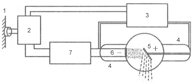 Rice. 3 - block diagram of a typical X-ray machine
 1 - network;
2 - autotransformer;
3 - step-up transformer;
4 - X-ray tube;
5 - anode;
6 - cathode;
7 - step-down transformer.
X-rays are formed at the moment of collision of a stream of accelerated electrons with the anode substance. When electrons interact with a target, 99% of their kinetic energy is converted into thermal energy and only 1% into X-ray radiation.
An X-ray tube consists of a glass cylinder into which 2 electrodes are soldered: a cathode and an anode. The air has been pumped out of the glass balloon: the movement of electrons from the cathode to the anode is possible only under conditions of relative vacuum (10 -7 –10 -8 mm Hg). The cathode has a filament, which is a tightly twisted tungsten spiral. When electric current is applied to the filament, electron emission occurs, in which electrons are separated from the filament and form an electron cloud near the cathode. This cloud is concentrated at the focusing cup of the cathode, which sets the direction of electron motion. The cup is a small depression in the cathode. The anode, in turn, contains a tungsten metal plate onto which electrons are focused - this is where X-rays are produced.
Rice. 4 - X-ray tube device: A - cathode; 
B - anode;
B - tungsten filament;
G - focusing cup of the cathode;
D - flow of accelerated electrons;
E - tungsten target;
F - glass flask;
Z - window made of beryllium;
And - formed x-rays;
K - aluminum filter.
There are 2 transformers connected to the electron tube: a step-down and a step-up. A step-down transformer heats the tungsten coil with low voltage (5-15 volts), resulting in electron emission. A step-up, or high-voltage, transformer fits directly to the cathode and anode, which are supplied with a voltage of 20–140 kilovolts. Both transformers are placed in the high-voltage block of the X-ray machine, which is filled with transformer oil, which ensures cooling of the transformers and their reliable insulation.
After an electron cloud has been formed using a step-down transformer, the step-up transformer is turned on, and a high-voltage voltage is applied to both poles of the electrical circuit: a positive pulse to the anode, and a negative pulse to the cathode. Negatively charged electrons are repelled from the negatively charged cathode and tend to the positively charged anode - due to this potential difference, a high speed of movement is achieved - 100 thousand km/s. At this speed, electrons bombard the tungsten plate of the anode, completing an electrical circuit, resulting in x-rays and thermal energy.
X-ray radiation is divided into bremsstrahlung and characteristic. Bremsstrahlung occurs due to a sharp slowdown in the speed of electrons emitted by a tungsten helix. Characteristic radiation occurs at the moment of restructuring of the electronic shells of atoms. Both of these types are formed in the X-ray tube at the moment of collision of accelerated electrons with atoms of the anode substance. The emission spectrum of an X-ray tube is a superposition of bremsstrahlung and characteristic X-rays.
 Rice. 5 - principle of formation of bremsstrahlung X-ray radiation.
 Rice. 6 - principle of formation of characteristic x-ray radiation.
X-rays have a specific wavelength and vibration frequency. The wavelength (λ) and oscillation frequency (ν) are related by the relation: λ ν = c, where c is the speed of light, rounded to 300,000 km per second. The energy of X-rays is determined by the formula E = h ν, where h is Planck's constant, a universal constant equal to 6.626 10 -34 J⋅s. The wavelength of the rays (λ) is related to their energy (E) by the ratio: λ = 12.4 / E.
X-ray radiation differs from other types of electromagnetic oscillations in wavelength (see table) and quantum energy. The shorter the wavelength, the higher its frequency, energy and penetrating power. The X-ray wavelength is in the range
. By changing the wavelength of X-ray radiation, its penetrating ability can be adjusted. X-rays have a very short wavelength but a high oscillation frequency and are therefore invisible to the human eye. Due to their enormous energy, quanta have great penetrating power, which is one of the main properties that ensure the use of X-ray radiation in medicine and other sciences.Intensity- a quantitative characteristic of X-ray radiation, which is expressed by the number of rays emitted by the tube per unit time. The intensity of X-ray radiation is measured in milliamps. Comparing it with the intensity of visible light from a conventional incandescent lamp, we can draw an analogy: for example, a 20-watt lamp will shine with one intensity, or strength, and a 200-watt lamp will shine with another, while the quality of the light itself (its spectrum) is the same . The intensity of x-ray radiation is essentially the amount of it. Each electron creates one or more quanta of radiation at the anode, therefore, the number of X-rays when exposing an object is regulated by changing the number of electrons tending to the anode and the number of interactions of electrons with atoms of the tungsten target, which can be done in two ways:
The X-ray intensity (anode current) multiplied by the exposure time (tube operating time) corresponds to the X-ray exposure, which is measured in mAs (milliamperes per second). Exposure is a parameter that, like intensity, characterizes the number of rays emitted by the X-ray tube. The only difference is that the exposure also takes into account the operating time of the tube (for example, if the tube works for 0.01 seconds, then the number of rays will be one, and if 0.02 seconds, then the number of rays will be different - twice more). The radiation exposure is set by the radiologist on the control panel of the X-ray machine, depending on the type of examination, the size of the object being examined and the diagnostic task.
Rigidity- qualitative characteristics of x-ray radiation. It is measured by the magnitude of the high voltage on the tube - in kilovolts. Determines the penetrating power of x-rays. It is regulated by the high voltage supplied to the X-ray tube by a step-up transformer. The higher the potential difference is created across the electrodes of the tube, the more force the electrons are repelled from the cathode and rush to the anode and the stronger their collision with the anode. The stronger their collision, the shorter the wavelength of the resulting X-ray radiation and the higher the penetrating ability of this wave (or the hardness of the radiation, which, like the intensity, is regulated on the control panel by the voltage parameter on the tube - kilovoltage).
λ - wavelength;  Rice. 7 - Dependence of wavelength on wave energy:
E - wave energy  Rice. 8 - The relationship between the voltage on the X-ray tube and the wavelength of the resulting X-ray radiation:
X-rays are used not only for x-ray diagnostic purposes, but also for therapeutic purposes. As noted above, the ability of X-ray radiation to suppress the growth of tumor cells makes it possible to use it in radiation therapy for cancer. In addition to the medical field of application, X-ray radiation has found wide application in engineering, materials science, crystallography, chemistry and biochemistry: for example, it is possible to identify structural defects in various products (rails, welds, etc.) using X-ray radiation. This type of research is called flaw detection. And at airports, train stations and other crowded places, X-ray television introscopes are actively used to scan hand luggage and baggage for security purposes.
Depending on the type of anode, X-ray tubes vary in design. Due to the fact that 99% of the kinetic energy of electrons is converted into thermal energy, during operation of the tube, significant heating of the anode occurs - the sensitive tungsten target often burns out. The anode is cooled in modern X-ray tubes by rotating it. The rotating anode has the shape of a disk, which distributes heat evenly over its entire surface, preventing local overheating of the tungsten target.
The design of X-ray tubes also differs in terms of focus. The focal spot is the area of the anode where the working X-ray beam is generated. Divided into real focal spot and effective focal spot ( rice. 12). Because the anode is angled, the effective focal spot is smaller than the actual one. Different focal spot sizes are used depending on the size of the image area. The larger the image area, the wider the focal spot must be to cover the entire area of the image. However, a smaller focal spot produces better image clarity. Therefore, when producing small images, a short filament is used and electrons are directed to a small target area of the anode, creating a smaller focal spot.
 Rice. 9 - X-ray tube with a stationary anode.
 Rice. 10 - X-ray tube with a rotating anode.
 Rice. 11 - X-ray tube device with a rotating anode.
 Rice. 12 is a diagram of the formation of a real and effective focal spot.

One of the oldest churches in the Moscow Kremlin stands on the edge of Cathedral Square on the edge of Borovitsky Hill. Many centuries...

On August 10, at the age of 63, Alexander Korovnikov, a State Duma deputy from the Novgorod region, a member of the United...

How delicious and unusual it is to prepare pears for the winter, so that you can treat the whole family to aromatic and vitamin-rich summer on frosty days...

[aram. ; Greek Βαρνάβας] († c. 57), ap. from 70 (memorial on June 11 and there are 70 apostles in the Cathedral). Considered the founder of the Cyprus...

Well, is it time to start preparing for the New Year holidays? I don’t even know if there is a more Christmas staple...

A bride seen in a dream symbolizes naivety and purity of thoughts in real life. She may be wearing shoes, a veil,...

I want to give you the most powerful prayer against witchcraft and corruption that I have. I have never seen anywhere...

several interpretations. the church is an icon for you, which means it’s yours for fun and life experience. (For example, and...

When talking about buying real estate abroad, people often mention the sights of the country, its history and...

Smoked cheese goes well with raw carrots, and it is with this cheese that the salad turns out more tender....

Culinary professionals rightly believe that onion sauce is a universal gravy for meat,...

Calories: 1450 Protein/100g: 11 Carbohydrates/100g: 15 If you love cheesecake, but the calorie content of this...

An extract from the Unified State Register of Legal Entities (hereinafter referred to as the Unified State Register of Legal Entities) is a document containing...

This is a very successful person, showing promise. To the heights Vyacheslav Lysakov is the most ordinary, from the people, with a kind...

On August 10, at the age of 63, Alexander Korovnikov, a State Duma deputy from the Novgorod region, a member of the faction...

How tasty and unusual it is to prepare pears for the winter, to treat the whole family on frosty days with aromatic and vitamin...