Philosophical vision of the problem
The meaning of life is related to the question “What to live for”, and not to the question of how to maintain life. A person's attitude is...
K.Yu. Bryushkovsky, Ph.D., A.G. Klyavin Ph.D.
Veterinary Oncology Center "Pride", St. Petersburg
Soft tissue sarcomas are one of the least studied groups of malignant tumors in dogs and cats. They are very diverse in their histological structure, growth rate, ability to metastasize, and response to treatment. Their incidence is approximately 15% of all malignant neoplasms in domestic animals. However, they rank 4th as the cause of death among cancers in dogs and cats. This suggests that the effectiveness of treatment of soft tissue sarcomas in veterinary medicine is at a very low level.
From the very beginning, it is necessary to determine the types of malignant neoplasms that belong to the large group of soft tissue sarcomas. Soft tissue sarcomas are mesenchymal tumors located outside the skeleton and internal organs. In 2002, a revised WHO classification of skin and soft tissue tumors in domestic animals was published.
Soft tissue sarcomas include the following neoplasms.
1. Fibrosarcoma:
a) post-vaccination cats;
b) highly differentiated upper and lower jaws of dogs.
2. Myxosarcoma:
3. Malignant fibrous histiocytoma:
a) spindle-pleomorphic cell type;
b) inflammatory;
c) giant cell.
Liposarcoma:
a) highly differentiated;
b) pleomorphic;
c) myxoid
Leiomyosarcoma.
Rhabdomyosarcoma
a) angiosarcoma of the ventral abdominal wall of cats
Malignant tumor of the peripheral nerve sheath of the skin and subcutaneous tissue (neurofibrosarcoma, malignant schwannoma)
Synovial sarcoma.
Malignant histiocytosis.
1. Canine hemangiopericytoma;
2. Malignant mesenchymoma.
The basis for successful treatment in oncology is its correct and advance planning. This is especially true in the case of soft tissue sarcomas. To determine the optimal treatment, you need to know the stage of the process:
TNMclassification
Size tumors T
T 1 < or = 5 cm
T 1 a superficial tumor with clear boundaries
T 1 b tumor without clear boundaries
T 2 >5cm T 2 a / T 2 b
Metastases V regional lymph nodes
N o - no metastases
N 1 - there are metastases
Remote metastases
M o - no metastases
M 1 - presence of metastases
At stage 4 of the process, surgical removal of the tumor is justified only if it significantly improves the patient’s quality of life, for example, it removes pain. Before planning an operation, we always carefully diagnose the presence of distant metastases in the body of a sick animal. To do this, it is necessary to conduct an X-ray diagnosis of the chest and ultrasound of the abdominal cavity. The metastatic ability of sarcomas depends on the tumor histotype:

In general, it should be noted the predominance of the hematogenous route of metastasis over the lymphogenous one. Before starting treatment planning, it is necessary to assess the factors influencing the aggressiveness of the oncological process.
For soft tissue sarcomas, you need to pay attention to the following factors:
Tumors in dogs larger than 5 cm are 3 times more likely to metastasize;
Tumor location: The average life expectancy of dogs with skin invasion was almost 3 times longer than that of dogs with muscle tissue invasion. Also, sarcomas on the extremities have a more aggressive growth than sarcomas in the head;
Mobility relative to surrounding tissues is a favorable prognosis factor.
After conducting a morphological study, the doctor receives valuable prognostic information:
The degree of differentiation of tumor cells - the lower the differentiation, the more likely distant metastasis and rapid local invasive tumor growth are;
The more foci of necrosis in a tumor, the worse its sensitivity to radiation and chemotherapy;
The number of mitoses in a tumor indicates the degree of its malignancy; the most malignant tumors have more than 20 mitoses in the field of view.
The main treatment method for sarcomas is surgery. In this case, it is very important to remove all tumor tissue, that is, to perform a radical operation. To do this, the following principles must be observed:
Ablasticity is the complete removal of tumor cells from the body and preventing them from entering the surgical wound during surgery. The most important thing during ablastic removal of soft tissue sarcoma is to correctly determine the boundaries of tumor resection in healthy tissues. As the sarcoma grows, it compresses the surrounding tissues, forming a so-called pseudocapsule - an area of compacted tissue around the tumor. This pseudocapsule is not a barrier to the passage of tumor cells, therefore, when removing a tumor, the resection margin should be no closer than 3 cm from the borders of the pseudocapsule. For post-vaccination feline sarcoma, the minimum distance to the edge of the tumor is 5 cm. It is unacceptable to damage the capsule when removing the tumor. The biopsy site must be within the area of tissue being removed. Often, when planning an operation to remove a sarcoma, it is necessary to plan a reconstructive part to close the resulting defect after removal of the tumor. It should be remembered that after completing the oncological part of the operation, it is necessary to change gloves and instruments to avoid contamination of the surgical wound with tumor cells. If there are ulcers or other damage to the skin on the tumor, they must be covered with sterile wipes so that gloves and instruments do not touch the tumor tissue. During surgery, the tumor should not be picked up, squeezed, or pressed on it, as all this stimulates the release of tumor cells into the body’s bloodstream.
The principle of sheathing: soft tissue sarcomas spread through the interfascial spaces, therefore, when removing them, it is necessary to remove all anatomical structures and tissues included in the common fascial sheath, that is, all the muscles and the fascia covering them.

If the tumor extends beyond the musculofascial boundaries, the surgeon should be guided by the principles of zonation and blocking. This is especially true when removing sarcomas that have a lymphatic metastasis route, primarily rhabdomyosarcoma, histiocytic sarcoma and hemangiosarcoma. Such tumors should be removed en bloc, including all tissues in the area of regional lymphatic drainage. The presence of tumor cells in regional lymph nodes is a poor prognostic factor. However, enlargement of regional lymph nodes does not indicate the presence of tumor cells in them. We encountered a case where, after a histological examination of removed enlarged lymph nodes in dogs with soft tissue sarcoma, no tumor cells were found and a diagnosis of reactive hyperplasia was made. We did not prescribe systemic chemotherapy to such patients.
When surgically removing soft tissue sarcomas, antiblastic techniques can be used. In our practice, we tried intraoperative irradiation of the surgical wound and the intraoperative use of photodynamic therapy. The use of ionizing radiation intraoperatively is associated with great technical difficulties, since the source of ionizing radiation is located outside our clinic. We also encountered a lengthening of the postoperative period and complications during the healing of the surgical suture.
When using photodynamic therapy intraoperatively, we administered to the patient a dose of photoditazine 1 mg/kg body weight 1 hour before surgery. The tumor was removed and the tumor bed was irradiated with a laser with a wavelength of 661 nm. Of the postoperative complications, only swelling of the surgical suture on days 3-7 and the presence of seroma were noticed.
Among the technical difficulties, it should be noted that the patient needs to stay in a dark room for 24 hours after photodynamic therapy. After surgery, the removed material should be sent for histological examination.
The main prognostic factor is the presence of tumor cells at the resection margin. In order for a morphologist to be able to reliably determine their presence, it is necessary to paint all surfaces of the specimen that came into contact with body tissues before fixation with a special paint. When it is impossible to present all the removed material for examination, the most suspicious areas should be marked with paint. If tumor cells are found in the stained areas, the operation is considered non-radical and the animal requires additional treatment. The most effective is a repeat operation, with excision of the surgical scar and capture of 5 cm of tissue in each direction; postoperative irradiation of the resection margins and surrounding tissues can also be used. We use adjuvant radiation therapy for positive resection margins, for rhabdomyosarcoma, and for high-grade sarcomas - G 3. We begin radiation therapy no later than 10-14 days after surgery at a dose of SOD 50-60 Gy. Dose per fraction - 5 Gy. Wide irradiation fields are used, departing 5-7 cm from the resection boundaries. Radiation therapy sessions are carried out 3-5 times a week, using sedation. The session time is usually 5-10 minutes; short-acting drugs are used for sedation: pofol and domitor with antisedan. We had no complications associated with anesthesia.
In humane medicine, preoperative irradiation is widely used in the treatment of soft tissue sarcomas. Its tasks are:
Reducing the malignant potential of the tumor due to the death of the most aggressive cells;
Total damage to subclinical tumor foci;
Reducing tumor volume.
The interval between the course of radiation therapy and surgery should be no more than 2-3 weeks. Because of this, after neoadjuvant radiation therapy, a large number of postoperative complications are recorded, up to 40%. When comparing preoperative and postoperative radiotherapy for soft tissue sarcomas, no statistically significant difference in effectiveness was found. In our practice, we use only adjuvant radiation therapy.
In the treatment of high-grade (G 3) soft tissue sarcomas, especially in the case of histologically confirmed histiocytic sarcoma, lymphangiosarcoma, synovial sarcoma, hemangiosarcoma and rhabdomyosarcoma, we use adjuvant chemotherapy. Doxorubicin alone or in combination with cyclophosphamide is used as a chemotherapeutic agent. According to a meta-analysis of randomized trials in humane medicine, doxorubicin reduces the risk of local and systemic recurrence with a trend towards increased survival, which was better observed when the tumor was localized to the extremity. However, such studies have not been conducted in veterinary medicine. Other combinations of doxorubicin have not shown greater effectiveness compared to doxorubicin alone.
Doxorubicin - 30 mg/m2 intravenously once every 3 weeks, 3-5 courses.
Doxorubicin - 30 mg/m2
Cyclophosphamide - 300 mg/m2 - once every 3 weeks - 3-5 courses.
We begin chemotherapy on the 10th to 14th day after surgery. It should be remembered that doxorubicin is a fairly toxic chemotherapy drug. It causes various anaphylactic reactions, myelosuppression, cardiotoxicity in dogs at a cumulative dose of more than 180 mg/m2 and nephrotoxicity in cats. All this must be taken into account when conducting a course of chemotherapy. As an additional drug treatment after surgery, metronomic chemotherapy can be used, which is aimed at slowing angiogenesis in the tumor and suppressing regulatory T cells that are necessary for tumor growth. In this protocol, chemotherapy drugs are given in reduced doses on a daily basis for an extended period of time. We use a combination of piroxicam at a dose of 0.3 mg/kg and cyclophosphamide at a dose of 15 mg/m2 daily. It is still premature to draw conclusions about the effectiveness, however, positive reviews can be found in the specialized foreign literature.
In the complex treatment of soft tissue sarcomas, rhabdomyosarcoma should be especially emphasized. This tumor is one of the most aggressive among soft tissue neoplasms. However, it responds better than other sarcomas to radiation and chemotherapy. In animals, it is most often localized on the limbs, but can also appear in other parts of the body (breast, lower jaw). For the treatment of rhabdomyosarcoma, we always use adjuvant radiation therapy, regardless of the grade of the tumor and the condition of the resection margins. Rhabdomyosarcoma actively metastasizes, so adjuvant chemotherapy should be part of complex treatment.
Dactinomycin - 0.5 mg/m2 once every 3 weeks.
Vincristine - 0.5 mg/m2 on days 8 and 15.
Cyclophosphamide - 250 mg/m2 once every 3 weeks. We repeat this course at intervals of 21 days. If owners cannot use dactinomycin, we administer chemotherapy with doxorubicin and cyclophosphamide.
In cats, one of the most aggressive soft tissue sarcomas is post-vaccination fibrosarcoma. Its name is associated with the hypothesis that the cause of this tumor is the adjuvant included in many vaccines. Causing chronic inflammation with proliferation in the injection area, it becomes a trigger for the development of sarcoma. There is also data on the viral nature of the disease and on the genetic predisposition of certain lines of cats to the development of this neoplasm. This tumor exhibits aggressive invasive growth and has a minimum tumor doubling time of 9 days; for comparison, the most aggressive breast tumor has a tumor doubling rate of 30 days. Post-vaccination sarcoma metastasizes infrequently, in less than 20% of cases and, as a rule, in advanced cases or after non-radical surgery when a relapse occurs. Therefore, to cure an animal, it is necessary to diagnose the disease as early as possible and carry out radical surgery. Any veterinarian should develop an oncological alertness and conduct a cytological examination of lumps in cats at the site of vaccination or injection of drugs. Warning signs of the development of fibrosarcoma are:
Swelling that persists for more than 3 months after vaccination;
The seal is more than 2 cm in diameter;
The lump increases in size 4 weeks after vaccination.
For ablastic removal of this tumor, it is necessary to perform a wide excision of the tumor. The surgical margins should be at least 2 cm from the edge of the tumor, however, this may not be sufficient. Among some veterinary oncologists, there is currently an opinion that a distance of 5 cm from the visible border of the tumor should be considered safe. The effectiveness of radiation and chemotherapy in addition to surgery for post-vaccination feline fibrosarcoma is currently being studied. In our opinion, adjuvant chemotherapy is justified in the presence of a positive resection margin. There are studies that show an increase in life expectancy in cats using adjuvant chemotherapy with doxorubicin alone, but these data require further study. As a preventive measure and to improve the possible resectability of the tumor, the following measures can be proposed:
Do not inject the vaccine into the area between the shoulder blades;
The rabies vaccine is administered under the skin of the right leg;
The FeLV vaccine is administered under the skin of the left leg;
The remaining vaccines are administered into the right shoulder.
To summarize, we would like to dwell on our own mistakes encountered in the treatment of soft tissue sarcomas in dogs and cats. Firstly, this is an incorrectly calculated volume of the operation. As practice shows, sometimes a surgeon, following the instructions of the owners, may sacrifice the radical nature of the operation in order to reduce the invasiveness of the intervention. Such cowardice can cost the patient’s life, because a recurrent tumor, as a rule, has a higher degree of malignancy and more often metastasizes. Secondly, it is not correct to refuse chemotherapy in the case of high-grade sarcomas (G 3) or in the presence of a diagnosis of rhabdomyosarcoma. We know from our own experience how painful it is to discover distant metastases after complex surgery and successful rehabilitation of an animal. Adjuvant chemotherapy should not be delayed, as this allows tumor cells to successfully divide and metastasize. And, in conclusion, I would like to warn against making decisions about euthanizing an animal only on the basis of a cytological diagnosis. In our practice, there were enough cases when, after removing a tumor and conducting a histological examination, the prognosis significantly improved, and the patient lived happily ever after. I hope our experience will help our colleagues and they will successfully treat their patients with this complex and aggressive neoplasm.
1. Davydov M.I. and others. Encyclopedia of Clinical Oncology. M. 2004 pp. 364-374
2. Aliev M.D. Modern approaches to the treatment of soft tissue sarcomas // Practical Oncology -2004 Vol. 5 No. 4 - pp. 250-253
Z. Henderson Ralph A. Rules of oncology // Abstracts of the report. XX Moscow International Veterinary Congress M.2012
4. Richard A.S.White. Oncological diseases of small domestic animals. M. 2003 - pp. 253 -258.
5. Shugabeiner P.H., Malauer M.M. Surgery of soft tissue sarcomas. M. 1996.
6. Joanna Morris, Jane Pobson. Small Animal Oncology. Blackwell Science 2001.P 69-78
7. Stephea J. Withrow. David M. Vail. Small Animal Clinical Oncology 2007. P 425-455
8. McGlennon NJ, Houlton JEF, Gorman NT: Synovial cell sarcoma: a review, J Small Anim Pract 29:139-152, 1988.
9. Duda RB: biology of mesenchymal tumors, Cancer J 7:52-62, 1994.
10. Thrall DE, Gillette EL: Soft-tissue sarcomas, Semin Vet Med Surg Small Anim 10:173-179, 1995.
11. Kuntz CA, Dernell WS, Powers BE et al: Prognostic factors for surgical treatment of soft-tissue sarcomas in dogs: 75 cases (1986 - 1996), J Am Vet Med Assoc 21: 1147 -1151, 1997.
12. Baez JL, Hendrick MJ, Shofer FS et al: Liposarcomas in dogs: 56 cases (1989-2000), J Am Vet Med Assoc 224:887-891, 2004.
13. Ward H, Fox LE, Calderwood-Mays MB et al: Cutaneous hemangiosarcoma in 25 dogs: a retrospective study, J Vet Intern Med
14. McAbee KP, Ludwig LL, Bergman PJ et al: Feline cutaneous hemangiosarcoma: a retrospective study of 18 cases (1998-2003),
J Am Anim Hosp Assoc 41:110–116, 2005.
15. Baker-Gabb M, Hunt GB, France MP: Soft tissue sarcomas and mast cell tumors in dogs clinical behavior and response to surgery, Aust Vet J 81:732-738,2003.
16. Bregazzi VS, LaRue SM, McNiel E et al: Treatment with a combination of doxorubicin, surgery, and radiation versus surgery and radiation alone for cats with vaccine-associated sarcomas: 25 cases (1995-2000), J Am Vet Med Assoc 218:547-550, 2001.
Oncological diseases among domestic animals, including cats, are the least studied area in veterinary medicine. Every year the situation is improving: new medications, technologies, and treatment regimens are appearing that can make a pet’s life easier, and in some cases lead to a complete recovery. In many ways, the quality of treatment depends on the owner and how early he notices the onset of the cancer process.
The term “cancer” is a collective definition of a disease that is caused by a neoplasm – a tumor. A tumor is a collection (accumulation) of abnormal cells in the body that divide and grow uncontrollably, resulting in an increase in the mass of an organ consisting of abnormally dividing cells.
What exactly causes cancer in cats can only be guessed at. It is generally accepted that genetic (inherited) predisposition to certain diseases comes first in cats. Acquired factors include risk factors - chemicals (carcinogens), exposure to sunlight and much more.
Some viral infections, such as oncornovirus and immunodeficiency virus, also cause cancers such as leukemia and lymphoma. Cats infected with these viruses develop cancer 5 to 50 times more often than those not infected. Fortunately, these viruses are now relatively rare in most places.
But still, in most cases, the genesis of cancer (the appearance of abnormal cells) remains an open question.
 Cancer occurs when the regulatory processes in a cell are abstracted and it begins to divide rapidly and uncontrollably.
The organ to which the original cell belonged is destroyed as cancer cells destroy its structure.
Surrounding tissues can be involved in this process, as a result of which the tumor inexorably grows in them.
The cancer cells eventually rupture the primary tumor and travel to a nearby lymph node, from where they are carried by the blood through the lymphatic vessels to new areas of the body. Where they stop, which is often far from the original tumor, they also begin to divide rapidly until there is enough normal tissue to support their growth. This form of cancer is called metastasis.
Tumors that do not spread to other parts of the body and, as a rule, do not penetrate into surrounding tissues are classified as “benign” neoplasms.
The term cancer in diagnosis is used when a “malignant” tumor is confirmed by research results. Due to the highly aggressive and invasive nature of cancer cells, malignant tumors (cancer) cause more widespread and serious illness and are more difficult to treat than benign tumors.
And although cats suffer from neoplasia (tumor development) less often than other domestic animals, nevertheless, when a cat has a neoplasm, the risk that the tumor is malignant is 3-4 times higher than, for example, in dogs. And the likelihood of serious consequences of such neoplasms also exists. The most common sites of cancer in cats are skin, blood (leukemia and lymphoma), mouth, stomach and intestines, and mammary glands.
The TNM classification, used to describe the anatomical distribution of the tumor process, is based on three components: 
Due to the huge variety of cancers that cats (and any other animal) can have, it is impossible to list all the different types and their manifestations.
However, some of the most common malignant tumors are:
There are no clearly defined signs of cancer development (abnormal cells). Older cats get cancer more often than younger ones. In most cases, the tumor will grow over a long period of time and signs such as poor appetite, lack of energy and weight loss are common in older cats and the development of cancer.
Only at a certain stage do obvious changes appear:
 Additional complications appear as the disease progresses and tend to develop based on which organs or tissues are affected.
Early diagnosis is important for positive cancer treatment. Any changes in the behavior and health of the animal (especially in older cats) allows cancer to be diagnosed in the early stages of development.
Many diseases have a set of standard symptoms, like cancer:
That is why it is especially important to consult a veterinary oncologist and undergo an examination in order to select treatment options and manage the disease for some time.
If cancer is suspected, a comprehensive examination is carried out (biochemical analysis of blood, urine, x-ray, ultrasound). To determine the location and size of the tumor, X-rays and ultrasound are used, but the conclusion “cancer” is confirmed only by macroscopic examination of tissues. To do this, a biopsy is performed (surgical removal of a small piece from the affected tissue or a smear of cells).
 X-ray of a cat, 13 years old, prominent
multiple metastatic foci of cancer
mammary gland in the lungs
X-ray diagnostics is one of the main methods for diagnosing tumors. An X-ray examination of the chest and skeleton (primary tumor, metastases), as well as hollow organs, blood vessels (angiography) and lymphatic vessels (lymphography) is performed. An X-ray examination of the breast is called mammography.
For the ultrasound method, tumors located no deeper than 10-12 cm are available. Under ultrasound control, tumor puncture and biopsy can be performed, which dramatically increases the accuracy of the research.
A biopsy is performed to determine the histological, in some cases enzymatic-chemical or immuno-histological nature of the tumor in the form of excision or taking material with a special needle. Urgent (during surgery) histological examination of biopsy material is often used. Chemical testing of tumor tissue can be performed for steroid receptors (breast tumors). The accuracy of a puncture biopsy increases if it is performed under the guidance of ultrasound or computed tomography.
A surgical biopsy involves opening the abdomen and removing pieces of tissue for analysis.
Pros: maximum access to the abdominal organs, taking a high-quality (reliable) sample of the affected tissue, complete information about the spread of the tumor and the presence of metastases.
Minuses: invasive. Requires general anesthesia and hospitalization. In addition, chemotherapy should be delayed until the wound has healed.
Research is carried out using an endoscope, the technical options of which allow for pinch biopsy, brush cytology, loop excision and aspiration biopsy. Endoscopes are inserted through natural openings (mouth, anus).
Aspiration biopsy using a thin needle under endoscopic ultrasound control allows you to take a biopsy from subepithelial lesions, as well as objects located outside the gastrointestinal tract (lymph nodes, pancreatic tumors).
Pros: the ability to carry out the procedure without hospitalization.
Disadvantages: does not always provide high-quality samples, so several are required.
While your cat's cancer diagnosis is bad news, it isn't necessarily a death sentence. A specific concept has been developed for each disease and the methods that are used in our time can not only alleviate, but also significantly prolong the life of the animal. Some of them are available in general medical practices, and some are only available in specialized cancer centers.
There are three main forms of cancer therapy:
Which treatment is used (or suggested) in each case will depend on factors such as:
Surgery for cats and cats with cancer is the most common form of cancer treatment and is curative in most cases. However, complete removal of the tumor by surgery is not always possible due to spread to surrounding tissues and other organs (metastases). The method of “surgical edges” when removing a tumor is generally accepted in oncology surgery. The reason is that there are abnormal cells in the healthy tissue around the tumor that can create a problem in the future if not removed as much as possible. Therefore, early diagnosis of the disease at an early stage significantly improves long-term prognosis.
In addition to “radical surgery” (an attempt to completely remove the tumor), chemotherapy and radiation therapy are used to improve quality and length of life.
 Linear accelerator Radiation therapy is used to target cancer tumors, eliminate them, or prevent cancer relapses.
Radiation therapy is a method of treating tumor and a number of non-tumor diseases using ionizing radiation.
This radiation is created using special devices that use a radioactive source (similar to X-rays).
There are several options for radiation therapy (radiotherapy). First of all, they are divided by type of radiation:
According to the location of the source relative to the body, there is remote irradiation (at a distance), contact (intracavitary), which can be delivered directly using thin needles (intracavity):
This therapy typically requires a short general anesthesia (so that the cat does not move during the procedure), and usually multiple treatments (each lasting only a few minutes) over several weeks.
The use of radiation therapy in combination with surgery and/or drugs (chemotherapy) enhances the effectiveness of cancer treatment.
Radiation therapy is often scary for pet owners, but using this method helps stop the growth and control the tumor. The treatment sessions are painless and in cases where cancer causes pain, it is the most effective way to relieve this pain. In most cases, damage to surrounding tissue is minimal. Skin irritation at the radiation treatment site and hair loss are some of the most common side effects.
Chemotherapy – This is the treatment of any malignant disease with the help of poisons and toxins that have a detrimental effect on the cells of malignant tumors with a relatively less negative effect on the host’s body.
As a rule, the procedure involves drip administration of drugs or taking pills. And although chemotherapy causes a number of side effects in humans (hair loss, gastrointestinal reactions, etc.), cats tolerate the course of treatment relatively calmly, in 20% of cats no side effects were observed, possibly because lower doses are used than in of people. Massive hair loss is isolated cases in sick cats, most often manifested by loss of whiskers. Gastrointestinal disorders that occur after chemotherapy are treated with antiemetic drugs.
Treatment of cancer (malignant tumors) is essentially a long-term remission, it is an opportunity to make it easier and prolong the life of an animal, but it is not a panacea.
If there is no longer any hope of recovery, you can save the animal from suffering by ordering the humane euthanasia of a cat from our 24-hour service. Don't let the animal suffer before the inevitable end.
/koshki-i-koty/usyplenie-koshek/
Veterinarians note that cancer in cats is quite common. By analogy with treatment in humans, it does not always have a positive effect in the case of pets. The most difficult case is sarcoma in cats, which ultimately leads to the inevitable death of the animal.
Some types of cancer can be treated with drugs. But their impact has a negative impact on all functions of the cat’s body. Sarcoma, unfortunately, is practically incurable due to its too rapid development.
Sarcoma is a malignant neoplasm, built primarily from connective tissue cells, most often the synovial membrane. The aggressiveness of the disease, the rapid spread of metastases and the virtual absence of symptoms in the early stages are noted. Therefore, more than half of the cats and cats that are diagnosed with sarcoma cannot be saved.
Sarcoma is being studied by scientists and veterinarians and many types have already been identified. However, the greatest danger is posed by:
Synovial tissue lines the joints and can regenerate quite quickly. Damage to them by diseased cells leads to the spread of the disease into the connective tissues. Therefore, soft tissue sarcoma in cats and paw bones are equally dangerous. Such malignant formations can arise suddenly and anywhere, without any connection to organs, metastasize instantly, and even surgical intervention may be delayed.
Another difficulty lies in the fact that the disease does not manifest itself in any way in the early period. And rapid spread will not give a clear picture of which organ the sarcoma began to destroy the cat’s body. The exact location from which the tumor started and metastasized, for example, to the kidneys of an animal, is completely impossible to determine.
Veterinarians find it difficult to name the exact cause of sarcoma in cats, but suggest that it may be a consequence of:
Sarcoma can be called a “silent” cancer, the symptoms of which do not appear until the destruction of the affected organs becomes critical. External manifestations include the following::
Any of the listed symptoms is a reason to contact a veterinary clinic. Cat owners should know that delay here can be fatal and lead to death for the pet.
There have been cases where, after standard vaccination, animals developed cancer. A seemingly harmless preventive measure became the cause of post-injection sarcoma in cats. It is difficult for veterinarians to explain the reason and mechanism of such a reaction; this is only a feature of the animal’s body, its response to the administration of the drug or inflammation at the injection site.
The most complete statistics on the disease were collected by American doctors, since much attention is paid to vaccination of pets there. However, there is no exact answer or conclusion about the cause. It is difficult to answer why after a regular injection a tumor can form - post-vaccination sarcoma in cats, from which it dies quickly and painfully.
Of course, only the owner of the animal can detect the symptoms. Only from his words will the veterinarian draw conclusions about the purpose of clinical studies. taken for analysis:
Only studying the material will give an accurate answer about the nature of the neoplasm. In addition, an x-ray examination of the affected area is prescribed to draw conclusions to what extent the sarcoma has affected nearby tissues.
Note that in some cases, when the disease has not yet deeply penetrated the cat’s body, surgical intervention aimed at excision of the affected area is possible. This will significantly reduce the cat’s quality of life, but will protect it from death. Agree that you can live without one limb, but for a few more years.
After sarcoma has been identified in a cat, conclusions are drawn about its spread and prediction of treatment results. The feasibility of treatment is also decided and the veterinarian makes a decision on its tactics. It is noted that only correct determination of the type of tumor and competent treatment methods can give a successful result.
Doctors note the fact that the result of treatment for sarcoma directly depends on the time of its detection. If it was possible to “capture” the disease at an early stage, then surgical intervention can give a good result in defeating it.
If we talk about prevention, it is difficult to give recommendations that will definitely protect a cat from sarcoma. You can only reduce the risks to a minimum by reducing the impact of carcinogenic substances on the animal’s body, boosting the immune system with vitamin supplements and good nutrition.
Cat owners need to know that it is completely impossible to cure sarcoma with folk remedies or on their own. The rapid development of the disease can take away the pet’s precious health with every minute and therefore it is necessary to immediately take it to the clinic. Only a correct diagnosis and proper treatment will help prolong the cat’s life.
Oncological diseases of domestic animals are, unfortunately, not a rare phenomenon. The chief physician of the Biocontrol veterinary clinic, Candidate of Biological Sciences Evgeniy Aleksandrovich Kornyushenkov talks about what types of bone tumors there are, about the experience of the Biocontrol clinic in their treatment and what the owner should pay attention to first.
What is a bone tumor?
A bone tumor is a pathological process in bone tissue that leads to its destruction.
What types Are bone tumors found in animals?
Of course, tumors are different. Chondrosarcoma, synovial sarcoma, fibrosarcoma are not the most common types of tumor; most often, in 90% of cases, osteosarcoma occurs in animals.
Who is more likely to have bone tumors?
Bone tumors are a problem most often in large dogs in the second half of life, after 6-7 years of age.
What are the first symptoms of a bone tumor?
The leading symptom is lameness. Often, unfortunately, such patients take anti-inflammatory drugs for weeks. This is precisely the reason why the patient comes to us not at the initial, but at a later stage of the tumor process. From what? For lameness, a general veterinarian prescribes anti-inflammatory drugs purely empirically (without much analysis). Yes, there is a positive reaction to them, lameness decreases, but the process of bone destruction, nevertheless, continues, hiding from us the first symptom - lameness.
The next sign is a pathological fracture, after which it becomes clear that the lameness was something O greater. As symptoms develop, the following sequence can be characterized: lameness, then swelling, then increased bone contours, tenderness, and finally a pathological fracture.
What stages of tumor development do oncologists distinguish?
Unfortunately, bone tumors, in particular osteosarcoma, as the most common tumor, do not behave openly. Owners do not notice the first manifestations of diseases. At the appointment, the doctor notes lameness and arthrosis in the anamnesis, and on x-rays the initial stage of the development of osteosarcoma may be missed. Unfortunately, this means that the first stage is almost elusive and the patient comes to oncologists already from the second stage of osteosarcoma.
The classification itself is a complex question, but to understand what is happening there is a simplified version of the stages of development:
What examination is performed when an animal is admitted with a suspected bone tumor?
First, a medical history is taken (have there been any previous operations due to cancer, fractures), then a clinical examination, X-ray examination (we look for signs of osteodestruction (destruction of bone tissue), especially in the metaphysis areas - this is a common location for osteosarcoma). If all the data is available, a preliminary diagnosis is made. Only trephine biopsy will finally confirm our suspicion. If changes are visible in the image, that is, classic manifestations are already present, then distant metastasis must be excluded. According to statistics, animals that come to us in 10-20 percent of cases already have metastases to vital organs, in particular to the lungs.
A cytological and histological report is mandatory, but in general, the first stage of treatment can begin after radiography. Of course, done in a specialized clinic, and not in a general practice clinic. Here the experience of the person who watches the animal plays a big role. When this is an oncologist specializing in the treatment of osteosarcomas, the likelihood of error will be very small.
Blood tests are also taken - we are interested in the change (increase) in alkaline phosphatase, it increases in the blood during the breakdown of bone tissue. But again, this is a very specific method that is necessary for an established diagnosis and treatment, and not at the differential stage.
Treatment depending on the stage. Is it possible to cure this type of cancer?
First of all, the owner must understand that the treatment of bone tumors must be considered not from the position of a surgeon, but from the position of an oncologist. Sometimes I often hear: “we have a good surgeon, we will turn to him.” However, when treating osteosarcoma, surgery alone is not enough. This tumor has a high potential for metastasis, and therefore cannot be treated without chemotherapy or immunotherapy. The main task is not only to perform the operation, it is important to stop the oncological process, to prevent metastases, so that after the operation the animal lives not 1-2 months, but several years, which we still manage to do to this day.

If we are talking about the stages of treatment, then the first stage in stages I and II is, in some cases it is combined with (in the presence of swelling, a soft tissue component). For bone tumors alone, radiation therapy is not a specific therapy, but in combination with chemotherapy, it helps the animal improve the quality of life and cope with pain. This serves as a preparatory stage for surgery and prevention of metastasis.
The second stage is the use of our unique technique, namely allogeneic bone marrow transfusion. Bone marrow is taken from the donor dog, divided into cells and transfused into the recipient (our patient). This procedure is performed a couple of days before organ-conserving surgery or amputation. What does it give? Delay of metastasis by several months compared with those groups that received chemotherapy alone.
The third stage is a radical operation using a method that has long been developed and used in our clinic - implantation of irradiated bone.
Irradiated bone implantation, how does it happen?
We perform a wide segmental resection of the bone along with healthy tissue, place it in liquid nitrogen and irradiate it at our source. Then in the operating room we return the irradiated bone without cancer cells to its place.
The second method of replacing defects during safe operations is transplantation of donor deimmunized bone. Within a week, the recipient's stem cells are injected into it so that they are distributed there and the fusion stage is easier to complete.
Dog Nyura, two weeks after placement of the bioimplant
How long does this operation take?
This operation lasts 4-6 hours, and can last up to 8. During bone irradiation, the animal lies under anesthesia in the operating room. The time depends on the complexity of the metal structure and skin grafting. The duration of the operation is not a big problem; we have a fairly powerful anesthesiology service that allows us to carry out long operations.
What happens after surgery?
We must achieve fusion of the bone segments. In our experiment we already had 10 animals in which we used similar methods for treating osteosarcoma. Rehabilitation begins immediately if we do not see complications. The animal walks for the first two weeks in a protective splint, then the stitches are removed, and from the first day without the splint the animal is already trying to lean on its limb. After surgery, in cases of osteosarcoma, chemotherapy courses are also carried out.
Does osteosarcoma occur in cats?
In cats, osteosarcoma behaves less aggressively, and its degree of malignancy is lower. Today we treat them without the use of chemotherapy. If it is chondrosarcoma, there is always surgery, no chemotherapy.

Patient: cat Tas. Placement of tibial bioimplant

Bioimplant of the tibia placed in the cat Tas
Cat Tas, 45 days after transplantation
Is home treatment carried out as prescribed by the doctor?
Specific home treatment for osteosarcoma, in addition to what is carried out in the clinic, is not required. We categorically disagree with colleagues who claim that it is necessary to discharge the animal from the hospital as quickly as possible and it will recover faster at home. Thus, we lose control over the patient. After all, complications may arise; the owner may not notice the problems that have arisen in time and will not promptly contact the clinic. And time will be lost.
Plus, in my opinion, the treatment of the animal should fall on the shoulders of the doctor, and not the owner, who actually becomes a nurse, performing injections and dressings, and not going about his daily life.
How often should I see a doctor after the postoperative rehabilitation period is over?
Immediately after the operation, we conduct 2-3 courses of chemotherapy, which means you need to prepare for two to three months of frequent visits to the clinic. It is best, both for the animal, and for the doctor and owner, to leave the animal in the clinic during the procedures and pick it up when there are breaks between courses. In the future, we let them go for a month or three, and they come only for medical examinations and control X-ray examinations. Here everything is decided individually for each patient.
How long do operated animals live?
From our experience, the maximum period is that the animal lived for 5 years and died from the onset of another cancer. The average life expectancy of an animal with osteosarcoma with complex treatment according to the literature is 1 year, however, we have many examples of long-livers, 2 - 3 - 4 years. Later, since these were most often older animals, they died, but not from osteosarcoma.
Is amputation always indicated for osteosarcoma?
, rather, discomfort and, of course, it does not lead to “disability” of the animal. However, for some animals, amputation may be an exit procedure. We perform amputations only in stage 3 cases, when the tumor size is very large or there is a pathological fracture.
Are there cases when surgery should not be performed?
Direct contraindications for generalization of the process (metastasis to vital organs), over 12-13 years of age, and in the presence of concomitant diseases. In cases of synovial sarcoma, and we currently have two such patients, there is a very good result with chemoradiotherapy. The observation period has already been 6 months - the animals were at risk of amputation, but for now we have suspended the process. One animal even underwent surgery for a mammary tumor.
Were there any cases when the implant did not take root?
Since we are discussing a new treatment method, we cannot yet talk about reliable statistics - we only had 10 animals. When it reaches 20, then we can talk about any reliable statistics. At the moment there were none. During bone irradiation, the issue of survival depended on the presence of infectious complications, the occurrence of the process at the implantation site itself, but we are also trying to solve this problem by installing an intravenous port.
Is an intravenous port also an intraclinical development?
A permanent intravenous port improves the quality of life of animals, because with numerous infusions, doctors do not have to injure the veins each time and “open the gates” of infection. The solution we found, taken from our medical colleagues, gives good results in the prevention of septic problems.
What should a general practitioner veterinarian know in order not to harm the patient??
As I said at the very beginning, the doctor should be alert to lameness of the animal, especially if it occurs over the age of 6 years. He must definitely order an X-ray examination and, if this doctor does not have experience in treating bone tumors, then refer such a patient to a specialized clinic where there is a team of oncologists, surgeons, anesthesiologists, chemotherapists and rehabilitation specialists with experience in working with this pathology.

Shrek the dog. Bioimplant placement

Shrek the dog - 30 days after surgery
Shrek the dog, one year after transplant

The meaning of life is related to the question “What to live for”, and not to the question of how to maintain life. A person's attitude is...
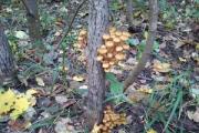
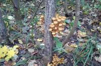
A mushroom is a living organism that forms a separate kingdom of the same name. For a long time they were classified as part of the plant kingdom. But in...
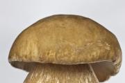
For lovers of “quiet” hunting, mushroom season begins in early summer and lasts until late autumn. And rarely do they...
Aleshnikova, V.I. Use of professional consultants. - M.: Infra-M, 1999. - 240 p. 2. Beich, E....

Orange juice. The symbolic meaning of orange juice in dream books is pleasure and temptation. Quite often we...

We most often overcome all kinds of difficulties encountered along the path of life. Of course, for this we make...

This year your patron Neptune will be in your constellation and this is a good sign, because you will...

1993 who? 1993 is the year of which animal? — According to the Chinese horoscope, 1873, 1933, 1993 belonged to the years of the Black...

Wave-particle duality of light means that light simultaneously has the properties of continuous...

The role of biology is enormous in our world. Although it is not one of the priority subjects, most schoolchildren and...

Amines are organic derivatives of ammonia containing an NH 2 amino group and an organic radical. In general...

How to answer questions in Part BThe second part of the social studies work consists of 7 tasks with a short answer....

Form TORG-15 is drawn up in the case when during transportation, movement between and within the warehouse, when...

Nutritionists say that for good health and a slim figure, you must include snacks in your...
A mushroom is a living organism that forms a separate kingdom of the same name. For a long time they were classified as a kingdom...
For lovers of “quiet” hunting, mushroom season begins in early summer and lasts until late autumn. And rarely do they...