Philosophical vision of the problem
The meaning of life is related to the question “What to live for”, and not to the question of how to maintain life. A person's attitude is...
Pellegrini - Shtidy's disease(A. Pellegrini, Italian surgeon; A. Stieda, German surgeon, 1869-1945; synonym: Pellegrini-Stied syndrome, calcifying periarthritis of the knee joint) - a disease characterized by foci of heterotopic bone formation in the periarticular tissues of the medial epicondyle of the femur due to previous trauma. For the first time, the clinical, radiological and pathomorphological picture of the disease was described by A. Pellegrini (1905), believing that it was characterized by calcification of the tibial collateral ligament of the knee joint. A. Shtida (1907) considered the formation of ossification to be a consequence of avulsion of the epicondyle of the femur, resulting from direct injury or sharp contraction of the adductor magnus muscle of the femur. Modern morphological and histochemical studies have shown that the disease is based on a perverted regeneration reaction in response to damage. It is believed that in the occurrence of this metaplasia, in addition to trauma, hemorrhage, necrosis, and prolonged tissue edema, the individual predisposition of the body plays a large role.
The disease develops more often in men aged 25-45 years, mainly among those involved in physical labor or sports. The triggering mechanism is usually a direct blow to the area of the medial epicondyle of the femur, forced abduction of the tibia, or a sharp uncoordinated contraction of the adductor magnus muscle of the thigh. As a result, hemorrhages occur at the site of attachment of the tibial collateral ligament and the tendon of the adductor magnus muscle of the thigh to the epicondyle and aseptic necrosis of tissue in the damaged area.
The main symptom of Pellegrini-Stied disease is pain, which always corresponds to the location of the ossification, and can spread along the obturator nerve or the infrapatellar branch of the saphenous nerve to the anterior surface of the leg. In 1/3 of patients, the pain intensifies at night and is of a burning, boring nature. In addition, there is a limitation of movements in the knee joint, both flexion and extension, which are usually combined with swelling of the soft tissues of the knee joint, local hyperthermia, hyperesthesia or hypoesthesia in the area of edema, as well as atrophy of the muscles of the thigh and lower leg.
In the early period after injury,
When the clinical picture is identical to that of the knee joint or distortion, as well as when combined with internal damage, its diagnosis is difficult. 3-4 weeks after the injury, ossification is determined on radiographs of the knee joint ( rice .), having the shape of a bracket, sickle or irregular shape, which is separated from the epicondyle of the femur by a strip of lucency. In case of negative radiological data, but in the presence of palpable ossification, it is necessary to take X-rays with internal or external rotation of the limb by 20° to eliminate the layering of ossification on the contour of the femoral condyle. Radionuclide testing can provide information about the degree of maturity of the ossificate. Differential diagnosis is made with avulsion of the medial epicondyle of the femur, which is revealed on an x-ray immediately after the injury. In addition, the marginal defect of the epicondyle corresponds in shape and size to the torn bone fragment. A similar x-ray picture can be observed with a restructured process in the epicondyle of traction origin, resulting from repeated forced strains of the adductor magnus muscle of the thigh, for example in football players. However, the gradual development of this disease, younger age, and association with sports help clarify the diagnosis.Treatment is often conservative. Physiotherapeutic methods are often ineffective, because provide only temporary improvement. The use of ultrasound therapy in combination with electrophoresis of a novocaine solution or 10% sodium chloride solution is effective only in the first 3 months after injury. A lasting positive effect is usually achieved by injections of hydrocortisone with novocaine into the focus of ossification.
Surgical interventions are performed if there is no effect from conservative measures, but not earlier than 3 months after the injury if there are signs of ossification maturity. To do this, the density, structure of the ossification, and the clarity of the sclerotic border are determined on radiographs. Radionuclide data are also used. When combining P. - Sh. b. with internal injuries of the knee joint, especially if the ossification is located near the nerve trunks or when it is introduced into the joint cavity, treatment is only surgical. To prevent recurrence of ossification after surgery, the scars surrounding the ossification are carefully removed,
Pellegrini-Stied disease occurs as a result of frequent injury or severe trauma to the knee joint. In this case, excessive growth of bone tissue occurs, which replaces the cartilage of the internal epicondyle. For a long time, the pathology is asymptomatic and is discovered accidentally during radiography.
The predisposition to developing the disease is inherited and affects mainly young people.
Pellegrini-Stied disease is a pathological growth of bone, or in scientific language - ossification of para-aticular tissues in the area of the internal epicondyle of the knee joint. It occurs as a result of injury or frequent microtrauma with significant loads on the joint. The disease does not cause symptoms for a long time, and subsequently leads to ankylosis and immobilization of the knee joint.
Pellegrini-Stied pathology develops as a result of exposure to the following factors on the human body:
 The high risk of developing pathology is due to human heredity.
 Exposure to one or more of these factors provokes increased ossification in the area of the medial epicondyles of the knee. Heredity and the presence of the disease in one of the closest relatives play a huge role in the development of the disease. This is due to the genetically determined characteristics of bone growth. But even in this case, the development of Pellegrini-Stied disease will require severe trauma or prolonged microtrauma.
For a long time, the pathology is asymptomatic. In this case, the patient may complain of minor pain in the knee, which intensifies after significant exercise or when the weather changes. Later, symptoms of arthropathy appear in the form of swelling, redness of the skin around the joint, increased temperature and disruption of the normal functioning of the limb. The patient may also experience gait disturbances and lameness. With a long course of the process, the volume of physical activity decreases significantly. At the final stages of the development of the disease, a person is not able to make movements in the knee joint, and develops.
 Based on the results of a survey and examination of the patient, the doctor can assume such a disease.
 A traumatologist may suspect the development of Pellegrini-Stied disease during a general examination and when asking about the patient’s well-being. To confirm the diagnosis, an X-ray examination is performed in several projections, which clearly visualizes excess bone growths in the area of the knee joint. To more accurately determine the extent of the lesion, magnetic resonance and computed tomography are used. It is also necessary to take a general blood test and determine the level of calcium in the body. To exclude endocrinological pathology, testing for the content of the main parathyroid hormones is indicated.
Therapy for Pellegrini-Stida disease should be comprehensive and include therapeutic techniques that are used in the early stages of the development of the process. In severe cases, surgery is used. Traditional methods of treatment are used as an additional method before the main therapy. At the initial stages of the disease, non-steroidal anti-inflammatory drugs are used, which relieve signs of inflammation and reduce pain. Muscle relaxants, chondroprotectors and vitamin complexes are also indicated.
Herbal medicine using decoctions of anti-inflammatory herbs will be useful.
If the volume of motor activity of the limb decreases, surgery is performed. During its course, the overgrown bone is removed, and the altered cartilage is removed and replaced with its own cartilage tissue taken from other parts of the body. After surgery, the patient requires a long period of rehabilitation. For this purpose, a complex of physiotherapeutic measures is carried out, therapeutic massage and restorative gymnastics are prescribed.
A. Pellegrini described the disease based on clinical and radiological studies. According to his assumption, the pathological morphology represents calcification of the tibial ligaments of the knee joint. A. Shtida described the formation of heterotopic ossifications as a result of separation of bone fragments of the femoral epicondyle due to injury or unintentional sharp contraction of the thigh muscles.
According to Russian doctors, the disease is based on a distorted reaction of restorative resources to the need to restore tissues calcified and damaged by heterotopic bone growths.
Metaplasia also occurs based on genetic predisposition, due to injuries, internal hemorrhages and tissue death.
The pathology is characterized by the appearance of foci of heterotopic bone formations that grow independently of the main bone tissues on the periarticular soft tissues.
In ICD-10, in classes M70-M79, combining “Other soft tissue diseases,” Pellegrini-Stida disease is designated by code M76.4 under the name “Tibial collateral bursitis.”
With the simple development of periarthritis, the pain is minimal and is felt in the affected joint with sudden movements. A slight limitation of mobility begins, and a slight limp is possible.
Acute periarthritis develops after injury or in the absence of timely treatment.
Chronic periarthritis, or “blocked joint,” develops over several years, and irreversible changes form in the soft tissues.
There are primary and secondary forms of Pellegrini-Stida syndrome. Secondary syndrome develops against the background of another disease.
The onset of pathological changes is characterized by the formation of tendon ruptures and foci of necrosis in the periosteal muscle tissue. There are no external manifestations yet, so Pellegrini-Stida disease on the knee joints often remains untreated.
The progression of the disease is manifested by severe symptoms and reactive periarthritis. First, the tendon is affected, then the pathology affects the synovial tissue.
Periarthritis is accompanied by tendobursitis - soft tissues in the structure of the knee joint swell. Clinical manifestations are severe limitation of movements, acute prolonged pain.
Targeted therapy leads to the disappearance of pathological signs. Otherwise, residual manifestations of the disease are observed, which leads to its chronic form.
During physical and motor stress, frequent injuries against the background of acute periarthritis, some soft tissues are not supplied with blood and remain vascularized. Foci of necrosis develop on them. Subsequently, the affected tissues become calcified, sclerosed, and secondary reactive inflammation occurs in them.
Tibial collateral bursitis is also called Pellegrini-Stida syndrome in honor of the two scientists who first described its symptoms and clinical picture.
This pathology is post-traumatic in nature. In the area of the periarticular tissues of the medial epicondyle, foci of heterotropic bone tissue formation are formed. The main reason for the development of the disease is the abnormal recovery reaction of the body.
Development usually begins after:
The problem in diagnosing is that the first manifestations are very similar to a number of other diseases. It is differentiated from an avulsion fracture of the medial epicondyle of the femur, as well as from the process of restructuring in the epicondyle of traction origin.
The formation of bursitis can be determined after about a month using an x-ray. If ossification is felt during palpation, but is not yet visible on the image, it is carried out from several angles to accurately determine the location and nature of the formation.
To understand how mature the ossification is, a radionuclide study is carried out.
In most cases, conservative treatment is carried out. Physiotherapy in this case is ineffective and can only provide temporary improvement.
Ultrasound therapy and electrophoresis with a solution of novocaine or sodium chloride are used. This measure is most effective in the first three months after injury.
The best results are shown by injections into the site of bursitis with a mixture of hydrocortisone and novocaine.
If conservative treatment does not produce results, surgery is performed after three months. To do this, the neoplasm must be already formed and mature. This is determined by x-rays.
Be sure to remove ossification, which is accompanied by damage to the knee or if it is located next to the nerves, as well as when inserted into the joint cavity.
All ossification is carefully removed along with scars to avoid relapse. Cavities in soft tissues are also eliminated.
With timely treatment, the limb is fully restored and the person can lead a normal active life.
To avoid re-formation, you should protect the limb from injury and too much stress. If the slightest problems with bones or joints arise, they should be eliminated immediately, as they can provoke a relapse.
Important manifestations of pathology are tissue damage, compactions and nodules in muscle tissue, painful on palpation or during movement. The muscles themselves become tense. Signs of pathology depend on its location.
Pellegrini-Stida syndrome in the knee joint is characterized by pathology of the muscle tendons and is localized in the soft tissues surrounding the joint.
The disease significantly worsens the quality of life: it brings discomfort, sleep disturbance due to pain, suffering. In the absence of timely treatment, working capacity and the possibility of free movement are limited.

Experts use a combination of several therapy methods:
Surgery is used in cases where traditional treatment does not produce results.
Conservative treatment is the main method. Non-steroidal anti-inflammatory drugs are prescribed. As the disease progresses, hormonal drugs and novocaine blockades are added. For severe pain, codeine and intra-articular steroid injections are prescribed.
Physiotherapeutic treatment is prescribed simultaneously with medications to improve blood supply to the affected tissues:
The combination of medication and physiotherapeutic treatment helps improve blood supply to affected tissues and enhance regeneration processes.
Timely treatment of Pellegrini-Stida disease completely restores the function of the knee. Prevention of relapses and transition of the disease to the chronic stage is to prevent knee injuries, physical and motor overload.
The most complete answers to questions
Pellegrini - Stied's disease (m. Pellegrini - Stied), peritendinitis of the knee joint, Stied's shadow or fracture, Stied's disease - Pellegrini, post-traumatic paracondylar ossification of the thigh, paraosseous accompanying shadow of Pellegrini, Keller - Pellegrini - Stied disease. It was first described in 1905 by the Italian physician Pellegrini A., and in 1907 by the German surgeon Stied A. Ossification, located in the soft tissues in the area of the internal femoral condyle. What is it, what are the causes and symptoms, as well as treatment methods, we will consider further.
Pellegrini-Stida disease is a post-traumatic ossification of paraarticular tissues in the area of the medial epicondyle of the femur. Most authors consider it a disease of traumatic origin. When the internal ligament is torn from the femoral condyle at the site of its attachment, the periosteum rises and a subperiosteal hematoma is formed, which undergoes ossification. The disease develops more often in men aged 25-45 years, mainly among those involved in physical labor or sports.
Most patients are asymptomatic or with minor pain in the medial part of the knee joint.
In cases of timely contact with specialists and the correct prescription of therapy, the prognosis in most cases is favorable and the patient’s complete recovery is observed.
The triggering mechanism is usually a direct blow to the area of the medial epicondyle of the femur, forced abduction of the tibia, or a sharp uncoordinated contraction of the adductor magnus muscle of the thigh. As a result, hemorrhages occur at the site of attachment of the tibial collateral ligament and the tendon of the adductor magnus muscle of the thigh to the epicondyle and aseptic necrosis of tissue in the damaged area.
Factors that increase the risk of periarthritis:
Exposure to one or more of these factors provokes increased ossification in the area of the medial epicondyles of the knee. Heredity and the presence of the disease in one of the closest relatives play a huge role in the development of the disease. This is due to the genetically determined characteristics of bone growth. But even in this case, the development of Pellegrini-Stied disease will require severe trauma or prolonged microtrauma.
The onset of pathological changes is characterized by the formation of tendon ruptures and foci of necrosis in the periosteal muscle tissue. There are no external manifestations yet, so Pellegrini-Stida disease on the knee joints often remains untreated.
The progression of the disease is manifested by severe symptoms and reactive periarthritis. First, the tendon is affected, then the pathology affects the synovial tissue.
Characteristic signs of knee joint disease:
may have similar symptoms and signs to other knee diseases, so consult your doctor for an accurate diagnosis.Pellegrini Stied disease
A doctor may suspect Pellegrini-Stied disease of the knee joint after a physical examination and palpation of the affected area.

Pellegrini-Stied disease on x-ray (left knee joint)
Pellegrini Stide's disease of the knee joint can be treated with conservative methods and surgery, below we will look at these 2 methods.
Surgical interventions are performed if there is no effect from conservative measures, but not earlier than 3 months after the injury if there are signs of ossification maturity.
A very important factor is competent implementation of postoperative therapy, since there are direct risks of relapse. In the rehabilitation process, electrophoresis, hydrokinesitherapy and therapeutic gymnastics are used.
With timely and competent treatment, as well as the patient’s compliance with therapeutic recommendations, the prognosis for recovery is favorable. After a rehabilitation period, the affected limb regains its function over time. However, the possibility of the disease returning cannot be ruled out.
Tibial collateral bursitis is also called Pellegrini-Stida syndrome in honor of the two scientists who first described its symptoms and clinical picture.
This pathology is post-traumatic in nature. In the area of the periarticular tissues of the medial epicondyle, foci of heterotropic bone tissue formation are formed. The main reason for the development of the disease is the abnormal recovery reaction of the body.
Most often, such bursitis is diagnosed in young men 25-45 years old who engage in heavy physical labor or sports.
Development usually begins after:
The problem in diagnosing is that the first manifestations are very similar to a number of other diseases. It is differentiated from an avulsion fracture of the medial epicondyle of the femur, as well as from the process of restructuring in the epicondyle of traction origin.
The formation can be determined in about a month using an x-ray. If it is felt during palpation, but is not yet visible on the picture, it is carried out from several angles to accurately determine the location and nature of the formation.
To understand how mature the ossification is, a radionuclide study is carried out.
 In most cases, conservative treatment is carried out. Physiotherapy in this case is ineffective and can only provide temporary improvement.
Ultrasound therapy and electrophoresis with a solution of novocaine or sodium chloride are used. This measure is most effective in the first three months after injury.
The best results are shown by injections into the site of bursitis with a mixture of hydrocortisone and novocaine.
If conservative treatment does not produce results, surgery is performed after three months. To do this, the neoplasm must be already formed and mature. This is determined by x-rays.
Be sure to remove ossification, which is accompanied by damage to the knee or if it is located next to the nerves, as well as when inserted into the joint cavity.
All ossification is carefully removed along with scars to avoid relapse. Cavities in soft tissues are also eliminated.
With timely treatment, the limb is fully restored and the person can lead a normal active life.
To avoid re-formation, you should protect the limb from injury and too much stress. If the slightest problems with bones or joints arise, they should be eliminated immediately, as they can provoke a relapse.

The meaning of life is related to the question “What to live for”, and not to the question of how to maintain life. A person's attitude is...
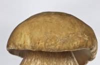
A mushroom is a living organism that forms a separate kingdom of the same name. For a long time they were classified as part of the plant kingdom. But in...
For lovers of “quiet” hunting, mushroom season begins in early summer and lasts until late autumn. And rarely do they...
Aleshnikova, V.I. Use of professional consultants. - M.: Infra-M, 1999. - 240 p. 2. Beich, E....

Orange juice. The symbolic meaning of orange juice in dream books is pleasure and temptation. Quite often we...

We most often overcome all kinds of difficulties encountered along the path of life. Of course, for this we make...

This year your patron Neptune will be in your constellation and this is a good sign, because you will...

1993 who? 1993 is the year of which animal? — According to the Chinese horoscope, 1873, 1933, 1993 belonged to the years of the Black...

Wave-particle duality of light means that light simultaneously has the properties of continuous...
The role of biology is enormous in our world. Although it is not one of the priority subjects, most schoolchildren and...

Amines are organic derivatives of ammonia containing an NH 2 amino group and an organic radical. In general...

How to answer questions in Part BThe second part of the social studies work consists of 7 tasks with a short answer....

Form TORG-15 is drawn up in the case when during transportation, movement between and within the warehouse, when...

Nutritionists say that for good health and a slim figure, you must include snacks in your...
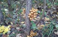
A mushroom is a living organism that forms a separate kingdom of the same name. For a long time they were classified as a kingdom...
For lovers of “quiet” hunting, mushroom season begins in early summer and lasts until late autumn. And rarely do they...