Philosophical vision of the problem
The meaning of life is related to the question “What to live for”, and not to the question of how to maintain life. A person's attitude is...
Content
Respiratory diseases are considered one of the most dangerous ailments of our time. In hospitals, a special method is used to examine the bronchi and trachea - bronchoscopy. This procedure is sometimes called tracheobronchoscopy. Timely diagnosis of the tracheobronchial tree helps to avoid the development of many diseases.
Bronchoscopy or fibrobronchoscopy (FBS) is usually called a method of visual examination of the bronchi and mucous membranes. A biopsy of the respiratory system is performed using an endoscopic examination. Fiberoptic bronchoscopy is also used to obtain samples of secretions from the lungs.
During the procedure, doctors use a special device – a bronchoscope.
It provides direct access to:

Bronchoscopy refers to invasive research methods, which implies the possibility of violating the external barriers of the body. During the procedure, the patient's mucous membrane, skin or blood vessels may be damaged. For this reason, FBS is allowed only as prescribed by a doctor. Indications for fibrobronchoscopy are:

Bronchoscopy is contraindicated in:
A modern bronchoscope is a hollow tube with a light source attached to the end. The device comes in two types - rigid and flexible.
The first option is a more outdated version; it is used only under anesthesia. A rigid bronchoscope is used in the following cases:
Main elements of the device:
A flexible bronchoscope is much safer and more convenient, and therefore causes less discomfort to the patient.
The device is able to penetrate the lower sections of the bronchi without injuring them. Due to the small diameter of the tubes, this type of equipment is often used in pediatrics. The procedure with a fiberoptic bronchoscope is performed without anesthesia. Components of the device:
A flexible bronchoscope is used:

Preparation for bronchoscopy is an important stage of the procedure. Some patients are prescribed additional tests before diagnosis, which may include:
It is important to promptly inform your doctor if you have allergies, chronic diseases, or are taking medications. All of the information listed can have a direct impact on preparation for the procedure. Experts recommend taking tranquilizers the day before bronchoscopy. This will help relieve anxiety.
The examination is carried out on an empty stomach.
Smoking is prohibited on the day of bronchoscopy. It is mandatory to have a bowel movement before visiting the hospital. For these purposes, you can use glycerin suppositories or an enema.
Bronchoscopy in children causes unpleasant associations. It is better not to reveal all the details of the procedure to your child to avoid a negative reaction. The examination takes place under general anesthesia, so children do not need to know all the intricacies of the process.
If the baby is very nervous, it is allowed to administer mild sedatives.

The procedure is performed in an endoscopy room with a high level of sterility. Fiberoptic bronchoscopy is performed by a doctor trained in bronchial examination. Depending on the type of equipment used, local anesthesia or general anesthesia is used. The procedure is carried out in several stages:
At the preparation stage, the doctor determines which type of anesthesia is preferable for the patient. Bronchoscopy under anesthesia is indicated when using rigid equipment. Not all people are able to endure the pain of inserting a bronchoscope, and local anesthesia is ineffective in such situations.
Under anesthesia, the patient will feel absolutely nothing.
This type of anesthesia is carried out using a 2-5% lidocaine solution. The drug causes the following reactions in patients:
Local anesthesia helps to successfully suppress the gag and cough reflex. The bronchoscope is introduced gradually, spraying with an anesthetic spray at each stage. First, the doctor irrigates the mucous membrane of the larynx, then goes lower to the vocal cords. Next, using screwing movements, the specialist advances the device to the bronchi and trachea.

According to doctors, following the recommendations below will help avoid complications:
Bronchoscopy is considered a safe procedure for human health. However, any research carries a risk of complications. The patient's condition may worsen both during and after the procedure. The most common complications are:
There are many factors that influence the occurrence of complications. These include not only medical errors, but also patients’ neglect of specialist recommendations. In order to avoid health problems, it is important to follow your doctor's instructions before and after bronchoscopy. If complications occur, you should immediately seek medical help.

|
Procedure name |
Medical facility |
||
|
Bronchoscopy |
Medical center "GUTA-CLINIC" |
Moscow, st. Fadeeva, house 2 |
|
|
Moscow, Entuziastom highway, building 62 |
|||
|
European Medical Center |
Moscow, Shchepkina street, building 35 |
||
|
Clinic "COSMETON" |
Moscow, 2nd Botkinsky proezd, building 8 |
||
|
OAO "Medicine" |
Moscow, 2nd Tverskoy-Yamskoy lane, building 10 |
||
|
Yusupov Hospital |
Moscow, Nagornaya street, building 17, building 6 |
||
|
Medical Center "Best Clinic" |
Moscow, Novocheremushkinskaya street, building 34, building 2 |
Found an error in the text?
Select it, press Ctrl + Enter and we will fix everything!
Bronchoscopy is a diagnostic procedure that allows you to visualize the inner surface of the bronchi and trachea. The study is carried out using a special device - a fiberoptic bronchoscope. The drug is an endoscope with a flexible hose, a flashlight, a camera and a control handle. The appointment for the procedure is given by the doctor, who also determines the frequency of the study depending on the diagnosis and age of the person.
 Indications for bronchoscopy:
During bronchoscopy, the following manipulations are possible:

If necessary, the procedure can be performed on newborns to assess the condition of the upper respiratory tract. For children, bronchoscopy is performed under general anesthesia.
It is important to properly prepare for bronchoscopy. First of all, it is necessary to undergo a series of diagnostic tests: a blood test, an X-ray examination of the lungs, a coagulogram (testing the blood for the rate of clotting). Additionally, the doctor prescribes laboratory tests for HIV, hepatitis A, B, C. Before the procedure, be sure to inform the doctor about any allergies to painkillers or other drugs, as well as chronic diseases.

Immediately before bronchoscopy, the following recommendations should be followed:
How is bronchoscopy done:
After bronchoscopy, complications may develop:
In medicine, there are a number of contraindications for which bronchoscopy is strictly prohibited:
In some cases, bronchoscopy is performed even in the presence of contraindications - when the risk of not performing it is much higher than performing it.

Bronchoscopy is a safe and very informative procedure that allows you to establish a diagnosis with 100% accuracy and select the correct treatment. It does not cause pain, but is accompanied by discomfort. After the procedure, complications and the appearance of unpleasant symptoms are possible, which should be reported to the doctor.
This is the most informative method for studying the tracheo-bronchial tree. It allows you to see minimal formations and tumors, but only in the trachea, large and medium bronchi. Bronchoscopy of the bronchi is also the optimal way to clean (lavage) the airways in those people who have to be on mechanical breathing for a long time.
Bronchoscopy is a procedure that is performed only in a hospital. Under local (treatment of mucous membranes with lidocaine) or general anesthesia, the doctor inserts a special device into the respiratory tract - a bronchoscope, which is either a flexible or rigid tube. At one end of the device there is an illuminator, the other ends with an optical system, where the doctor looks directly with his eyes.
There are holes on the side of the bronchoscope where you can connect:
For these instruments, there is a special channel in the body of the device through which they pass. In addition, the device can communicate with video equipment so that the doctor assesses the condition of the bronchi, looking not into the “tube” of the device itself, but by looking at the monitor.
Typically the bronchoscope is inserted through the mouth. Some doctors use a laryngoscope for this - a device that will simultaneously illuminate the path for the bronchoscope and squeeze the root of the tongue and the epiglottis - the cartilage into which the flexible bronchoscope can rest.
Since bronchoscopy is vital in many cases (for example, if there is an injury or malformation of the neck and breathing needs to be done using a breathing apparatus), the bronchoscope can be inserted through the nose.
Also, if the patient breathes through a tracheostomy (an opening in the trachea through which a special cannula connected to a breathing apparatus is inserted), the bronchoscope is inserted directly into the tracheostomy opening. In this case, separate anesthesia is not required.
What does bronchoscopy show:
The bronchoscope does not visualize smaller bronchi and bronchioles. If there is a suspicion that a tumor or inflammation is located there, a computed tomography or magnetic resonance imaging scan is performed.
We hope that it is clearly explained what it is - bronchoscopy of the lungs, although it is more correct to call this manipulation simply bronchoscopy (it means “visualization of the bronchi”).
You need to undergo a bronchoscopy if:
This is a diagnostic bronchoscopy and is used to make a diagnosis.
There is also a treatment procedure that is used when:
Basically, bronchoscopy is performed using a flexible bronchoscope – fiberoptic bronchoscope. It is quite thin and can bend in different directions. But in some cases, it is necessary to introduce a rigid (metal) device that does not bend and cannot be inserted into the bronchi that extend at an angle.
Indications for bronchoscopy with a rigid bronchoscope are the removal of foreign bodies, expansion of bronchi narrowed by inflammation or adhesions. It is more convenient to put a stent (an expanding tube made of rigid corrugated plastic) on a rigid bronchoscope and install the latter into the narrowed bronchus. It is best used during thoracic operations - in the treatment of conditions associated with the entry of pus, air or liquid into the pleural cavity, as well as pulmonary hemorrhage. Then, using a bronchoscope, you can block the bronchus on the painful side, where surgeons work, and ventilate the second lung with the device.
In addition to rigid and flexible bronchoscopy, another type of examination has been developed - virtual bronchoscopy. It is a computed tomography scan of the lungs and bronchi, which is processed by a special computer program that recreates a three-dimensional image of the bronchi.
The method is not as informative, but it is non-invasive. With it, you cannot take a sputum test, rinsing water or a biopsy of a suspicious area, you cannot remove a foreign body or rinse the bronchi from sputum.
No preparation is required for a virtual biopsy. According to the method of execution, it does not differ from computed tomography. The patient lies down on a couch that is placed inside the X-ray source.
Although X-ray radiation is low-dose, the method is not suitable for children and pregnant women.
Preparation for bronchoscopy is very important, since the manipulation is very serious, classified as invasive and requires only special equipment and special skills from the doctor.
Therefore, you need to start with a detailed conversation with your therapist. He will tell you what specialist consultations are needed. So, if a person has suffered a myocardial infarction, he needs, in agreement with the cardiologist, to increase the dose of beta blockers 2 weeks before the study. If a person suffers from arrhythmia, he needs to reconsider antiarrhythmic therapy and possibly increase the dose of drugs or add some other antiarrhythmic drug. The same applies to diabetes mellitus and arterial hypertension.
Also, everyone needs to undergo the following studies and show their results:
The last meal is no later than 8 pm. Then you can take your last scheduled pills. The need to take them in the morning is discussed separately.
Be sure to empty your bowels in the evening using an enema, Microlax microenema (Norgalax), glycerin suppositories.
You are not allowed to smoke on the day of the test. Immediately before the procedure, you need to empty your bladder. You must take a towel or diaper with you so that you can dry yourself after the examination; for those suffering from arrhythmia - antiarrhythmic drugs; for those suffering from bronchial asthma - an inhaler. Removable dentures will need to be removed.
It is imperative to familiarize the doctor who will perform the procedure with previous diseases and allergies, as well as medications that you are constantly taking.
Learn more about how bronchoscopy is performed. First, let's talk about how this procedure is performed without anesthesia - under local anesthesia:
During bronchoscopy, do not bend over so as not to damage the airways with the bronchoscope (especially if a rigid device is used).
If bronchoscopy with biopsy is performed, it is painless. There is only discomfort behind the sternum. The bronchial mucosa has virtually no pain receptors. The introduction of lidocaine before manipulation is due to the need to turn off vagal (from the word “nervus vagus” - “vagus nerve”) reflexes from the root of the tongue and vocal cords, which can lead to cardiac arrest.
If bronchoscopy is performed under anesthesia, it is performed with the patient lying down. Then the injections are performed intravenously, and the person falls asleep as a result. A rigid polypropylene tube is inserted into his trachea, which is connected to a breathing apparatus. For some time, air is pumped into the lungs with a breathing apparatus (exhalation occurs spontaneously), then a bronchoscope is inserted through the tube, and bronchoscopy is performed. A person does not feel how a bronchoscopy is done.
The procedure under anesthesia is performed in children, people who are very afraid of the procedure, and people with unstable mental health. It is performed on patients who have already been on mechanical breathing, as well as when surgical intervention is necessary.
After bronchoscopy you feel:
The following rules must be followed:
It is also necessary to monitor your condition. Must not be:
The doctor writes the first results of bronchoscopy immediately after the examination. These words could be:
There are such contraindications to bronchoscopy (namely diagnostic):
In these cases, virtual bronchoscopy can be performed.
The procedure should be postponed during an acute infectious disease, exacerbation of bronchial asthma, for women - during menstruation and from the 20th week of pregnancy.
When bronchoscopy is intended to assist intubation, or is needed to remove foreign bodies, bronchial stenting or other therapeutic purposes, there are no contraindications. This procedure is carried out jointly by an endoscopist and an anesthesiologist, under anesthesia, after proper intensive preparation.
With bronchoscopy, the consequences may be as follows:
Bronchoscopy can be performed in children from the neonatal period, provided that the hospital has a device of such a small diameter. The procedure is carried out only under anesthesia, and after it antibiotics are prescribed.
Bronchoscopy is performed in children when:
Children are more likely to develop laryngo- or bronchospasm due to the rich blood supply to the respiratory tract. Therefore, general anesthesia is often supplemented with local anesthesia.
In addition, complications may include collapse (a sharp decrease in blood pressure) and anaphylactic shock. Tracheal perforations are extremely rare, since bronchoscopy is performed with flexible bronchoscopes.
Bronchoscopy for tuberculosis is an important diagnostic and treatment procedure. It allows:
Do you know everything about colds and flu?
© 2013 ABC of Health // User Agreement // Personal Data Policy // Site Map The information on the site is intended for informational purposes only and does not encourage self-treatment. To establish a diagnosis and receive treatment recommendations, consultation with a qualified physician is necessary.
One of the most important research methods in pulmonology is bronchoscopy. In some cases, it is used not only as a diagnostic method, but also as a therapeutic method that allows one to effectively eliminate certain pathological changes. We will talk about what lung bronchoscopy is, what are the indications and contraindications for this study, what is the methodology for conducting it, in this article.
Bronchoscopy, or tracheobronchoscopy, is a method of examining the lumen and mucous membrane of the trachea and bronchi using a special device - a bronchoscope. The latter is a system of tubes - flexible or rigid - with a total length of up to 60 cm. At the end, this device is equipped with a video camera, the image from which, magnified many times, is displayed on the monitor, i.e. the specialist conducting the study personally observes the condition of the respiratory tract in the mode real time. In addition, the resulting image can be saved in the form of photographs or video recordings, so that in the future, comparing the results of the current study with the previous one, it is possible to assess the dynamics of the pathological process. (Read about bronchography in our other article.)
Bronchoscopy was first performed back in 1897 by the doctor G. Killian. The purpose of the procedure was to remove a foreign body from the respiratory tract, and since it was very traumatic and painful, cocaine was recommended to the patient as an anesthetic. Despite the large number of complications after bronchoscopy, it was used in this form for more than 50 years, and already in 1956, the scientist H. Fidel invented a safe diagnostic device - a rigid bronchoscope. Another 12 years later, in 1968, a fiberoptic bronchoscope, a flexible bronchoscope made from fiber optics, appeared. An electronic endoscope, which allows you to repeatedly enlarge the resulting image and save it to a computer, was invented not so long ago - in the late 1980s.
Currently, there are 2 types of bronchoscopes - rigid and flexible, and both models have their advantages and are indicated in certain clinical situations.
Consists of a smooth flexible tube with an optical cable and light guide inside, a video camera at the inner end and a control handle at the outer end. There is also a catheter for removing fluid from the respiratory tract or supplying a drug into it, and, if necessary, additional equipment for diagnostic and surgical procedures.
A rigid bronchoscope includes a system of rigid hollow tubes with a light source, video or photographic equipment at one end, and a manipulator for controlling the device at the other. The kit also includes various mechanisms for therapeutic and diagnostic procedures.
Indications for fibrobronchoscopy are:
Examination using a rigid bronchoscope is the method of choice in the following cases:
In some cases, bronchoscopy is necessary not as a planned, but as an emergency medical intervention necessary to quickly make a correct diagnosis and eliminate the problem. The main indications for this procedure are:
For most of the above pathologies, emergency bronchoscopy is performed in intensive care conditions through an endotracheal tube.
In some cases, bronchoscopy is dangerous for the patient. Absolute contraindications are:
The last 4 pathologies are contraindications only for rigid bronchoscopy, and fiber-optic bronchoscopy is acceptable in these cases.
In some conditions, bronchoscopy is not contraindicated, but it should be temporarily postponed until the pathological process resolves or clinical and laboratory parameters stabilize. So, relative contraindications are:
Before bronchoscopy, the patient must undergo a series of examinations prescribed by the doctor. As a rule, this is a general blood test, biochemical blood test, pulmonary function tests, chest x-ray or others, depending on the disease of the individual patient.
Immediately before the study, the patient will be asked to sign a consent for this procedure. It is important not to forget to inform the doctor about any allergies to medications, especially to anesthesia drugs, if any, pregnancy, medications taken, acute or chronic diseases, since in some cases (see above) bronchoscopy is absolutely contraindicated.
As a rule, a planned study is carried out in the morning. In this case, the patient has dinner the night before, and is prohibited from eating in the morning. At the time of the study, the stomach should be empty to reduce the risk of reflux of its contents into the trachea and bronchi.
If the patient is very worried about the upcoming bronchoscopy, he may be prescribed mild sedatives a few days before the examination.
Bronchoscopy is a serious procedure that is performed in a room specially equipped for this purpose in compliance with all sterile conditions. Bronchoscopy is performed by an endoscopist or pulmonologist who has been trained in this type of examination. An endoscopist assistant and an anesthesiologist also take part in the study.
Before the examination, the patient must remove glasses, contact lenses, dentures, hearing aids, jewelry, unbutton the top button of his shirt if the collar is tight enough, and empty his bladder.
During bronchoscopy, the patient is in a sitting or supine position. When the patient is sitting, his torso should be slightly tilted forward, his head slightly back, and his hands should be lowered between his legs.
When performing fibrobronchoscopy, local anesthesia is used, for which a lidocaine solution is used. When using a rigid bronchoscope, general anesthesia, or anesthesia, is required - the patient is put into a state of medicated sleep.
In order to dilate the bronchi for easy advancement of the bronchoscope, a solution of atropine, aminophylline or salbutamol is administered to the patient subcutaneously or by inhalation.
When the above drugs have taken effect, a bronchoscope is inserted through the nose or mouth. The patient takes a deep breath and at this moment the bronchoscope tube is passed through the glottis, after which it is inserted deeper into the bronchi with rotational movements. To reduce the gag reflex at the time of insertion of the bronchoscope, the patient is advised to breathe shallowly and as often as possible.
The doctor assesses the condition of the respiratory tract as the bronchoscope moves from top to bottom: first examines the larynx and glottis, then the trachea, after which the main bronchi. The examination with a rigid bronchoscope is completed at this level, and during fiberoptic bronchoscopy the underlying bronchi are also examined. The most distant bronchi, bronchioles and alveoli have a very small lumen diameter, so their examination with a bronchoscope is impossible.
If any pathological changes are detected during bronchoscopy, the doctor can carry out additional diagnostic or direct therapeutic manipulations: take swabs from the bronchi, sputum or a piece of pathologically altered tissue (biopsy) for examination, remove the contents blocking the bronchus, and rinse them with an antiseptic solution.
As a rule, the study lasts for 30–60 minutes. All this time, specialists monitor the level of blood pressure, heart rate and the degree of oxygen saturation of the patient’s blood.
Contrary to the anxious expectations of most patients, they do not feel any pain during bronchoscopy.
With local anesthesia, after administration of the drug, there is a feeling of a lump in the throat, nasal congestion, the palate becomes numb, and it becomes difficult to swallow. The bronchoscope tube has a very small diameter, so it does not interfere with the patient’s breathing. As the tube moves through the airways, slight pressure may be felt in them, but the patient does not experience any discomfort.
During general anesthesia, the patient sleeps and therefore does not feel anything.
Recovery after bronchoscopy takes no more than 2–3 hours. 30 minutes after the end of the study, the effect of the anesthetic will wear off - during this time the patient is in the endoscopy department under the supervision of medical personnel. You can eat and drink after 2 hours, and smoke no earlier than after 24 hours - such actions minimize the risk of bleeding from the respiratory tract after bronchoscopy. If the patient received certain sedatives before the study, he is strictly not recommended to drive a vehicle for 8 hours after taking them.
As a rule, this study is well tolerated by patients, but sometimes, extremely rarely, complications still arise, such as:
I would like to repeat that bronchoscopy is a very important diagnostic and therapeutic procedure, for which there are both indications and contraindications. The need and advisability of bronchoscopy is determined in each specific case by a pulmonologist or therapist, but it is performed exclusively with the consent of the patient after his written confirmation.
Lung bronchoscopy is an invasive procedure for examining and assessing the condition of the mucous membranes of the bronchi and trachea. The manipulation is carried out both in medical hospitals and on an outpatient basis using specialized endoscopes.
What is pulmonary bronchoscopy? It is an informative procedure that allows the doctor to visually assess the processes occurring in the human respiratory system.
For manipulation, 2 types of bronchofiberscopes are used - soft and hard. This device is a flexible tube with control handles, a light source, cameras for video recording, and manipulators for medical procedures and diagnostic studies.
Examination of the respiratory system using an endoscope is used for therapeutic purposes and as an auxiliary method to clarify the patient’s diagnosis.
Indications for diagnostic fibrobronchoscopy:
Indications for therapeutic bronchoscopy:
Depending on the purpose of the manipulation, different types of bronchoscopes are used - soft or bendable and hard.
A flexible endoscope is similar in appearance to a gastroscopy probe. The length of the tube does not exceed 60 cm, and the diameter is 0.5 cm. The introduction of a soft bronchoscope is possible both through the nasal passages and through the oral cavity. The diameter of the probe does not interfere with natural nasal breathing.
A rigid bronchoscope is an endoscope with a tube diameter of 9 to 13 mm and is equipped with a system for forced ventilation of the lungs. The probe is inserted only through the oral cavity. During the manipulation, the connection of the patient to the condition monitoring system is shown.
Bronchoscopy is an invasive procedure and has a number of contraindications. Manipulation is not prescribed in the following cases:
The success of the procedure depends on proper preparation for the examination. Bronchoscopy is performed exclusively in a hospital setting in a specially equipped operating room or manipulation room.
Preparation for a bronchoscopic examination is simple, but is carried out in several stages:
How to perform bronchoscopy of the lungs, the path of insertion of the endoscope is determined by the endoscopist. There are 2 ways to insert the instrument - through the nasal passages and through the mouth. The first method is mainly used, as this reduces the risk of vomiting.
General procedure for manipulation:
The procedure is invasive, so complications may develop. These include:
These symptoms last no more than a day after the procedure and do not require medical intervention. In severe cases, during bronchoscopy the following may occur:
Eating and drinking liquids is allowed only after the anesthetic drugs wear off. If a rigid bronchoscopy was performed with the collection of material for a biopsy, then eating is allowed only after examination by a doctor.
Bronchoscopy of the lungs is not the most pleasant therapeutic or diagnostic procedure. But this is an important step towards making the correct diagnosis, which allows you to select adequate treatment for a particular individual. Instrumental examination of the bronchi and trachea increases the patient's chances of recovery.
Bronchoscopy or tracheobronchoscopy is a method of direct examination and assessment of the condition of the mucous membranes of the tracheobronchial tree, trachea and bronchi. Bronchoscopy of the lungs is carried out using special devices: bronchofiberscope, rigid respiratory bronchoscope, and a variety of endoscopes.
Indications for bronchoscopy: determination of suspected inflammation or tumor; removal of foreign bodies; removal of blood and pus; administration of medications directly to the lesion; elimination of lung collapse; restoration of tracheal patency. Diagnostic bronchoscopy is inextricably linked with biopsy in cases where it is necessary to find out about the nature of the formation.
Preparation for bronchoscopy consists of a number of procedures: chest radiography, determination of urea levels and blood saturation, electrocardiography. Eating is allowed at least 10 hours before surgery, since bronchoscopy is performed on an empty stomach. Contraindications to bronchoscopy: stenosis of the larynx and trachea of II and III degrees; respiratory failure degree III; acute period of bronchial asthma; aortic aneurysm; myocardial infarction and cerebral infarction (stroke) suffered less than six months ago; blood clotting disorder; intolerance to anesthesia drugs; mental illness. Bronchoscopy of a child is performed only for compelling indications and under general anesthesia. There are no age restrictions for bronchoscopy.
Thank you
The site provides reference information for informational purposes only. Diagnosis and treatment of diseases must be carried out under the supervision of a specialist. All drugs have contraindications. Consultation with a specialist is required!
 Bronchoscopy is a research method with which the lumen and mucous membrane of the bronchi are examined. Bronchoscopy refers to endoscopic research methods and can be carried out for both therapeutic and diagnostic purposes.
 Endoscopic research methods are methods that allow us to examine organs that have at least some minimal cavity ( "endo" means inside, and copy means to explore.) Thus, the purpose of endoscopic methods is to examine the internal cavity of the organ. When carrying out these diagnostic methods, rigid and flexible instruments are used ( endoscopes). The first includes metal tubes of various diameters, and the second includes fiber optics devices. At the end of the endoscope there is a light bulb that illuminates the cavity being examined, and a video camera that is connected to the monitor. When performing bronchoscopy, flexible endoscopes are used ( synonym - fiberscope), which made a real revolution in medicine. They consist of many glass fibers ( LEDs) through which the image is transmitted. Due to the phenomenon of total reflection at the boundary of two media, the resulting picture is highly informative. During bronchoscopy, a fiberscope is inserted into the bronchi through natural openings, that is, through the nose or mouth.
 Bronchoscopy allows you to identify pathologies localized at the level of the lower respiratory tract - the trachea, main bronchi and bronchioles. In order to understand what exactly bronchoscopy examines, you need to know the structure of the bronchial tree.
 Anatomy of the bronchi and bronchial tree
The human lower respiratory tract consists of the trachea, the main ( right and left) bronchi and bronchial tree. The trachea or windpipe is divided into the right and left main bronchus. Secondary bronchi depart from them, which, in turn, are divided into small branches, and those into even smaller ones. The set of all secondary bronchi and their branches is called the bronchial tree. Thus, conditionally, the lower respiratory tract can be expressed as follows. Trachea – left and right main bronchus – secondary bronchi – bronchial tree. During bronchoscopy, the fiberscope examines the trachea, main and secondary bronchi, then it passes into the middle and small branches of the bronchi. However, the fiberscope cannot penetrate the smallest bronchioles due to their small diameter. To study smaller branches, other diagnostic methods are used, for example, virtual bronchoscopy.
A fiberscope is inserted through the nose or mouth, which passes into the larynx, and from it into the trachea and bronchi. Through an eyepiece connected at the other end, the doctor examines the passing paths. Further tactics depend on the purpose of bronchoscopy. For aspiration ( ventilation) pathological fluid in the bronchi or sanitation ( washing) of the purulent cavity, a special aspiration tip is inserted into the lumen of the bronchi, through which the liquid is sucked out. If the purpose of bronchoscopy is to wash the bronchial tree, then a solution for washing the bronchial tree is first introduced through the fiberscope tube ( it could be a solution of furatsilin). The liquid is introduced in small portions and then removed. By alternating the processes of fluid administration and aspiration, sanitation is carried out ( simple rinsing) bronchi.
When removing a foreign body from the bronchi, special forceps are used that grasp the object ( it could be a pea, bean) and remove it. For bronchial bleeding, a procedure called bronchial tamponade is used. In this case, take a piece of foam rubber, which should be twice the diameter of the bronchi. It is rolled up tightly, moistened in an antiseptic solution and placed in the cavity of the bronchus, thus closing its lumen. In order to insert this foam into the bronchus, hard forceps are used, which are passed through a fiberscope. When the fiberscope reaches the bleeding site, the forceps unclench, and the foam expands and fills the lumen. In this “compacted” state, the foam rubber remains in the lumen of the bronchial tree until the bleeding stops.
If the bleeding is minor, then instead of tamponade, irrigation of the bleeding vessel with a solution of adrenaline can be used. Adrenaline is a substance that causes a sharp constriction of blood vessels and stops bleeding ( if the vessel is small).
Preparation for bronchoscopy includes the following activities:
Preparation for bronchoscopy involves the following studies:
Psychological preparation of the patient
The emotional state has a great influence on the quality of bronchoscopy and the results obtained. During the procedure, the patient should be relaxed and calm, since otherwise it will be difficult for the doctor to carry out the necessary manipulations with the bronchoscope. The best way to help the patient calm down is to become familiar with all aspects of the procedure. To get a complete picture of how bronchoscopy is performed, the patient should, during the preliminary consultation, ask the doctor all the questions that concern him. The duration of the procedure, the nature of the sensations before and after bronchoscopy, the type of planned anesthesia - these and other questions that the patient may have must be discussed with the doctor.
In addition to medical consultation, the patient should also independently work on his emotional state. To put your mind at ease, it is recommended to think about the fact that bronchoscopy significantly speeds up the healing process, regardless of the purpose for which it is performed ( diagnostic or therapeutic). You should also take into account the fact that there are no pain receptors in the bronchial mucosa. Therefore, discomfort during bronchoscopy is more due to psychological than physical factors. The day before the examination, it is not recommended to watch films or programs of a negative nature. Also, if possible, you should limit the influence of various household or professional stress factors.
There are the following food products that cause gas formation:
When performing bronchoscopy, the patient's intestines must be empty. Otherwise, due to intra-abdominal pressure, involuntary emptying may occur during the procedure. Therefore, in the morning, before visiting the clinic, you should empty your bowels. Some patients, due to anxiety or characteristics of the gastrointestinal tract, have difficulty with morning bowel movements. In such cases, the patient is prescribed a cleansing enema.
Taking sedatives
To reduce anxiety, most patients are prescribed sedatives before bronchoscopy ( calming) actions. These medications should be taken in the evening, the day before the examination. In some cases, repeated use of sedatives is indicated, 1 to 2 hours before the procedure.
Performing a series of actions immediately before the procedure
Before the bronchoscopy, the patient must visit the toilet to empty the bladder. If a person has jewelry on the neck or on such parts of the body as the nose, tongue, lips, they must be removed, as they will prevent the doctor from carrying out the necessary manipulations. The bronchoscope can be obstructed by braces and other devices that are attached to the teeth, so if possible, these should also be removed.
Types of endobronchitis are:
In addition to inflammatory changes, bronchoscopy can diagnose a violation of the tone of the bronchial tree. As a rule, hypotonic dyskinesia is diagnosed, which is characterized by an increase in respiratory mobility and collapse of the bronchi during exhalation.
Due to the proliferation of tumor tissue or frequent inflammatory changes, the lumen of the bronchi may narrow. This is also visible on bronchoscopy. In this case, the doctor performing the bronchoscopy can assess the degree of narrowing. In the first degree, the lumen is narrowed by no more than one-eighth, in the second degree - by half, and in the third degree - by more than two-thirds.
 As already mentioned, bronchoscopy can be performed for therapeutic or diagnostic purposes. In the first case, the doctor may irrigate the bronchial tree, administer medications, or remove foreign objects. In the second case, bronchoscopy is performed to assess the condition of the mucosa or take a biopsy.
 Types of bronchoscopy include:
Indications for therapeutic bronchoscopy include:
Like any study, therapeutic bronchoscopy also has contraindications.
Contraindications to therapeutic bronchoscopy are:
Indications for diagnostic bronchoscopy are:
Contraindications for diagnostic bronchoscopy include:
Virtual bronchoscopy is based on the X-ray method. Rotating, the X-ray tube produces an image, which is subsequently converted into three-dimensional. Thus, using a special program, a complete image of the entire bronchial tree is reconstructed ( main and small bronchi). In this case, all layers of the bronchi, including the mucous membrane, are visible in the picture. The advantage of this method is the ability to examine even the smallest bronchi, which cannot always be seen with conventional bronchoscopy.
Pros and cons of virtual bronchoscopy
Minuses | pros |
The diagnostic value is lower than with conventional bronchoscopy - it is not possible to take a biopsy ( piece of material for research). | Highly informative - virtual bronchoscopy allows you to see small caliber bronchi, from 1 to 2 millimeters. |
The procedure cannot be carried out for therapeutic purposes, that is, it is impossible to pull out a foreign object or eliminate bleeding. | Much fewer contraindications. Contraindications include only third degree obesity and pregnancy. |
The cost of the procedure is 2–3 times higher than conventional bronchoscopy. | Painless, non-traumatic. |
Virtual bronchoscopy is limited in case of claustrophobia ( fear of closed spaces) and early childhood. | Does not require special preparation, duration is from 5 to 15 minutes ( the usual procedure takes about 30 minutes or more). |
When performing virtual bronchoscopy, the patient receives a certain dose of radiation. | Even seriously ill patients can be diagnosed. |
 Bronchoscopy in children can be performed as both a therapeutic and diagnostic procedure. Modern anesthesia drugs allow for painless and safe procedures. This significantly increases the list of pathologies in young patients for which examination of the lungs with a bronchoscope is indicated.
 The procedure is carried out to establish the true causes of certain diseases of the respiratory system. Using the device, the doctor can obtain a secretion ( slime) from deep-lying parts of the bronchial tree for further bacteriological research. This procedure can also involve tissue sampling ( biopsy) for subsequent analyses, removal of foreign objects or neoplasms. Bronchoscopy allows you to deliver drugs directly to the lesions, remove pathological mucus and carry out other treatment procedures with a high therapeutic effect.
Another common reason for bronchoscopy is tuberculosis. The procedure is prescribed in order to confirm or refute the presence of changes in the bronchi or lungs characteristic of tuberculosis. Bronchoscopy is also indicated for obtaining mucus in order to identify the causative agent of the disease. In older children, tuberculosis can cause bleeding in the lungs, and in such cases a procedure is prescribed to stop this process. There are other pathological conditions for which bronchoscopy in children is indicated.
There are the following indications for bronchoscopy in children:
List of preliminary examinations ( blood test, radiography, cardiogram) is determined by the doctor, who takes into account the child’s age, general condition and other factors. The child should not be fed 6–8 hours before bronchoscopy, and should not drink anything 3–4 hours before the bronchoscopy. Children who are breastfed can be fed for the last time 4 hours before the procedure.
During the procedure, the child is in a horizontal position, which increases the likelihood of bronchospasm. Therefore, before performing pediatric bronchoscopy, medical personnel prepare the necessary equipment for artificial ventilation. After manipulation of the bronchoscope, the child must be prescribed antibiotics in order to prevent the development of infection.
 Bronchoscopy is invasive ( violating the external barriers of the body - skin, mucous membranes) by research method, and therefore, despite its advantages, it is carried out according to strict indications. The main indications for bronchoscopy are pulmonary tuberculosis, bronchial cancer, and foreign bodies in the respiratory tract.
 Sometimes the test may involve inserting catheters ( straws) into the small bronchi to obtain a smear. This procedure is called catheterization and is necessary to diagnose peripheral cancer. If cancer has already been confirmed and bronchoscopy is performed for observation purposes, then a biopsy of the lymph nodes is also required. It is necessary to determine metastases.
 Bronchoscopy for bronchial asthma may be prescribed to diagnose or treat the disease. In acute stages of the disease, the procedure is not performed, as it can cause exacerbation and deterioration of the patient’s condition.
 Despite the heterogeneity of opinions, it should be emphasized that at the moment, pulmonary bronchoscopy is one of the most accurate methods for establishing the correct diagnosis for suspected bronchial asthma. Also, in some cases, bronchoscopy is the only possible method of performing a particular treatment procedure.
Therapeutic bronchoscopy is prescribed to reduce symptoms and improve the patient’s well-being.
The following indications for therapeutic bronchoscopy for asthma are distinguished:
 After bronchoscopy, the patient may experience a number of unpleasant sensations, the cause of which is the anesthesia and manipulations performed. In some rather rare cases, pulmonary endoscopy is accompanied by complications that can appear both during the procedure and after it.
 Consequences of bronchoscopy
Typically, patients complain of difficulties that arise in the process of swallowing, the sensation of a foreign body in the throat, and numbness of the pharynx. In some cases, after the procedure, small blood clots may be present in the coughed up mucus. Blood appears because during bronchoscopy the device injures the mucous membrane of the respiratory tract. Also, some patients have temporary nasal congestion. To reduce discomfort and prevent the development of more serious complications, people should follow certain rules after bronchoscopy.
Complications that occur during the procedure may be caused by the drugs used for anesthesia. If you are allergic to local or general anesthesia, the patient may experience seizures or develop anaphylactic shock. A sharp drop in blood pressure, breathing problems, and heart rhythm disturbances are also possible.
It should be noted that an allergic reaction to anesthesia occurs in rare cases, and the direct presence of a doctor can quickly normalize the patient’s condition. Another cause of complications during the procedure may be damaged blood vessels, which causes bleeding. The likelihood of bleeding is highest when a biopsy is performed during bronchoscopy ( pinch off a fragment of the lung or bronchi with forceps).
Factors that provoke complications after the procedure can be various infections or mistakes made during bronchoscopy.
There are the following complications that develop after bronchoscopy:
 The cost of bronchoscopy determines both the method of performing the procedure and the location in which it is performed.
 The following factors determine the cost of bronchoscopy:
To make an appointment with a doctor or diagnostics, you just need to call a single phone number
+7 495 488-20-52 in Moscow
+7 812 416-38-96 in St. Petersburg
The operator will listen to you and redirect the call to the desired clinic, or accept an order for an appointment with the specialist you need.
Facilities that offer bronchoscopy
City | Name of institution | Address | Telephone | Website |
Moscow | Clinic "Be Healthy" | Komsomolsky Prospekt, 28 | (495 ) 782-88-82 | clinicbudzdorov.ru |
Medical center "MEDLUX" | Sirenevy Boulevard, 32a | (499 ) 704-49-26 | ||
Center "Best Clinic" | Nizhnyaya Krasnoselskaya street, house 15/17 | (499 ) 519-34-75 | ||
Saint Petersburg | Clinic "Admiralty Shipyards" | Sadovaya street, house 126 | (812 ) 409-90-18 | |
Research Institute of Oncology named after Petrov | Pesochny village, Leningradskaya street, house 68 | (812 ) 243-19-60 | ||
Clinic named after Peter the Great | Piskarevsky prospect, house 47 | (812 ) 303-50-60 | ||
Novosibirsk | Almita Medical Center | Zheleznodorozhnaya street, building 12/1 | (383 ) 363-06-31 | |
Medical Center "A" | Rimsky-Korsakov street, building 19 | (383 ) 346-00-70 | ||
Clinic "Sanitas" | Vokzalnaya Magistral street, building 16 | (383 ) 233-66-00 | ||
Kazan | Republican Hospital | Orenburgsky tract, house 138 | (843 ) 231-21-09 | |
Hospital No. 7 | Marshal Chuikov street, building 54 | (843 ) 237-91-71 | ||
Maternity hospital No. 16 | Gagarina street, house 54 | (843 ) 560-66-52 | ||
Ufa | Emergency Hospital | Batyrskaya street, house 39/2 | (347 ) 255-66-71 | bsmp-ufa.rf |
Clinic of the Bashkir State Medical University | Shafieva street, building 2 | (347 ) 223-11-92 | ||
Republican Hospital named after Kuvatov | Dostoevsky street, house 132 | (347 ) 279-03-97 |

The meaning of life is related to the question “What to live for”, and not to the question of how to maintain life. A person's attitude is...
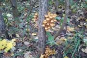
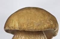
A mushroom is a living organism that forms a separate kingdom of the same name. For a long time they were classified as part of the plant kingdom. But in...
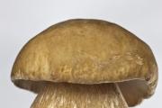
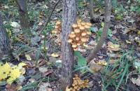
For lovers of “quiet” hunting, mushroom season begins in early summer and lasts until late autumn. And rarely do they...
Aleshnikova, V.I. Use of professional consultants. - M.: Infra-M, 1999. - 240 p. 2. Beich, E....

Orange juice. The symbolic meaning of orange juice in dream books is pleasure and temptation. Quite often we...

We most often overcome all kinds of difficulties encountered along the path of life. Of course, for this we make...

This year your patron Neptune will be in your constellation and this is a good sign, because you will...

1993 who? 1993 is the year of which animal? — According to the Chinese horoscope, 1873, 1933, 1993 belonged to the years of the Black...

Wave-particle duality of light means that light simultaneously has the properties of continuous...
The role of biology is enormous in our world. Although it is not one of the priority subjects, most schoolchildren and...

Amines are organic derivatives of ammonia containing an NH 2 amino group and an organic radical. In general...

How to answer questions in Part BThe second part of the social studies work consists of 7 tasks with a short answer....

Form TORG-15 is drawn up in the case when during transportation, movement between and within the warehouse, when...

Nutritionists say that for good health and a slim figure, you must include snacks in your...
A mushroom is a living organism that forms a separate kingdom of the same name. For a long time they were classified as a kingdom...
For lovers of “quiet” hunting, mushroom season begins in early summer and lasts until late autumn. And rarely do they...