Philosophical vision of the problem
The meaning of life is related to the question “What to live for”, and not to the question of how to maintain life. A person's attitude is...
The thymus (thymus or thymus gland) is the primary organ of immunity, which can be considered the main, central organ of immunogenesis. In addition, it is an organ of hematopoiesis.
The thymus gland is located near the thyroid gland, for which it received one of its names. More precisely, by going down 2 fingers below the jugular notch you can determine the location of the thymus. It has several names: the thymus gland is called because of its similar shape to a two-pronged fork. Its third name, thymus, translated from Greek means “life force.”
It received this name because it is where T-lymphocytes mature and train to fight pathogens. In addition, lymphocyte differentiation occurs here.
This learning of lymphocytes occurs most actively in the first 3 years of life, and by 5 years its function begins to decline. This is noted due to the fact that immunity by this time has already become independent due to its complete formation.
At the age of 30, the function is almost not felt and is absent; after 40 years, the smallest part of the thymus remains. Age-related involution of the thymus occurs.
In addition to fighting viruses in children, the thymus plays an equally important role in adulthood. Some researchers also call it the happiness point, since it is closely related to the production of endorphins.
If you can learn to activate it with the help of special exercises, then you will be quite capable of getting rid of stress and anxiety and managing your mood. Moreover, such an exercise for 2-3 hours will keep your mood feeling happy. When the thymus gland cannot function fully due to pathology, it increases in size, becoming like a butterfly.
The gland has a loose consistency of gray-pink color; its morphology consists entirely of epithelial cells. It is surrounded by a dense capsule that extends deep into the substance of the gland with separate partitions and divides it into lobules or segments.
There are only 2 large lobes, they are fused with each other or tightly close to each other. The gland appears to be widened at the top and narrowed at the bottom, resembling the Latin letter V.
Each lobe contains its own medulla and cortex. The cortex contains immune cells and epithelial cells.
The latter have 3 varieties:
Immune cells consist of immature lymphocytes, dendrites and macrophages. Already mature lymphocytes are all contained in the medulla; they are ready to enter the bloodstream. In addition to them, the medulla contains macrophages, supporting cells and stellate cells.
There are also small capillaries and lymph vessels. It is the capillaries that receive mature lymphocytes and carry them into the general bloodstream. And some lymphocytes are captured by lymphatic vessels and delivered to the lymph nodes and spleen.

The size of the gland in newborns is 5x4 cm and weighs 15 g. The gland grows before the start of puberty, and reaches 37 g. From 3 to 20 years, the weight of the gland remains relatively stable. Then regression or involution of the thymus gland begins.
In old age, the thymus is almost indistinguishable from the adipose tissue of the mediastinum and at 75 years old weighs only 6 grams. The white color is replaced by a yellowish color. It must be said that no other organ of the immune system undergoes age-related involution - this is a feature of the thymus. But even in a state of involution, the thymus continues to function in adults.
In children, the thymus plays a very important role. At the age of up to one year, it is he who protects the child’s body from infections. Often in children the thymus gland becomes enlarged, but this does not mean an increase in its strength. On the contrary, such a child becomes susceptible to frequent diseases.
Supports 2 types of immunity: cellular and humoral. Humoral identifies and rejects pathogens; This is carried out by proteins - antigens in the blood. Cellular immunity is responsible for the synthesis of antibodies.
The work of the thymus gland is regulated by GCS of the adrenal cortex and humoral immunity factors - interferons, lymphokines, interleukins; they are synthesized by other immune cells. GCS have the ability to suppress not only immunity, but also a number of functions of the thymus gland. In addition, they simply cause its atrophy.
Also, thymus atrophy increases under the influence of sex hormones. But pineal gland peptides slow down the process of involution of the thymus and can even cause its rejuvenation (this is melatonin).
The work and main concern of the thymus gland is to successfully bring T-lymphocytes to maturity and their proliferation, which improves immunity. The formation of lymphocytes is preceded by the so-called. precursor cells; they are produced in the red bone marrow and are the founders of lymphocytes. In addition, the thymus produces hormones.
During various shocks (hypothermia, hunger, stress), T-lymphocytes are destroyed in large quantities and the functionality of the gland decreases - this is a temporary or rapid involution of the thymus.

The thymus also provides: replenishment of the body's energy reserves along with the thyroid gland; accelerates the breakdown of carbohydrates; increases the function of the pituitary gland and thyroid gland; actively helps in the exchange of BZHU, regulates the functioning of minerals and vitamins.
Enhances protein synthesis and thereby accelerates the growth of osteoblasts. Slows down central nervous system processes; slows down the pulse. The thymus also performs a drainage function - it collects and deposits all the lymph coming from the lymphatic vessels.
T-lymphoblasts, under the influence of thymic hormones and nurse cells, mature and divide into the following fractions:
If the thymus begins to fade ahead of time, this leads to a decrease in immunity. The production of biologically active substances necessary for immunity ceases.
In the entire lymphoid system, the thymus is the most labile. Such possible reactive changes were noticed in 1929 by the Swedish anatomist A. Gammar and called by him accidental involution (from the Latin accidents - chance).
But we are not talking about random involution, but about the randomness of the cause, while the response of the thymus is natural and stereotypical. Thymus hormones do not take part in this response. The thymus response to stress is combined with the involvement of the adrenal glands in the process. It acts indirectly through the hypothalamic-pituitary-adrenal system.
Its participation in this case boils down to the fact that mature T-lymphocytes are released into the blood against the background of increasing decay of immature cortical lymphocytes. The functionality of the thymus decreases and this is due to the effects of corticosteroids.
In 1969, J. Lashene and E. Stalioraityte proposed the term “accidental transformation” of the thymus, which is more successful and is used in domestic medicine. This term reflects the ability of the thymus to regenerate.
This phenomenon of accidental involution can occur for the reasons indicated above, as well as during irradiation, taking hormones and cytostatics; for childhood infections; hematological malignancies and oncology.
Accidental involution of the thymus differs from physiological age-related; With it, the thymus lobules become smaller, which means the volume of the gland decreases.
The number of lymphocytes in the gland's cortex drops so much that organ collapse occurs. But such phenomena are temporary - this is also different from age-related involution. Conventionally, this entire process in iron fits into 5 phases:
During accidental involution, a developing decline in the mass and volume of the thymus occurs; its activity also drops even to the point of complete exhaustion.

The pathogenesis of accidental involution is very complex, and even today is not fully understood. But the fact of possible regeneration of the gland after such a phenomenon has already been revealed today.
In parallel with this, there is a growth of T-lymphocytes in the blood during the period of convalescence in any patient. In other words, accidental involution of the thymus gland with all its decline is reversible.
Regeneration of the thymus after accidental involution begins quite quickly, after 3-4 days and is accompanied by an increase in mitosis, resulting in complete and rapid recovery.
The thymus is populated with lymphocytes from the bone marrow. When part of its tissue is lost, the thymus loses its ability to recover completely. It not only cannot regenerate, but also cannot hypertrophy. For thymus regeneration, preservation of the reticuloepithelial stroma is mandatory.
Content
People don't know everything about their body. Many people know where the heart, stomach, brain and liver are located, but few people know the location of the pituitary gland, hypothalamus or thymus. However, the thymus or thymus gland is the central organ and is located in the very center of the sternum.
The iron got its name due to its shape resembling a two-pronged fork. However, this is what a healthy thymus looks like, while a sick one takes on the appearance of a sail or butterfly. Because of its proximity to the thyroid gland, doctors used to call it the thymus gland. What is the thymus? This is the main organ of vertebrate immunity, in which the production, development and training of T-cells of the immune system occurs. The gland begins to grow in a newborn baby before the age of 10, and after the 18th birthday it gradually decreases. The thymus is one of the main organs for the formation and activity of the immune system.
You can detect the thymus gland by placing two folded fingers on the upper part of the sternum below the clavicular notch. The location of the thymus is the same in children and adults, but the anatomy of the organ has age-related characteristics. At birth, the weight of the thymus organ of the immune system is 12 grams, and by puberty it reaches 35-40 g. Atrophy begins at approximately 15-16 years. By the age of 25, the thymus weighs about 25 grams, and by 60 it weighs less than 15 grams.
By the age of 80, the weight of the thymus gland is only 6 grams. By this time, the thymus becomes elongated, the lower and lateral sections of the organ atrophy, which are replaced by adipose tissue. Official science does not explain this phenomenon. This is the biggest mystery in biology today. It is believed that lifting this veil will allow people to defy the aging process.
We have already found out where the thymus is located. We will consider the structure of the thymus gland separately. This small-sized organ has a pinkish-gray color, soft consistency, and lobular structure. The two lobes of the thymus are completely fused or tightly adjacent to each other. The upper part of the organ is wide, and the lower part is narrower. The entire thymus gland is covered with a capsule of connective tissue, under which there are dividing T-lymphoblasts. The bridges that extend from it divide the thymus into lobules.
The blood supply to the lobular surface of the gland comes from the internal mammary artery, thymic branches of the aorta, branches of the thyroid arteries and the brachiocephalic trunk. Venous outflow of blood occurs through the internal mammary arteries and branches of the brachiocephalic veins. The growth of various blood cells occurs in the tissues of the thymus. The lobular structure of the organ contains the cortex and medulla. The first appears as a dark substance and is located on the periphery. Also, the cortex of the thymus gland contains:
In addition, the thymus secretes the following substances into the bloodstream:
Thymus forms all body systems in a child, and maintains good immunity in an adult. What is the thymus responsible for in the human body? The thymus gland performs three important functions: lymphopoietic, endocrine, and immunoregulatory. It produces T-lymphocytes, which are the main regulators of the immune system, that is, the thymus kills aggressive cells. In addition to this function, it filters the blood and monitors the outflow of lymph. If any malfunction occurs in the functioning of the organ, this leads to the formation of oncological and autoimmune pathologies.

In a child, the formation of the thymus begins in the sixth week of pregnancy. The thymus gland in children under one year of age is responsible for the production of T-lymphocytes by the bone marrow, which protect the child’s body from bacteria, infections, and viruses. An enlarged thymus gland (hyperfunction) in a child does not have the best effect on health, as it leads to a decrease in immunity. Children with this diagnosis are susceptible to various allergic manifestations, viral and infectious diseases.
The thymus gland begins to involute as a person ages, so it is important to maintain its functions in a timely manner. Rejuvenation of the thymus is possible with a low-calorie diet, taking the drug Ghrelin and using other methods. The thymus gland in adults takes part in modeling two types of immunity: a cellular type response and a humoral response. The first forms the rejection of foreign elements, and the second manifests itself in the production of antibodies.
The main polypeptides produced by the thymus gland are thymalin, thymopoietin, and thymosin. They are proteins by nature. When lymphoid tissue develops, lymphocytes are able to take part in immunological processes. Thymus hormones and their functions have a regulatory effect on all physiological processes occurring in the human body:

Under the influence of thymosin, lymphocytes are formed in the thymus, then, with the help of thymopoietin, the blood cells partially change their structure to ensure maximum protection for the body. Timulin activates T-helper and T-killer cells, increases the intensity of phagocytosis, and accelerates regeneration processes. Thymus hormones are involved in the functioning of the adrenal glands and genital organs. Estrogens activate the production of polypeptides, while progesterone and androgens inhibit the process. A glucocorticoid produced by the adrenal cortex has a similar effect.
In the tissues of the thymus gland, blood cells proliferate, which enhances the body’s immune response. The resulting T-lymphocytes enter the lymph, then colonize the spleen and lymph nodes. Under stressors (hypothermia, starvation, severe injury, etc.), the functions of the thymus gland weaken due to the massive death of T-lymphocytes. After this, they undergo positive selection, then negative selection of lymphocytes, then regenerate. The functions of the thymus begin to decline by the age of 18, and fade almost completely by the age of 30.
As practice shows, diseases of the thymus gland are rare, but are always accompanied by characteristic symptoms. The main manifestations include severe weakness, enlarged lymph nodes, and a decrease in the body’s protective functions. Under the influence of developing diseases of the thymus, lymphoid tissue grows, tumors form, which cause swelling of the extremities, compression of the trachea, borderline sympathetic trunk or vagus nerve. Malfunctions of the organ appear when the function decreases (hypofunction) or when the thymus functions increase (hyperfunction).
If the ultrasound photo showed that the central organ of lymphopoiesis is enlarged, then the patient has thymic hyperfunction. Pathology leads to the formation of autoimmune diseases (lupus erythematosus, rheumatoid arthritis, scleroderma, myasthenia gravis). Thymus hyperplasia in infants manifests itself in the following symptoms:

The central organ of human lymphopoiesis may have congenital or primary aplasia (hypofunction), which is characterized by the absence or weak development of thymic parenchyma. Combined immunological deficiency is diagnosed as congenital DiGeorge disease, in which children experience heart defects, seizures, and abnormalities of the facial skeleton. Hypofunction or hypoplasia of the thymus gland can develop against the background of diabetes mellitus, viral diseases, or alcohol consumption by a woman during pregnancy.
Thymomas (tumors of the thymus) occur at any age, but most often such pathologies affect people from 40 to 60 years of age. The cause of the disease is not established, but it is believed that a malignant tumor of the thymus gland arises from epithelial cells. It was noticed that this phenomenon occurs if a person suffered from chronic inflammation or viral infections or was exposed to ionizing radiation. Depending on which cells are involved in the pathological process, the following types of thymus gland tumors are distinguished:
When the functioning of the thymus changes, an adult feels breathing problems, heaviness in the eyelids, and muscle fatigue. The first signs of thymus disease are long-term recovery from the simplest infectious diseases. When cellular immunity is impaired, symptoms of a developing disease begin to appear, for example, multiple sclerosis, Graves' disease. If there is any decrease in immunity and corresponding symptoms, you should immediately contact a doctor.

If a child has frequent colds that turn into severe pathologies, there is a high predisposition to allergic processes, or the lymph nodes are enlarged, then a diagnosis of the thymus gland is needed. For this purpose, a sensitive ultrasound machine with high resolution is required, since the thymus is located near the pulmonary trunk and atrium, and is covered by the sternum.
If hyperplasia or aplasia is suspected after a histological examination, the doctor may refer you for a computed tomography scan and examination by an endocrinologist. A tomograph will help identify the following pathologies of the thymus gland:
In a newborn baby, the size of the thymus gland is on average 3 cm wide, 4 cm long and 2 cm thick. The normal average size of the thymus is presented in the table:
Width(cm) | Length(cm) | Thickness(cm) |
|
1-3 months | |||
10 months - 1 year | |||
When immunogenesis is disrupted, changes in the gland are observed, which are represented by diseases such as dysplasia, aplasia, accidental involution, atrophy, hyperplasia with lymphoid follicles, thymomegaly. Often, thymus pathology is associated either with an endocrine disorder, or with the presence of an autoimmune or oncological disease. The most common cause of a decline in cellular immunity is age-related involution, in which there is a deficiency of melatonin in the pineal gland.
As a rule, thymus pathologies are observed up to 6 years of age. Then they disappear or develop into more serious diseases. If a child has an enlarged thymus gland, then he should be observed by a phthisiatrician, immunologist, pediatrician, endocrinologist and otolaryngologist. Parents should monitor the prevention of respiratory diseases. If symptoms such as bradycardia, weakness and/or apathy are present, urgent medical attention is required. Treatment of the thymus gland in children and adults is carried out with medication or surgery.
When the immune system is weakened, biologically active substances must be administered to maintain the body. These are the so-called immunomodulators that thymus therapy offers. Treatment of the thymus gland in most cases is carried out on an outpatient basis and consists of 15-20 injections, which are administered into the gluteal muscle. The treatment regimen for thymus pathologies may vary depending on the clinical picture. In the presence of chronic diseases, therapy can be carried out for 2-3 months, 2 injections per week.
5 ml of thymus extract isolated from animal thymus gland peptides is injected intramuscularly or subcutaneously. This is a natural biological raw material without preservatives or additives. After just 2 weeks, improvements in the patient’s general condition are noticeable, since protective blood cells are activated during the treatment process. Thymus therapy has a long-term effect on the body after therapy. A repeat course can be carried out after 4-6 months.

Thymectomy or removal of the thymus is prescribed if the gland has a tumor (thymoma). The operation is performed under general anesthesia, which keeps the patient asleep throughout the operation. There are three methods of thymectomy:
Diet therapy plays an important role in the treatment of thymus pathologies. Your diet should include foods rich in vitamin D: egg yolk, brewer's yeast, dairy products, fish oil. It is recommended to eat walnuts, beef, and liver. When developing a diet, doctors advise including in the diet:

To improve immunity, children's doctor Komarovsky advises warming up the thymus gland with the help of a special massage. If an adult has an unreduced gland, then he should support the immune system for prevention by taking herbal infusions with rose hips, black currants, raspberries, and lingonberries. Treatment of the thymus with folk remedies is not recommended, since the pathology requires strict medical supervision.
Attention! The information presented in the article is for informational purposes only. The materials in the article do not encourage self-treatment. Only a qualified doctor can make a diagnosis and make recommendations for treatment based on the individual characteristics of a particular patient.
Found an error in the text? Select it, press Ctrl + Enter and we will fix everything!The thymus in children is one of the main organs of the immune system. It is located behind the sternum between the lungs, above the heart.
Synonymous names - thymus gland, thymus gland, gland of “childhood”. Thymus - because it is similar in shape to the Latin letter V. Goiter - presumably because it is located near the thyroid gland.
The main task of the thymus in the body is to ensure the maturation, differentiation and immunological “training” of T-lymphocytes.
T-lymphocytes are blood cells responsible for the body's immune response to the introduction of antigens. Antigens are organisms (bacteria, protozoa, viruses, etc.), bodies or substances foreign to the body, which can cause harm to the body and which need to be neutralized.
Enlargement of the thymus gland or thymomegaly is a pathology in which the size and weight exceed the normal values typical for a certain age of the child.
Before we talk about enlargement of the thymus, we will find out what size of this gland is considered normal. By the way, it changes in size and weight in a person with age.
When a child is just born, the weight of the thymus gland is on average about 12 g. Then the child grows, and the thymus gradually increases. By the age of puberty (approximately 15 years), his normal weight is about 30 g.
Since the main task of the thymus is to educate and train immunological cells, it is in childhood that it is most needed, when children first encounter bacteria and viruses.
Once puberty ends, the thymus gland begins to involute (atrophy). Already by the age of 25 years, the weight of the thymus is on average 25 g. By the age of 60, it weighs approximately the same as a newborn child weighed - 12-15 g. And in a 70-year-old person it weighs only about 6-7 g.

Now let's return to the enlargement of the thymus gland in young children.
This problem occurs mainly in children under one year of age. Cases of thymomegaly are reported more often in boys than in girls.
Various factors can provoke an enlargement of the thymus. These can be exogenous (external environmental factors) and/or endogenous (internal factors of the body) factors.
Such factors may be:
In a slightly simplified way, the fact that the thymus gland is enlarged can be explained by the fact that it has to actively perform the function of immune defense even before birth or immediately after birth.
The load on the gland exceeds its capabilities. Therefore, it increases in size to cope with the task. But, as a rule, even the enlarged gland fails to cope with the task, and its lymphoid tissue degenerates...
Since, in fact, an enlargement of the thymus gland is not the cause of the pathology, but a consequence, it is clear that, in addition to the enlargement of the thymus, there may be many other problems in the body of such a child.
Babies with an enlarged thymus gland are characterized by high birth weight, increased appetite and rapid weight gain in the future. At the same time, their muscle tone is reduced and the muscles themselves are poorly developed. Such children usually have large facial features, a curvy body and broad shoulders.
Patients with thymomegaly have weak pigmentation, so they have pale skin, very light eyes and hair. When screaming or crying, infants experience blueness of the lips or nasolabial triangle. Doctors call this skin cyanosis.
A bright vascular pattern can be seen on the skin. In other words, a venous network is visible on the chest, abdomen, and back, which gives the skin a so-called marble pattern.
Such children are characterized by increased sweating, so their feet and palms are often wet and cold.
If the thymus is significantly enlarged, it puts pressure on neighboring organs. Therefore, newborn babies with thymomegaly often regurgitate. They also cough without symptoms of a cold, since the thymus puts pressure on the trachea.
In children with an enlarged thymus gland, there is hypertrophy of other types of lymphoid tissue involved in providing immunity: tonsils, adenoids, lymph nodes.
Often, girls with thymomegaly experience hypoplasia or underdevelopment of the genital organs, and in boys, the testicles do not descend into the scrotum at the time of birth.
First of all, the pediatrician or specialists study the mother’s medical history and how the pregnancy proceeded. The characteristics of the baby’s newborn period and its anthropometric data (weight, height, monthly weight gain and height) are also analyzed.
The doctor will be able to verify the diagnosis only after conducting an examination. The following diagnostic methods will help him with this.
It allows you to determine the size of the thymus gland and the degree of its enlargement. The doctor can do this by calculating the cardiothymycothoracic index (CTTI) on the image.
So, with 1st degree thymomegaly, the CCTI is 0.33 – 0.37. The second degree of increase is indicated by CCTI from 0.37 to 0.42. The range of CCTI for the third degree of thymomegaly is 0.42 – 3.
This method allows you to determine the mass, volume and location (using a 3D sensor) of the thymus gland. During an ultrasound, not only the thymus gland is examined, but also the abdominal organs and adrenal glands.
With thymomegaly, there is a reduced number of mature T-lymphocytes and a weakening of their functional activity, a decrease in immunoglobulins of class G and A.
Children with suspected thymomegaly should be examined by a pediatrician together with an immunologist or endocrinologist.

A significant increase in the thymus gland can provoke the development of autoimmune and allergic diseases. Also, this pathology can become an impetus for the development of endocrine disorders in the child’s body, which can manifest itself as obesity and diabetes.
It has also been proven that children with an enlarged thymus gland have a much higher risk of sudden infant death syndrome.
Due to hypertrophy of lymphoid tissue with thymomegaly, infectious diseases can be complicated by otitis media or abdominal pain (enlarged intra-abdominal lymph nodes). Children are often diagnosed with heart rhythm disturbances.
Due to reduced immunity, such children are more likely to experience colds and various infectious diseases.
A slight enlargement of the thymus gland does not require specific treatment. With the first and second degrees of thymomegaly, parents and doctors need to provide dynamic monitoring of the child and simply be vigilant regarding the baby’s health.
Parents are advised to adhere to all the rules of a healthy lifestyle: breastfeeding at least in the first year of the baby’s life, sufficient physical activity of the child, limiting contact with infectious patients.
Drug treatment of thymomegaly in children is prescribed when there is a significant enlargement of the gland, when complications of this disease are observed. In severe cases of the disease, the issue of surgical intervention is decided individually.
In preparation for surgery and in case of serious health problems associated with thymomegaly, a course of glucocorticosteroids is prescribed.
For children with thymomegaly, an individual course of adaptogens and natural immune stimulants (eleutherococcus or ginseng) is recommended to correct immunity.
Children with an enlarged thymus gland are monitored by a pediatrician, endocrinologist and immunologist. And only specialists, based on an individual approach, decide on treatment tactics for a particular patient. Do not self-medicate.
Most often, with proper treatment and good care for the child, the problem is solved and recovery occurs.
With thymomegaly, the child’s immune system is greatly weakened. Therefore, parents are concerned about the issue of vaccination of such children.
On the one hand, they cannot be left without protection from common infections. On the other hand, vaccination is a temporary additional burden on the child’s immune system.
That is, after the vaccine is administered, some minimal manifestations of the disease for which the vaccine was vaccinated appear in the body. A healthy baby can easily cope with these minimal manifestations, gain immunity and will not suffer from this disease in the future.
Is there a guarantee that the introduction of the vaccine will not provoke a deterioration in the health of an already weakened baby, that his body will cope with the load?
In all other cases, vaccination is acceptable. But the question of the possibility of vaccination in a particular patient should always be decided by the attending physician.
Parents cannot and should not decide this issue on their own. And even the most experienced doctor cannot give recommendations on this matter in absentia, without a comprehensive examination of the child.
Summary: enlargement of the thymus gland is a serious pathology that has a favorable prognosis with timely detection and properly selected treatment.
As a rule, by the age of six years of a child’s life, all signs of pathology disappear without a trace. But these 6 years are a serious test for a child with thymomegaly and his parents. And all these years, attention, common sense and patience are required from the parents and the pediatrician observing the child.
A practicing pediatrician and twice-mother, Elena Borisova-Tsarenok, told you about thymomegaly in children.
The thymus gland belongs to the central organs of the immune system, at the same time it is also an endocrine gland, therefore it is called a connecting link, a “switch” between the immune and endocrine systems.
Main functions thymus gland (lymphopoietic, immunoregulatory and endocrine) are carried out mainly due to the secretion of hormones by its epithelial cells, mainly of a polypeptide nature - thymosin, thymopoietin, thymic serum factor, etc. The influence of the thymus gland on the processes of immunogenesis is also mediated by the endocrine system and regulatory T-lymphocytes - T -effectors, helpers, suppressors.
During life, the thymus gland undergoes age-related involution, which is characterized by the gradual replacement of its tissue with adipose tissue. However, at any age, islands of thymus parenchyma remain in the fatty tissue of the anterior mediastinum and the secretion of thymic hormones and the production of T-lymphocytes are partially preserved. Age-related involution of the thymus gland is one of the reasons for the decline in the activity of cellular immunity and the increase in infectious, autoimmune and oncological diseases in older people.
Pathology The thymus gland is represented by its aplasia, hypo- and dysplasia, accidental involution, atrophy, thymomegaly and hyperplasia with lymphoid follicles. The development of a number of immunodeficiency syndromes, autoimmune diseases and some endocrine disorders is associated with pathology of the thymus gland.
Aplasia, Hypo- and dysplasia of the thymus are congenital anomalies of the development of the thymus and are accompanied by a deficiency of cellular immunity or combined immune deficiency. Thymic hormones are not produced or their production is minimal. With aplasia (agenesis), the thymus gland is completely absent; with hypo- and dysplasia (dysgenesis), its size is reduced, division into the cortex and medulla is impaired, and the number of lymphocytes is sharply reduced.
Accidental involution of the thymus represents a rapid decrease in its mass and volume under the influence primarily of glucocorticosteroids in various stressful situations, including infectious diseases, intoxication, and injuries. At the same time, the production of thymic hormones progressively decreases, the emigration of T-lymphocytes from the thymus increases, although the bulk of them undergo decay at the site (apoptosis). The functional significance of accidental involution of the thymus remains unclear, but its delay (the “immobile” thymus) is accompanied by a decrease in the activity of the cellular and humoral components of immunity. Accidental involution of the thymus gland is reversible, but in case of an unfavorable outcome it leads to atrophy of the thymus gland.
Thymus atrophy develops as an unfavorable outcome of accidental involution of the thymus and is the cause of some acquired immunodeficiency syndromes (in chronic infectious diseases, immunosuppressive therapy). Due to the loss of lymphocytes and the collapse of the network of epithelial cells, the lobules of the thymus parenchyma decrease in volume, thymic bodies calcify, and connective and adipose tissue grows in the perivascular spaces. The production of thymic hormones is significantly reduced.
Timomegaly characterized by an increase in the mass and volume of the thymus parenchyma above the age norm while maintaining its normal structure. It can be congenital or acquired. Congenital thymomegaly is detected more often in children, less often in adults, and is often combined with malformations of the nervous and cardiovascular systems, congenital dysfunction of the endocrine system, especially chronic insufficiency of the adrenal glands and gonads. Congenital thymomegaly, especially often in infectious diseases, is accompanied by generalized hyperplasia of lymphoid tissue. The production of thymic hormones is reduced, and disturbances of the predominantly cellular component of immunity, similar to congenital immunodeficiency syndrome, are noted. Acquired thymomegaly occurs in young adults with the development of chronic adrenal insufficiency and is accompanied by immune disorders similar to congenital thymomegaly.
Cause of death Patients with thymomegaly may have infectious and infectious-allergic diseases. Due to endocrine disorders under the influence of stress factors (medical procedures, surgical interventions), sudden death can occur.
Previously, cases of thymomegaly were united by the concept of “thymic-lymphatic condition,” the basis of which was considered to be congenital hyperfunction of the thymus gland. This interpretation is essentially incorrect, therefore the concept of “thymic-lymphatic condition” has been removed from medical use. Currently, it has acquired a different meaning, reflecting immunoendocrine dysfunction of a different nature.
Thymic hyperplasia with lymphoid follicles is characteristic of autoimmune diseases. In the sharply expanded intralobular perivascular spaces of the thymus parenchyma, B-lymphocytes and plasma cells accumulate, and lymphoid follicles appear, which are not normally found there. The production of thymic hormones may be increased or decreased. The role of thymic hyperplasia with lymphoid follicles in the pathogenesis of autoimmune diseases is unclear. It is assumed that damage to the thymus gland may be one of the reasons for the development of the autoimmune process, but secondary damage is possible.
The thymus gland plays a vital role, especially in early life, in the processes of lymphopoiesis and immunological activity. It is believed that it is the thymus gland that produces lymphocytes that participate in most immunological processes in the body. The thymus gland is the immunologically most active organ during the neonatal period; later this function is assumed by other lymphoid organs. In the thymus, the main cells of the bone marrow become immunocompetent. Epithelial cells of the thymus gland also produce a hormone that gives lymphocytes in any part of the body the ability to respond to antigenic stimulation. It was believed that all primary immunological reactions occur in the thymus gland, from which the cells involved migrate to the lymph nodes and spleen, where they multiply in accordance with immunological needs.
The size of the gland at the time of birth is relatively large and its hyperplasia is a fairly common change in infancy and early childhood. The tissue of the thymus gland is soft and jelly-like and cannot cause symptoms of compression in hyperplasia, although in some cases wheezing in infants is associated with a significant increase in the size of this organ. In young children, the size and shape of the enlarged thymus undergo pronounced changes; its size is greatest during exhalation and during crying. It is reasonable to assume that the thymus gland is enlarged in the presence of a suspicious formation in the upper or anterior mediastinum in children under 2 years of age. Radiologically, the hyperplastic thymus gland appears as a bilateral spherical expansion of the shadow of the upper mediastinum, sometimes the picture resembles the sole and is defined as a “windmill wing shadow”. A simple enlargement of the gland should be distinguished from its tumors, retrosternal goiter, dermoid cysts, teratomas, lipomas, enlarged mediastinal lymph nodes and cystic hygroma. In case of doubtful diagnosis, diagnostic pneumomediastinum was used in some cases.
If the most likely diagnosis is simple enlargement of the thymus gland, then no treatment is required as spontaneous regression usually occurs within a few months.
The processes occurring with the thymus gland after infancy are very interesting. In the first two years of life, its intensive growth is noted, until the age of 7 it remains almost unchanged, then it grows again until puberty, when its regression begins. Subsequently, processes of involution of the gland occur, so that in older people it is normally represented by only a small area of adipose tissue. However, facts indicating extremely rapid post-mortem changes in the gland and its significant shrinkage during long-term debilitating diseases may to some extent determine the opinion of anatomists and pathologists about atrophy of the thymus gland in adults.
Embryogenesis of the thymus gland. The thymus gland consists of the right and left lobes, closely connected by fibrous tissue, and is located in the upper part of the anterior mediastinum, immediately behind the sternum, between both pleural sacs above the pericardium, partially covering it and extending to the base of the neck. In young children, it is divided by fibrous septa into lobules - follicles. It is possible to distinguish the cortical and medullary zones of the gland. The cortical zone is filled with lymphocytes with single epithelial cells. It is known that in some cases of thyrotoxicosis, the thymus gland is as enlarged as all the lymphoid structures of the body. Thymic hyperplasia can also be observed in systemic lupus erythematosus. The medullary zone contains a larger number of epithelial cells, which in some places are grouped to form distinct concentric Hasl bodies. These bodies and the reticular formations of branching cells that form the framework of the follicles are derived from both the ectoderm and the endoderm in their development from a pair of dense primordia growing from the 3rd branchial arch in the 6th week of embryonic development.
Pathology associated with the thymus gland. Given the complex embryological processes, it should not be surprising that the thymus is a common site of tumors and cysts. Sometimes additional lobes of the gland are located in the back of the neck, more often in the lower parathyroid glands and very rarely in the upper parathyroid glands. Other changes in the thymus gland may include hypoplasia, pathological involution, hyperplasia (often with germinal centers in lymphoid follicles) and infiltration, the development of which can be caused by a number of reasons: hormonal, associated with immunological deficiency or with autoimmune diseases.
Several clinical syndromes are associated with dysfunction of the thymus gland. The most common example is myasthenia gravis. Thyrotoxicosis and systemic lupus erythematosus have already been mentioned. In addition, it should be said about hypogammaglobulinemia, myositis, myocarditis, acquired hemolytic and erythroblastic anemia. Recently, a disease of immunological deficiency in infants, known as congenital lymphocytophytis, has been added to this list, in which thymic aplasia is combined with generalized lymphoid aplasia and hypogammaglobulinemia.
Thymus cysts. They are rare, usually small in size, multiple and included in the substance of the gland. Sometimes cystic formations reach several centimeters in diameter and are connected to the gland through a wide stalk. Larger cysts have a thin fibrous wall and are lined with flattened cells of epithelial or reticular origin. The more commonly observed small cysts are lined by ciliated or columnar mucus-producing epithelium, which is believed to be derived from degenerated Hasl bodies. There is no consensus among pathologists as to whether the cysts originate from the branchial sac or represent postembryonic differentiation of the reticular stroma of the gland. Some animal studies have shown that at least some cysts develop after birth.
In most cases, thymus cysts are asymptomatic. If they are large in size, they can cause chest pain or coughing. A chest x-ray reveals an enlarged thymus gland. Since thymus cysts cannot be differentiated with certainty from tumors, surgical treatment seems to be the method of choice.
Tumors of the thymus gland. It is generally believed that thymomas are the third most common among mediastinal tumors, although it should be remembered that their exact frequency is unknown. The pathological anatomy of thymomas is so diverse that, despite numerous attempts to classify them, none of them can be considered satisfactory. There is one firm rule regarding their evaluation: all these tumors should be considered malignant, regardless of macroscopic and microscopic findings.
Pathological anatomy. Tumors may be solid or cystic, with well-demarcated edges or with diffuse ingrowth into other tissues, soft or with areas of calcification. Neither the presence of a capsule nor calcification are signs of a benign tumor. A characteristic feature of thymomas is their combination with pronounced fibrosis in adjacent tissues, but this does not prevent their tendency to break through these boundaries and penetrate into surrounding formations. Therefore, local infiltration develops very quickly with corresponding severe consequences. Distant metastases are rare, but mediastinal lymph node involvement is common. Invasion of the pleura, pericardium and lung, as well as obstruction of large veins, are quite common.
In cases of enlargement of the thymus gland (hypertrophy or thymoma) in adults with myasthenia gravis, the disease often progresses rapidly and is not treatable. In myasthenia gravis, tumors are more common than just thymic hyperplasia.
The histological picture of thymomas is extremely diverse. Some tumors give a picture resembling the structure of a normal gland and should be classified as adenomas; others consist of reticular cells with varying levels of lymphocytes. In both of these types, the tumor may be surrounded by a well-defined fibrous capsule. Other forms of thymus tumors have been described - lymphosarcoma, myxosarcoma, cancer and spindle cell carcinoma - depending on the predominance of histological features. Mitosis and the formation of giant cells are common, in contrast to Hasl bodies, which are rarely observed.
Clinical picture (symptoms and signs). In the absence of symptoms of myasthenia gravis, the lesion may only be detected by random x-ray examination or due to signs of compression. The latter include stridor, wheezing, respiratory problems caused by bronchial or tracheal compression, and dysphagia due to esophageal compression. When the trachea is compressed, severe hypoxia with cyanosis is observed. Clinical symptoms may also include chest pain. There may be occlusion of the superior vena cava. Occasionally, coughing attacks and bronchospasm may occur when pressure is placed on the vagus nerve.
X-ray data. In any patient with myasthenia gravis, a tumor of the thymus gland should be suspected, as a result of which he should undergo an x-ray examination. Thymomas appear as irregular masses located on one or both sides of the superior mediastinal shadow on conventional posteroanterior radiography, but they may only be visible on lateral radiographs as round or oblong shadows in the anterior aspect of the superior mediastinum. The upper pole of thymoma is clearly visible on direct radiographs, which distinguishes these tumors from substernal goiter. In approximately 10% of thymomas, areas of calcification are visible on routine examination or tomography. Thymoma combined with myasthenia is always detected on plain or lateral films, and pneumomediastinum is completely unnecessary to identify it.
Treatment. Thymomas always require surgical treatment, regardless of whether they are combined with myasthenia gravis or not. In some centers, radiation therapy is routinely given before surgery, primarily as a therapeutic test. Radiation therapy always causes rapid shrinkage of the tumor. However, it does not improve the results of surgical treatment, since it acts specifically on lymphocytes that do not play a decisive role in malignancy or in the tendency to develop myasthenia gravis, which, apparently, are associated mainly with the epithelial cells of the thymus gland, which are known to be radioresistant even with megavoltage therapy. Thymomas are detected in 10-15% of cases with myasthenia gravis, and the frequency of myasthenia gravis (at the time of visiting a doctor or later) in patients with thymus tumors ranges from 25 to 75%.
As for thymusectomy for myasthenia gravis, it is definitely indicated in the presence of a tumor, since all thymomas are malignant in any case. Myasthenia gravis in combination with malignant thymoma in most patients should be subjected to persistent treatment. Thymusectomy in the absence of evidence of tumor appears to have the best results in young women with generalized, recent-onset myasthenia gravis and in men with generalized muscle weakness who have failed drug treatment. Localized or bulbar-type myasthenia gravis usually does not affect all muscle groups, and thymusectomy is not indicated. It is also unnecessary when the full effect of cholinergic drugs is achieved.
The reader is referred to a review by Simpson, whose work is consistent with that of most investigators in the field, and a review of surgical cases by Holmes Sellors et al.

The meaning of life is related to the question “What to live for”, and not to the question of how to maintain life. A person's attitude is...

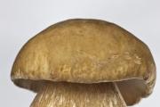
A mushroom is a living organism that forms a separate kingdom of the same name. For a long time they were classified as part of the plant kingdom. But in...
For lovers of “quiet” hunting, mushroom season begins in early summer and lasts until late autumn. And rarely do they...

Aleshnikova, V.I. Use of professional consultants. - M.: Infra-M, 1999. - 240 p. 2. Beich, E....

Orange juice. The symbolic meaning of orange juice in dream books is pleasure and temptation. Quite often we...

We most often overcome all kinds of difficulties encountered along the path of life. Of course, for this we make...

This year your patron Neptune will be in your constellation and this is a good sign, because you will...

1993 who? 1993 is the year of which animal? — According to the Chinese horoscope, 1873, 1933, 1993 belonged to the years of the Black...

Wave-particle duality of light means that light simultaneously has the properties of continuous...
The role of biology is enormous in our world. Although it is not one of the priority subjects, most schoolchildren and...

Amines are organic derivatives of ammonia containing an NH 2 amino group and an organic radical. In general...

How to answer questions in Part BThe second part of the social studies work consists of 7 tasks with a short answer....

Form TORG-15 is drawn up in the case when during transportation, movement between and within the warehouse, when...

Nutritionists say that for good health and a slim figure, you must include snacks in your...

A mushroom is a living organism that forms a separate kingdom of the same name. For a long time they were classified as a kingdom...
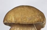
For lovers of “quiet” hunting, mushroom season begins in early summer and lasts until late autumn. And rarely do they...