Procedure for filling out an income tax return
An example of a correct income tax return in 2017, download a new current form for free in Excel. What...
)
the general name for paired sections of white matter of the spinal cord, limited by its fissures and grooves. Lateral cord of the spinal cord(f. lateralis) - with brain Lateral cord .
Posterior cord of the spinal cord(f. posterior) - with brain Posterior cord .
Anterior cord of the spinal cord(f. anterior) - with brain Anterior cord .
1. Small medical encyclopedia. - M.: Medical encyclopedia. 1991-96 2. First aid. - M.: Great Russian Encyclopedia. 1994 3. Encyclopedic Dictionary of Medical Terms. - M.: Soviet Encyclopedia. - 1982-1984.
- (funiculus medullae spinalis, PNA, BNA; fasciculus medullae spinalis, JNA) the general name for paired sections of the white matter of the spinal cord, limited by its fissures and grooves... Large medical dictionary
- (f. lateralis) see Lateral funiculus... Large medical dictionary
- (f. posterior) see Posterior funiculus... Large medical dictionary
- (f. anterior) see Anterior funiculus... Large medical dictionary
Medical terms
CORD- 1. A set of fibers forming any of the three columns of white matter of the spinal cord. 2. A bundle of nerve fibers covered with a sheath; bun. 3. (Obsolete) spermatic or umbilical cord. 4. (Funis) (in anatomy) any chord-like structure, in ... Explanatory dictionary of medicine
- (funiculus lateralis, PNA, JNA; funiculus lateralis (medullae spinalis), BNA: synonym: lateral cord of the spinal cord, lateral cord) a set of longitudinally running bundles of nerve fibers of the spinal cord located between the exit line... ... Large medical dictionary
- (funiculus posterior, PNA: funiculus posterior (medullae) spinalis, BNA; synonym: dorsal cord, spinal cord posterior cord) a paired bundle of nerve fibers in the white matter of the spinal cord, located between the posterior median and posterior ... Large medical dictionary
- (funiculus anterior, PNA, BNA; fasciculus ventralis, JNA; anterior synonymous cord of the spinal cord) a paired bundle of nerve fibers located in the white matter of the spinal cord between the anterior median fissure and the anterior lateral sulcus; contains... ... Large medical dictionary
The system of efferent neurons, the bodies of which are located in the cerebral cortex, terminate in the motor nuclei of the cranial nerves and the gray matter of the spinal cord. As part of the pyramidal tract (tractus pyramidalis), cortical nuclear fibers are distinguished... ... Medical encyclopedia
A set of longitudinally running bundles of nerve fibers of the spinal cord located between the exit line... ... Medical encyclopedia
All systems and organs in the human body are interconnected. And all functions are controlled by two centers: . Today we will talk about and the white formation it contains. The white matter of the spinal cord (substantia alba) is a complex system of unmyelinated nerve fibers of varying thickness and length. This system includes both supporting nervous tissue and blood vessels surrounded by connective tissue.
What does white matter consist of? The substance contains many processes of nerve cells; they make up the conductive tracts of the spinal cord:
The spinal cord is a special substance located inside the bone tissue. This important system is located in the human spine. In cross-section, the structural unit resembles a butterfly; the white and gray matter in it are evenly distributed. Inside the spinal cord, a white substance is covered with sulfur and forms the center of the structure.
The white matter is divided into segments, separated by the lateral, anterior and posterior grooves. They form the spinal cords:
The structure of the white matter is a complex system of pulpy fibers of different thicknesses; together with the supporting tissue, it is called neuroglia. It contains small blood vessels that have almost no connective tissue. The two halves of the white matter are connected by a commissure. The white commissure also extends in the region of the transversely extending spinal canal, located in front of the central canal. The fibers are connected into bundles that conduct nerve impulses.

The task of the ascending pathways is to transmit impulses from peripheral nerves to the brain, most often to the cortical and cerebellar regions of the central nervous system. There are ascending paths that are too welded together; they cannot be assessed separately from each other. Let us identify six fused and independent ascending bundles of white matter.
Descending pathways are associated with ganglia and the gray matter region. Nerve impulses are transmitted through bundles, they come from the human nervous system and are sent to the periphery. These pathways have not yet been sufficiently studied. They often intertwine with each other, forming monolithic structures. Some paths cannot be considered without separation:
We looked at the main ways that have been more or less studied by scientists at the moment. It is worth noting that there are also local bundles that perform a conductive function, which also connect different segments of different levels of the spinal cord.

The white matter connective system acts as a conductor in the spinal cord. There is no contact between the gray matter of the spinal cord and the main brain, they do not contact each other, do not transmit impulses to each other and affect the functioning of the body. These are all functions of the white matter of the spinal cord. The body, due to the connecting capabilities of the spinal cord, works as an integral mechanism. The transmission of nerve impulses and information flows occurs according to a certain pattern:
This is all a rather complex structure, but the processes are actually instantaneous, a person can lower or raise his hand, feel pain, sit down or stand up.
The brain includes several zones. The human skull contains the medulla oblongata, telencephalon, midbrain, diencephalon and cerebellum. The white matter of the spinal cord is in good contact with these structures; it can establish contact with a specific part of the spine. When there are signals associated with speech development, motor and reflex activity, taste, auditory, visual sensations, speech development, the white matter of the telencephalon is activated. The white substance of the medulla oblongata is responsible for conduction and reflex function, activating complex and simple functions of the whole organism.
The gray and white matter of the midbrain, which interacts with the spinal connections, are responsible for various processes in the human body. The white matter of the midbrain has the ability to enter into the active phase the following processes:
In order for information to quickly travel through the spinal cord to the central nervous system, its path lies through the diencephalon, so the body’s work is more coordinated and accurate.
More than 13 million neurons are contained in the gray matter of the spinal cord; they make up entire centers. From these centers, signals are sent to the white matter every fraction of a second, and from it to the main brain. It is thanks to this that a person can live a full life: smell, distinguish sounds, rest and move.
Information moves along the descending and ascending tracts of white matter. Ascending pathways move information that is encoded in nerve impulses to the cerebellum and large centers of the main brain. The processed data is returned in downstream directions.
The white matter is located under three membranes, they protect the entire spinal cord from damage. It is also protected by a solid spine frame. But there is still a risk of injury. The possibility of infection cannot be ignored, although these are not common cases in medical practice. More often, spinal injuries are observed, in which the white matter is primarily affected.
Functional impairment may be reversible, partially reversible, or have irreversible consequences. It all depends on the nature of the damage or injury.
Any injury can lead to the loss of the most important functions of the human body. When an extensive rupture or damage to the spinal cord occurs, irreversible consequences appear and the conduction function is disrupted. When a spinal bruise occurs, when the spinal cord is compressed, damage occurs to the connections between the nerve cells of the white matter. The consequences may vary depending on the nature of the injury.

Sometimes certain fibers are torn, but the possibility of restoration and healing of nerve impulses remains. This may take considerable time, because nerve fibers grow together very poorly, and the possibility of conducting nerve impulses depends on their integrity. The conductivity of electrical impulses can be partially restored with some damage, then sensitivity will be restored, but not completely.
The likelihood of recovery is affected not only by the degree of injury, but also by how professionally first aid was provided, how resuscitation and rehabilitation were carried out. After all, after damage, it is necessary to teach the nerve endings to conduct electrical impulses again. The recovery process is also affected by age, the presence of chronic diseases, and metabolic rate.
The spinal cord is fraught with many mysteries, so scientists around the world are constantly conducting research to study it.
Myelinated nerve fibers are grouped into tracts according to the specific direction - to or from the brain - and the type of impulse they receive or transmit. The ascending tracts transmit nerve impulses about all sensations arising in the body up to the . The descending tracts carry impulses from the brain to the skeletal muscles, causing voluntary and involuntary movements.
Pathways of the posterior funiculus:
1. Thin bun ( fasciculus gracilis) is located medially, it contains fibers coming from the lower half of the body, lower extremities through 19 lower spinal nodes and further to the medulla oblongata.
2. Wedge-shaped bundle ( fasciculus cuneatus) is located laterally, in it fibers pass from the upper part of the body through the upper 12 spinal ganglia to the medulla oblongata. Both bundles conduct conscious tactile, proprioceptive sensitivity and a sense of stereognosis.
3. Back own beam ( fasciculus proprius posterior).
Pathways of the lateral funiculus:
4. Side own beam ( fasciculus proprius lateralis).
5. Anterior spinocerebellar tract ( tr. spinocerebellaris anterior).
6. Posterior spinocerebellar tract ( tr. spinocerebellaris posterior).
Both conduct unconscious proprioceptive sensation.
7. Dorsal opercular tract ( tr. spinotectalis).
8. Lateral spinothalamic tract ( tr. spinothalamicus lateralis) - conducts conscious temperature and pain sensitivity.
9. Lateral corticospinal tract ( tr. corticospinalis lateralis) - conscious motor, pyramidal pathway.
10. Red nuclear spinal tract ( tr. rubrospinalis).
11. Olive-spinal fibers ( fibrae olivospinales).
12. Thalamospinal ( tr. thalamospinalis).
Pathways 10 - 12 are unconscious, motor, extrapyramidal.
Paths of the anterior funiculus:
14. Front own bundle ( fasciculus proprius anterior).
15. Anterior corticospinal tract ( tr. corticospinalis anterior) - conscious, motor pyramidal pathway.
16. Roof-spinal tract ( tr. tectospinalis).
17. Reticulospinal fibers ( fibrae reticulospinalis).
18. vestibulospinal tract ( tr. vestibulospinalis).
Pathways 16 - 18 are unconscious, motor, extrapyramidal.
19. Anterior spinothalamic tract ( tr. spinothalamicus anterior) - conducts conscious tactile sensitivity.
20. Medial longitudinal fasciculus ( fasciculus longitudinalis medialis) is present only in the cervical segments.
Segmental apparatus of the spinal cord- this is a set of nerve structures that ensure the execution of innate reflexes, it includes: dorsal root fibers, intrinsic bundles, nuclei of the anterior horns, scattered cells, cells of the gelatinous substance, spongy and terminal zones.
The conduction apparatus of the spinal cord provides two-way communication between the spinal cord and the integration centers of the brain (cerebellar cortex, cerebral cortex, superior colliculi). This apparatus is represented by sensory and motor pathways.
The integration (suprasegmental) apparatus of the spinal cord includes the ascending and descending tracts, as well as the nuclei: proper, thoracic and medial intermediate.
The spinal cord (medulla spinalis) is located in the spinal canal. At the level of the 1st cervical vertebra and occipital bone, the spinal cord passes into the medulla oblongata, and extends downwards to the level of the 1st–2nd lumbar vertebrae, where it thins out and turns into a thin filament terminale. The length of the spinal cord is 40–45 cm, thickness 1 cm. The spinal cord has cervical and lumbosacral thickenings, where the nerve cells that provide innervation to the upper and lower extremities are localized.
The spinal cord consists of 31–32 segments. A segment is a section of the spinal cord that contains one pair of spinal roots (anterior and posterior).
The anterior root of the spinal cord contains motor fibers, the posterior root contains sensory fibers. Connecting in the area of the intervertebral node, they form a mixed spinal nerve.
The spinal cord is divided into five parts:
Cervical (8 segments);
Thoracic (12 segments);
Lumbar (5 segments);
Sacral (5 segments);
Coccygeal (1–2 rudimentary segments).
The spinal cord is slightly shorter than the spinal canal. In this regard, in the upper parts of the spinal cord, its roots run horizontally. Then, starting from the thoracic region, they descend somewhat downwards before emerging from the corresponding intervertebral foramina. In the lower sections, the roots go straight down, forming the so-called ponytail.
On the surface of the spinal cord, the anterior median fissure, posterior median sulcus, and symmetrically located anterior and posterior lateral sulci are visible. Between the anterior median fissure and the anterior lateral groove is the anterior cord (funiculus anterior), between the anterior and posterior lateral grooves - the lateral cord (funiculus lateralis), between the posterior lateral groove and the posterior median sulcus - the posterior cord (funiculus posterior), which is in the cervical part The spinal cord is divided by a shallow intermediate groove into a thin fasciculus gracilis. adjacent to the posterior median sulcus, and located outward from it, a wedge-shaped bundle (fasciculus cuneatus). The funiculi contain pathways.
The anterior roots emerge from the anterior lateral sulcus, and the dorsal roots enter the spinal cord in the region of the posterior lateral sulcus.
In a cross-section of the spinal cord, the gray matter located in the central parts of the spinal cord and the white matter lying on its periphery are clearly distinguished. Gray matter in a cross section resembles the shape of a butterfly with open wings or the letter “H”. In the gray matter of the spinal cord, more massive ones are distinguished. wide and short anterior horns and thinner, elongated posterior horns. In the thoracic regions, a lateral horn is detected, which is also less pronounced in the lumbar and cervical regions of the spinal cord. The right and left halves of the spinal cord are symmetrical and connected by commissures of gray and white matter. Anterior to the central canal is the anterior gray commissure (comissura grisea anterior), followed by the anterior white commissure (comissura alba anterior); posterior to the central canal, the posterior gray commissure and the posterior white commissure are located successively.
Large motor nerve cells are localized in the anterior horns of the spinal cord, the axons of which go to the anterior roots and innervate the striated muscles of the neck, trunk and limbs. The motor cells of the anterior horns are the final authority in the implementation of any motor act, and also have trophic effects on the striated muscles.
Primary sensory cells are located in the spinal (intervertebral) nodes. Such a nerve cell has one process, which, moving away from it, is divided into two branches. One of them goes to the periphery, where it receives irritation from the skin, muscles, tendons or internal organs. and along another branch these impulses are transmitted to the spinal cord. Depending on the type of irritation and, therefore, the pathway along which it is transmitted, the fibers entering the spinal cord through the dorsal root may end on the cells of the dorsal or lateral horns or directly pass into the white matter of the spinal cord. Thus, the cells of the anterior horns carry out motor functions, the cells of the posterior horns carry out the sensitivity function, and the spinal vegetative centers are localized in the lateral horns.
The white matter of the spinal cord consists of fibers of the pathways that interconnect both the different levels of the spinal cord with each other, and all overlying parts of the central nervous system with the spinal cord.
The anterior cords of the spinal cord contain mainly pathways involved in motor functions:
1) anterior corticospinal (pyramidal) tract (uncrossed) coming mainly from the motor area of the cerebral cortex and ending on the cells of the anterior horns;
2) vestibulospinal tract, coming from the lateral vestibular nucleus of the same side and ending on the cells of the anterior horns;
3) tegmental-spinal tract, starting in the upper colliculi of the quadrigeminal tract of the opposite side and ending on the cells of the anterior horns;
4) the anterior reticular-spinal tract, coming from the cells of the reticular formation of the brain stem of the same side and ending on the cells of the anterior horn.
In addition, near the gray matter there are fibers that connect different segments of the spinal cord with each other.
The lateral cords of the spinal cord contain both motor and sensory PATHWAYS. Motor pathways include:
Lateral corticospinal (pyramidal) tract (crossed) coming mainly from the motor area of the cerebral cortex and ending on the cells of the anterior horns of the opposite side;
The spinal tract, coming from the red nucleus and ending on the cells of the anterior horns of the opposite side;
Reticular-spinal cord tracts, coming predominantly from the giant cell nucleus of the reticular formation of the opposite side and ending on the cells of the anterior horns;
The olivospinal tract connects the inferior olive to the motor neuron of the anterior horn.
The afferent, ascending conductors include the following paths of the lateral cord:
1) posterior (dorsal uncrossed) spinocerebellar tract, coming from the cells of the dorsal horn and ending in the cortex of the superior cerebellar vermis;
2) anterior (crossed) spinal-cerebellar tract, coming from the cells of the dorsal horns and ending in the cerebellar vermis;
3) the lateral spinothalamic tract, coming from the cells of the dorsal horns and ending in the thalamus.
In addition, the dorsal tegmental tract, spinal reticular tract, spino-olive tract and some other conduction systems pass through the lateral cord.
The afferent thin and cuneate fasciculi are located in the posterior cords of the spinal cord. The fibers included in them begin in the intervertebral nodes and end, respectively, in the nuclei of the thin and wedge-shaped fasciculi, located in the lower part of the medulla oblongata.
Thus, part of the reflex arcs is closed in the spinal cord and the excitation coming through the fibers of the dorsal roots is subjected to a certain analysis and then transmitted to the cells of the anterior horn; the spinal cord transmits impulses to all overlying parts of the central nervous system up to the cerebral cortex.
The reflex can be carried out in the presence of three successive links: 1) the afferent part, which includes receptors and pathways that transmit excitation to the nerve centers; 2) the central part of the reflex arc, where the analysis and synthesis of incoming stimuli occurs and a response to them is developed; 3) the effector part of the reflex arc, where the response is carried out through skeletal muscles, smooth muscles and glands. The spinal cord is thus one of the first stages at which the analysis and synthesis of stimuli both from internal organs and from receptors of the skin and muscles are carried out.
The spinal cord carries out trophic influences, i.e. damage to the nerve cells of the anterior horns leads to disruption of not only movements, but also the trophism of the corresponding muscles, which leads to their degeneration.
One of the important functions of the spinal cord is the regulation of the activity of the pelvic organs. Damage to the spinal centers of these organs or the corresponding roots and nerves leads to persistent disturbances in urination and defecation.

An example of a correct income tax return in 2017, download a new current form for free in Excel. What...
P. S. Pallas (1741 - 1811) - naturalist and traveler-encyclopedist, who glorified his name with major contributions to...
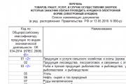
Today, all issues related to the placement of government orders are regulated by the Law on the Contract System -...
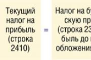
Accounting Regulations Accounting for calculations of income tax of organizations PBU 18/02 (as amended by Orders of the Ministry of Finance of the Russian Federation...
A trainee salesperson is usually called those salespeople who are not yet ready to work completely independently. Process...
Educational institution "Gomel State Medical University" Department of Neurology and Neurosurgery...

One of the most controversial and controversial methods for the early development of children was developed in the 80s by a sociologist...

Contents Dietary supplement based on an extract obtained from the fly beetle (or...
Ekaterina MirimanovaSystem minus 60. RevolutionSystem minus 60 with Ekaterina Mirimanova“System minus 60....
Heaviness and bloating are the causes of both ordinary overeating and more serious digestive problems...

The second blood group, Rh-negative, appeared many years ago, when a person ceased to be...

PSYCHOLOGICAL ASPECTS OF ANOREXIA PHENOMENON (EXPERIMENTAL STUDY) T. V. Tarasova, E. V. Arsentieva...
Contents Since the skin in this area is thin, it is more prone to the appearance of various types of spots....

vseslav Sat, 10/17/2015 - 20:50 Vasileostrovskaya station is one of the oldest stations...
P. S. Pallas (1741 - 1811) - naturalist and traveler-encyclopedist, who glorified his name with major contributions...
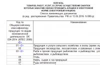
Today, all issues related to the placement of government orders are regulated by the Law on Contract...