Act on damage, damage, scrap of inventory items
Form TORG-15 is drawn up in the case when during transportation, movement between and within a warehouse, during storage...
The cardiovascular system, by ensuring a constant flow of blood, supplies all internal organs of a person with oxygen and nutrients every second, and therefore its importance is undeniably high. And that is why, when the slightest disturbance occurs in it, cascade reactions of failures are caused in all other systems, and therefore symptoms always appear. But how is the heart and blood vessels examined? There are many methods for this.
When a patient first turns to a therapist either for preventive purposes (physical examination) or with specific complaints, the specialist must necessarily examine the heart area and conduct simple studies of this organ and its branches. So, first of all, the doctor conducts a general examination of the patient, paying attention to his skin (with diseases of this system, pallor and even cyanosis, dense cold swelling, small hemorrhages are possible), the condition of the visible mucous membranes (injection of the sclera, white coating at the root of the tongue) , development of the musculoskeletal system (hypotonia, weakness, dystrophy or, on the contrary, obesity), the nature of the pulse (its presence and synchronicity in both arms, conduction of the pulse into the neck veins). Next, the doctor must conduct an examination of the heart, such as percussion of its borders, which can reveal hypertrophy of individual chambers. It is imperative to auscultate it, counting the number of heartbeats, detailing its tones, rhythm, and possible pathological noises.

Finally, blood pressure is measured because it is an important indicator of cardiovascular health. Next, the doctor must detail the complaints, because a complete examination of the heart includes a detailed medical history. Thus, diseases of the cardiovascular system are characterized by pain in the chest (often of a pressing, squeezing nature) or, more precisely, behind the sternum, shortness of breath (appears with increased physical activity normally, and in pathology - with a slight load or even at rest), and a feeling of what - “interruptions” in the functioning of the heart, manifestations of high blood pressure (headaches, dizziness, heaviness in the body). Be sure to find out the time of their appearance, the factors that provoke and eliminate them, and their intensity.

The heart examination also includes asking the patient what he associates with the development of his disease, thereby identifying risk factors. So, this could be a strong emotional shock the day before (the death of a loved one, stress at work), lifting heavy objects or performing difficult physical work. Symptoms also appear when weather conditions change. Heredity is also an important criterion, because most diseases (diabetes mellitus, arterial hypertension) tend to be transmitted to the next generation. As a rule, a correctly collected anamnesis provides 50% of the patient’s clinical diagnosis. After talking with the patient and examining him, the doctor must refer his ward for a heart examination.We should remember the anatomy and physiology of this organ.

So, it is, roughly speaking, a pump consisting mainly of muscles and a complex system of blood vessels. Inside it there are four chambers that communicate with each other in a strictly defined way and ensure constant movement of blood. And in order for the heart itself to continuously contract and relax, its tissues contain conductive structures through which a nerve impulse passes, thereby causing alternating tension in the muscles of each chamber and the opening and closing of the valves between them. Therefore, all methods of examining the heart can be aimed either at visualizing the anatomy of this organ (ultrasound, Doppler mapping, computed tomography, chest radiography, radioisotope methods) and directly at the arteries and veins (probing of the great vessels, angiography, coronary angiography), or at examining the condition its conduction system (electrocardiography, bicycle ergometry), or to auditory its tones and noises (phonocardiography).

As you can see, the examination of the heart must certainly be detailed, detailed, and not lose sight of anything. Because damage to the cardiovascular system can be either a manifestation of an independent disease or a consequence of the pathology of another system. If we talk about visual ones, the first thing that comes to mind is Echo-CG or, as it is also called. What the device shows during this important study can be guessed logically. By penetrating the ultrasound deep into the tissues and returning them back, an image appears on the screen that allows you to evaluate the structure of the heart, the size of its cavities, the condition of the valves and great vessels. Plus, this method is non-invasive and does not involve radiation, and therefore can be used even by pregnant, lactating and children. Although more effective, it still cannot replace ultrasound as a diagnostic tool.

At different stages of gestation, a woman periodically undergoes an ultrasound of the heart for the fetus, which shows a patent ductus arteriosus, stenosis of the ostia of vessels, prolapses or valve insufficiency, the condition of the interventricular and interatrial septum and other congenital malformations. Another important advantage of this method for the patient and the medical institution is its relative cheapness, the possibility of it being carried out on an outpatient basis, the short duration of the study, as well as the instantaneous acquisition of images and interpretation of all data. That is why ultrasound of the heart is so popular for diagnostic purposes.
In obese people, as well as patients with diabetes, the most common lesions of the cardiovascular system are atherosclerotic lesions of blood vessels, as well as hyalinosis of their walls. Therefore, it is so necessary to examine the vessels of the heart, because only they nourish this important organ, and its work requires a colossal amount of energy and nutrient substrates. So, first a catheter is inserted into the femoral catheter, through which the vessels are filled with a contrast agent, clearly visible on the X-ray screen. The most important method for atherosclerosis and ischemic myocardial disease is a coronary examination of the heart vessels. It reveals their passability, the correctness of their progress. Also, many operations on this important organ are carried out under his supervision.
Thus, there are currently a lot of methods for studying cardiac and vascular pathology, but each of them has strict indications and contraindications, and therefore it is economically unrealistic and diagnostically pointless to carry them out to everyone. Therefore, the key link is a competent doctor who will carefully supervise the patient and prescribe him the necessary treatment or send him to a more competent institution.
Today, the most common, taking lives more often than any other disease, are diseases associated with disruption of the cardiovascular system.
Fortunately, modern cardiology has great diagnostic capabilities, which allows timely detection of one or another abnormality in the cardiovascular system. Methods are very diverse, but they are used only after a palpation examination by a cardiologist, who first conducts a survey of the patient, focusing on complaints, listens to the sounds and sounds of the heart muscle, measures the pulse rate and blood pressure.
1. Electrocadiography (ECG).
1.1 ECG mapping.
1.2 Holter monitoring.
1.3 Bicycle ergometry and treadmill test.
2. Ultrasound examination of the heart and blood vessels.
3. Doppler examination of the heart and blood vessels.
4. Duplex study of blood vessels and heart.
5. Triplex study of blood vessels.
6. X-ray examination of the heart and blood vessels.
6.1 Angiocardiography.
6.2 Vasography.
6.3 Coronography.
7. Radioisotope methods for studying the heart.
8. Phonocardiography (PCG).
9. Electrophysiological study of the heart and blood vessels (EPS).
To finalize the diagnosis and confirm it, after a preliminary examination by a doctor, various instrumental research methods are used on the patient, the main one of which is ECG.
This mandatory diagnostic method takes a short period of time and allows you to:
Thanks to this research method, a heart attack, ischemic diseases, angina pectoris, myocarditis, endocarditis and pericarditis, pathological changes in the size of the atria or ventricles are promptly detected, however, for other cardiovascular diseases, the ECG does not give a complete picture, therefore, if necessary, additional diagnostic methods are used, for example , electrophysiological mapping of the heart (ECG mapping).
1.1 ECG mapping
Such research is based on the use of a significant number of wires (electrodes), which makes it time-consuming and impractical. However, using this method it is determined:
1.2 Holter monitoring
Holter monitoring is a long-term research method - heart function is recorded throughout the whole day. This method helps in diagnosing hidden disorders of the heart, which may not be noticeable during a regular ECG.
1.3 Bicycle ergometry and treadmill test
These research methods are based on recording the work of the heart muscle during dosed physical activity. During the testing process, the patient is under the supervision of a doctor who monitors the pressure, work and condition of the patient’s heart using an ECG.
For bicycle ergometry, an exercise bike is used, and for treadmill testing, a treadmill is installed at a certain angle to increase the load.
The purpose of such diagnostic methods is to identify hidden cardiovascular diseases and establish the limits of physical activity, beyond which the work of the heart is at risk.

Echocardiographic examination of the heart (Echocardiography) is an examination method in which the heart is examined using ultrasound. Modern ultrasound examination of the heart and blood vessels helps to combine:
EchoCG allows you to examine the heart in motion, evaluate its functioning as a whole and its individual sections. Often, this research method is used after a heart attack to determine the degree of myocardial damage from scars.
Doppler examination of the heart and blood vessels is carried out, like EchoCG, using ultrasound, the difference is that with such an ultrasound examination an additional change in the frequency of waves occurs when reflected from red blood cells, which makes it possible to accurately determine:
Doppler examination of blood vessels makes it possible to assess the risk of vascular rupture or thrombosis. Doppler ultrasound is successfully used in the diagnosis of varicose veins and various disorders caused by blockage or narrowing of the arteries. Modern systems make it possible to reproduce, using color Doppler mapping (CDC), even a multi-colored cartogram of blood flow in the vessel under study, where the color reflects the intensity and direction of blood flow.

Duplex examination of blood vessels and the heart is a method that combines two ultrasound modes - B-mode and Doppler mode.
B-mode involves the use of a sensor with many crystals emitting ultrasonic waves of a certain frequency. Such waves, penetrating through tissue at different angles and with different time delays, instantly scan the organ under study and, upon returning, reproduce a two-dimensional reconstruction of the heart and blood vessels on the screen.
The Doppler mode, when studying moving elements in blood vessels, along with the B-mode, makes it possible to obtain data on:
Using duplex scanning, atherosclerotic plaques, occlusions, stenoses, vascular malformations and other pathologies are successfully identified.
Triplex vascular examination is a diagnostic method based on the use of the Doppler effect and display of the studied organs in a configuration extremely close to their anatomical structure.
This study of the heart vessels allows for a detailed examination of the blood flow passing through individual sections of the vascular system. This diagnostic method is supplemented by colorectal dosage, which makes it more effective than the duplex study on which this study is based.
Thus, thanks to the triplex diagnostic method, the following is thoroughly examined at the same time:
Thanks to the accurate information received, the doctor determines the most effective treatment.

X-ray examination of the heart and blood vessels is a diagnostic method that allows you to find out the location of the heart. A change in the location of the heart may indicate the presence of pleurisy, mediastinal tumors, and all kinds of adhesions, which makes this research method very popular in medical practice.
6.1 Angiocardiography
This x-ray research method involves the use of a special contrast agent in the great vessels.
Angiocardiography makes it possible to diagnose the condition of large vessels and is therefore practically irreplaceable in determining the presence of congenital heart defects. In addition, this method is a basic examination before performing cardiac surgery.
6.2 Vasography
An X-ray of blood vessels is called vasography.
This procedure is carried out along with the introduction of a special substance that quickly spreads through the bloodstream, as a result of which the vessels are stained and become visible on the X-ray machine.
Vasography has many varieties, each of which has its own specifics. The main types of such x-ray examination include:
This method of studying the heart and blood vessels, such as coronography, requires special attention, since this technique is one of the most effective in identifying cardiovascular pathologies.
6.3 Coronography
This additional diagnostic method is used not only to confirm the diagnosis, but also to determine the location of pathologies. The result of the study of the coronary vessels is displayed on an angiograph, a device that gives a complete picture of the heart disease. Thanks to coronagraphy, it is clearly determined:
This study helps the cardiologist decide on the treatment method, since today it is the most accurate method for diagnosing the condition of the coronary arteries.

These diagnostic methods use a radioactive isotope, which is introduced into the body and accumulates in the heart, reflecting its condition at a given time. The substance accumulates in different quantities depending on the integrity or damage of areas of the myocardium, so this method is very effective in establishing:

FCG helps to register heart murmurs that cannot be detected with a phonendoscope. This method is very effective in situations where the question arises of establishing the correct functioning of the heart.

Electrophysiological examination of the heart and blood vessels is based on recording potentials arising on the inside of the heart. To carry out this diagnosis, special catheter tubes and a device for recording pathological findings are used. EPI helps to accurately determine the source and cause of the arrhythmia, as well as establish the location of its localization.
EPI is very effective in diagnosing and treating heart diseases, as it helps to monitor and regulate the effectiveness of prescribed therapy.
Only cardiologists have extensive practical experience, allowing them to accurately diagnose diseases of the heart and blood vessels, based on data from a complex of diagnostic methods. All methods of examining the heart and blood vessels are effective for identifying a particular cardiovascular disease, therefore only the attending physician, after reading the patient’s complaints and conducting a preliminary examination, can determine which method will be most rational in a particular case. However, over the years of practice, experts have become convinced that the most effective are x-ray research methods, in particular coronagraphy, and complex diagnostic methods, such as duplex and triplex examinations.
Modern people often develop heart and vascular diseases due to stress, fast pace of life, environment and other factors. They may not know about the pathology until a critical situation arises. In this case, it will be difficult for doctors to help and cure the patient, so to maintain heart health, it is important to carry out preventive diagnostics, and we will tell you how to check the heart in this article.
Many people do not go to the doctor when heart problems appear, since the symptoms are often vague and can be confused with other diseases, for example, the lungs or stomach. Even if the problem is obvious, and the patient understands that something is wrong with the heart, he often goes to the pharmacy to buy drugs that can relieve the symptoms, but the disease itself is not treated and progresses.
The main symptoms of heart disease, which require medical help and a thorough examination, are as follows:
Important! In addition to heart rhythm problems, you should consult a doctor if your resting heart rate is more than 90 or less than 60 beats per minute.
The cardiovascular system leads in the number of diseases. A common problem is vascular atherosclerosis. It develops slowly. throughout life. Therefore, doctors advise undergoing diagnostics once a year. If the disease is detected early, treatment will be quick and effective.
Methods for studying the heart in medical practice are divided into two types:
During the initial examination in the hospital, doctors use an objective examination of the cardiovascular system. After the examination, the doctor makes a presumptive diagnosis, then uses instrumental diagnostics.
Objective methods for examining the heart include:
 Palpation
 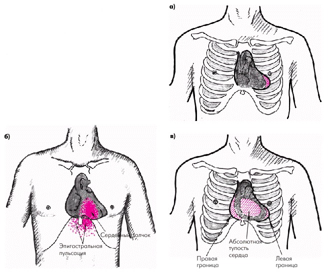 Percussion
  Auscultation
 The described methods of studying the cardiovascular system are carried out only during the initial examination; if the doctor finds certain deviations from the norm, he prescribes additional methods to check the activity of the heart; in this case, a full examination with special equipment is used.
This diagnostic method allows you to record and then study the electrical impulses that the heart muscle produces during operation. If the heart is without pathologies, then electrical excitation passes through different parts of the heart with a certain sequence. If the excitability of the heart muscle fails, this indicates pathologies and possible diseases.
When the myocardium contracts and relaxes, all data is recorded and written in the form of teeth, after which the doctor receives a curve or graph.
 ECG curve
 The data is recorded by a special device called an electrocardiograph. This diagnostic method allows you to evaluate the frequency and uniformity of the heart rhythm, various electrical processes occurring in the organ. An ECG is performed to detect arrhythmia, ischemia, and heart attack.
Important! Shifts in the ECG curve occur not only due to improper functioning of the heart. The reason may be diseases not related to this organ: pneumonia, pleurisy, obesity, etc.
Electrocardiography may be included in a comprehensive examination of the heart along with other methods.
In addition to taking a cardiogram at rest, other ECG techniques are used:
In the first case, the study continues for a day. Equipment and sensors are connected to the patient, after which round-the-clock recording of indicators of changes in excitability begins. Often, this method is used for severe patients, or if the problem appears periodically, for example, with short-term arrhythmia.
In the second case, an ECG is taken before and after stress on the body. This method allows us to identify the patient’s sensitivity to physical activity. Bicycle ergometry is often used for ischemia, namely exertional angina.
Phonocardiography allows you to record all sounds and murmurs of the heart. Recording is performed through a phonocardiograph, which is usually an additional device to an electrocardiograph. This method of instrumental diagnosis allows you to evaluate the symptoms of diseases by sound.
 Phonocardiography
 Echocardiography is performed using ultrasound. Today there are several methods for conducting echocardiography:
Methods for studying the heart and blood vessels using X-rays allow us to evaluate the size and shape of the heart, large vessels, and the volume of fluid in the pericardial part. When using this method, a person receives a dose of radiation, so there is no point in using it unnecessarily. It is used when other methods do not provide adequate information about the condition of a person and his organ.
X-rays cannot be used to examine pregnant women. One of the types of radiography is tomography. The latter method is more informative, since the picture is displayed on the monitor screen, simulating the patient’s organ, however, the radiation exposure in this case is higher than with x-rays.
An isotope study of the heart, namely the radionuclide method, is carried out by introducing radioisotopes into the blood, which make it possible to further evaluate their distribution. This method helps determine the formation of blood clots in blood vessels, as well as myocardial infarction. In this case, the patient also receives radiation.
Angiocardiography involves injecting a radiopaque contrast agent directly into the heart. With its help, doctors can study many parameters of the heart chambers and blood vessels. A procedure is used to determine the feasibility of surgery on an organ. This method is one of the main ones when examining for blood clots. Angiocardiography is performed by catheterization.
 Cardiac thrombosis
 Important! Only the doctor chooses how to check the blood vessels of the heart, by Dopplerography or angiography. The choice of method is influenced by many parameters, including the purpose of the study.
For each person and specific case, a certain type of diagnosis can be used, although in some situations more than one method may be used, but several at once. It depends on the state of health, the age of the patient and the reason why the heart hurts, that is, the existing pathology.
You can check your heart function at home, and people over 40 years of age are recommended to do this more often in order to detect deterioration of the condition in time. For home diagnostics, a tonometer is used, which can measure blood pressure and pulse rate.
A tonometer can be used of any type, for which you have enough money. Measurements are taken only in a sitting or lying position, at rest. You can do them on both arms, but only on the elbow. If during measurements the pressure is more or less than 110/70-140/90 and the indicator persists for a long time, it is recommended to visit a cardiologist.
Diagnosis of heart disease is very important, because more than 40% of deaths worldwide are caused by pathologies of the cardiovascular system.
The risk group includes not only older people, but also children, adolescents, and young people under 30 years of age.
To prevent the occurrence of heart diseases, an annual cardiological examination is carried out, modern methods of which make it possible to identify diseases at the initial stage and select the necessary treatment.
It is quite difficult to independently determine diseases of the cardiovascular system.
They manifest their characteristic features from time to time, alternating with an improvement in general condition.
Symptoms that may indicate heart problems include:
Manifestations of heart disease are preceded by heavy physical exertion, stress, emotional stress, fatigue, climate change or taking medications.
Diagnosis of heart disease is necessary to obtain data and confirm the diagnosis, which assumes that the patient has pathologies of the cardiovascular system.
For this purpose, the doctor selects and prescribes suitable methods for studying the condition of the heart:
The results obtained are processed in the laboratory or announced by the cardiologist at a subsequent appointment. Further treatment is prescribed depending on the age, gender of the patient, stage of the disease and its form.
People are forced to consult a specialist by pain and tingling in the chest area, an increase or decrease in blood pressure.
What you need to know before your first visit to a cardiologist:
 At the initial examination, the cardiologist will listen to all complaints and measure blood pressure and pulse, and listen to the heartbeat using a stethoscope. Based on the results obtained, the doctor will prescribe a complete cardiac examination.
Tapping or percussion is widely used at the initial stage of examining diseases and pathological processes in the heart. The essence of percussion is quite simple.
Through physical influence on the outer surface of the chest, the doctor provokes the appearance of various sounds, by which he determines the natural location of the muscular organ. The beating of the heart is accompanied by a dull sound.
The cardiac percussion procedure determines the boundaries of its dullness:
Allows you to determine the procedure for tapping the chest and quantitative values corresponding to the norm of the heart border:
A displacement of the heart to the right side indicates an increase in myocardium, hypertrophy, and pathology of the right ventricle. Removal to the left side indicates arterial hypertension and changes in the functioning of the left ventricle. If a uniform increase in the boundaries is observed, the patient is referred for additional examination. Possible causes: pericarditis, liver cirrhosis, pneumonia.
Cardiac auscultation is a traditional diagnosis of heart disease by indirectly listening to the heart with a special device - a stethoscope. It is used during the initial examination before hardware diagnostics and laboratory tests.
The cardiac auscultation procedure involves listening to the tone during myocardial contraction using the corresponding points on the body:
In a healthy person, the doctor determines only two tones, quickly replacing each other; the alternation of a pause and the appearance of a sound takes equal periods of time. A weak first tone indicates myocardial damage, heart failure, an increase in the mass of the organ or dysfunction of its valves.
Increased initial sound may be caused by right ventricular stenosis and tachycardia. A too quiet second tone is caused by aortic insufficiency and low blood pressure. Loud and clear sound - acceleration of valves, hypertension.
Disorders of the cardiovascular system are observed not only in adults. Pathological changes are increasingly affecting young patients.
What heart diseases are observed in children:
They require examination and consultation with a cardiologist, followed by diagnosis and treatment; if necessary, the child is placed in a hospital.
Diagnosis of heart and vascular diseases is very important, since heart and vascular diseases occupy a leading position in the list of the most common diseases among the population.
In addition, according to statistical indicators, cardiovascular diseases rank first in terms of mortality. Fortunately, the level of modern medicine makes it possible to quickly and accurately diagnose and, thereby, prevent the development of the disease at an early stage.
 Instrumental methods when examining a patient are:
Electrocardiography is a method of determining the electrical impulses of the heart using an electrocardiograph. The impulses are marked on a special moving tape.
At the end of the electrocardiography, the doctor receives an electrocardiogram (ECG), after studying which he can determine the pacemaker of the heart (sinus node or His bundle), assess the general condition of the conduction system, heart rate, and also identify certain heart diseases (for example, myocardial infarction).
Diagnosis of heart and vascular diseases by electrocardiography is completely safe for the patient and is carried out as follows. Small electrodes are placed on the chest and limbs of the subject, which determine the intensity and direction of heart currents. As a result, the ECG displays curves with teeth, segments and intervals, which can be used to judge the work of the heart.
Physical activity can be an indirect indicator of coronary heart disease or coronary circulatory disorders (for example, people with angina pectoris do not tolerate physical activity well). Based on the results of stress tests, we can talk about diseases that do not manifest themselves at rest.
During physical activity, the heart may lack oxygen due to narrowed coronary arteries, which normally deliver oxygen-rich blood to the organ. If you examine the lungs in a timely manner, you can tell whether the lack of oxygen is due to a disease on the part of the respiratory system, or on the part of the cardiovascular system, or a disease of a mixed nature.
During a stress test, the subject engages in certain physical exercises, and the doctor continuously monitors the patient and records the necessary indicators. The patient moves along a special path, the pace of which gradually increases, or pedals a bicycle.
As a rule, the speed is increased until the heart rate reaches 70-90% of the maximum permissible value characteristic of a person of a particular age and gender.
However, if the subject develops severe shortness of breath or acute pain in the chest area, and sudden surges in pressure are observed, the stress test is stopped before the set time.
A computed tomography (CT) scan can reveal anatomical changes in the chest cavity. Using a computer, so-called X-ray “slices” of the chest are made, which make it possible to detect pathologies of any nature.
An improved method has now been developed, called cine computed tomography. With its help, you can see an image of the heart in three-dimensional space and evaluate not only anatomical changes, but also disorders of the contractile function of the heart.
 Diagnosis of coronary heart disease is often associated with risk for the patient; the more complex the heart examination procedure, the greater the risk. When a catheter is inserted into the heart to conduct a coronary vascular study, there is a high probability of adverse consequences in the form of a heart attack, heart attack, or stroke. The incidence of death with angiography is 1:1000.
In stress tests, the risk of heart attack or death is 1:5000.
IN radionuclide examination the patient may receive a small dose of radioactive radiation, but no more than X-ray exposure.
In e electrophysiological examination Rhythm disturbances and the flow of nerve impulses into the heart are assessed. The examination takes place through the introduction of microelectrodes into the chambers of the heart, arteries and veins. By the electrical signals coming from the electrodes, you can find out the location of the nerve fibers through which the impulses travel.
Sometimes during a study, a doctor may deliberately induce an arrhythmia in a patient - this is necessary in order to determine the ability of the medicine to stop attacks and make sure that the operation is advisable. The pulse rate is brought back to normal using a shock. Despite the introduction of various instruments, this research method is safe, with a fatality rate of 1:5000.
In case of heart diseases, x-rays of the chest cavity are necessarily taken from the side, from the front.
The image allows you to study the vascular system in the lungs, assess the size of the heart, its chambers and the general structure of the organ. Using an image, you can detect pathological conditions of the heart - changes in shape, increased calcium content in the vessels. X-ray examination can also reveal abnormalities in the structure of the lungs and blood vessels, as well as detect fluid in the lung tissue.
The size of the heart may change due to impaired cardiac function or changes in the valves. But there are diseases in which the size of the heart remains the same, this can happen, for example, with constructive pericarditis. The heart is enveloped in an additional layer, but the dimensions remain normal.
X-ray examination characterized by the creation of a small X-ray “film” from several images, where the organs of the chest cavity and blood vessels are photographed in dynamics.
This research method is used to clarify diagnoses, to identify heart defects, and acts as an auxiliary method for electrophysiological studies and for vascular catheterization. Since the radiation dose during the examination is high, it is replaced by other safer research methods (ECHO and others).
Electrocardiography- This is the most common method of examination for heart disease. The main advantage of the method is the absence of radiation, as well as the ability to provide a clear image of the condition of the heart and blood vessels.
Diagnostics uses ultrasonic high-frequency waves, which transmit an image through a special sensor to the device screen. On the screen you can see the heartbeat and blood circulation. During the examination, the doctor can move the sensor along the patient’s chest, change the angle of inclination, which allows him to see a complete picture of the patient’s cardiac activity.
The image is recorded on a cassette; in order to obtain a clearer and higher-quality image, as well as to see small structural components, a special sensor is inserted into the patient’s esophagus and through it the image is transmitted to the screen.
The examination procedure does not cause harm to the patient; it is available for all age categories.
Magnetic tomography (MRI)- a method in which magnetic field energy is used to examine the chest organs.
 For this purpose, the patient is placed in a large electromagnetic box, where the atoms of the entire body are vibrated.
The atoms, in turn, transmit signals to a recording device, in which an image of all cardiac structures is formed.
This method usually does not require the introduction of additional contrast agents, but if the heart disease is associated with impaired vascular activity, then a substance with paramagnetic properties is injected to determine the location of weak vascular circulation.
MRI is the most difficult of all diagnostic methods, which is in the process of development, its disadvantage is the high cost of the examination and the difficulty in making the correct diagnosis.
At radionuclide examination Radioactive substances are injected into the arteries, which are contrast agents to identify painful areas in the heart. Radioactive tracers are distributed throughout the body at high speed; a specialized gamma camera records the radiation of substances.
The camera records the image and stores it on disk for later study. Despite the fact that during the examination the body receives a small dose of radiation, it is safe, unlike x-ray irradiation.
Positron emission tomography. This method has similarities with radionuclide examination; the difference is that with PET, one substance labeled with a radioactive agent is injected, which moves only in the area of the heart and its structures. Sensors on the device register areas with increased agent activity, and a high-quality 3D image appears on the computer screen. The selected area shows the active areas of the heart that absorb radioactive material.
PET is an expensive research method, therefore it is used to clarify the diagnosis or when reliable data are not obtained in other diagnostic methods.
Catheterization. This diagnosis of coronary heart disease is carried out as follows. The catheter is inserted into the veins or arteries, and gradually moves along with the bloodstream to the main areas of the heart. If it is necessary to examine the right area of the atrium and its chambers, a catheter is inserted into a vein. To examine the left atrium and chambers, a catheter is placed into the artery.
Catheterization is a universal tool for diagnosing and treating the heart; sometimes microscopic devices are installed in catheters to measure pressure.
Catheters are divided into types, depending on their purpose:
The advantage of using catheters is that they avoid surgery during treatment. Catheterization takes place in a hospital using painkillers.
Angiographic examination involves studying the activity and structure of the coronary vessels through catheterization and x-ray imaging. The catheter is inserted into a vein in the arm or groin area and advanced into the heart area; the movement of the catheter is monitored by a doctor using x-rays.
To obtain an image of the vessels, a contrast agent is injected into the catheter and, thus, any defects in the activity or structure of the coronary arteries can be seen on the screen.
This method allows you to detect coronary artery disease and begin treatment in a timely manner.
“Our heart is a treasure: waste it at once, and you are a beggar,” wrote Honore de Balzac more than 150 years ago. But we think about the heart more often when it begins to hurt, interruptions appear, and due to shortness of breath many familiar and favorite things become inaccessible. But even then, a visit to a doctor is often postponed until “later” and, unfortunately, some meet with a specialist already in the hospital. But today’s medical capabilities make it possible to conduct a complete examination of the body even in many clinics. It is often very important to make a diagnosis in time, then treatment will be more effective.
A heart examination should be performed if:
- You feel pain in the heart area or behind the sternum
- Feelings of palpitations, irregularities, “freezing”, lightheadedness or fainting occur
- Waist circumference in women is more than 80 cm, and in men more than 94 cm, which indicates abdominal obesity, which adversely affects the heart
- You have diabetes
- Increased blood pressure
- Some of your relatives suffered (or are suffering) from heart disease
- You just monitor your health and want to know about the state of your cardiovascular system
The most correct decision in this case is to consult a cardiologist or therapist so that you can be prescribed the necessary heart examinations.
What studies are performed on an outpatient basis?
The first stage of the examination will prescribe you:
- Resting ECG (a short recording allows you to see the main problems with rhythm or ischemia)
- Lipidogram (lipid spectrum): cholesterol, triglycerides, various fractions of lipoproteins, which makes it possible to determine the degree of lipid metabolism disorders, the risk of developing atherosclerosis
- Blood glucose (diabetes mellitus diagnosis)
- Complete blood count (hemoglobin level is important here to determine the presence of anemia, which can aggravate or simulate ischemic pain)
- Echocardiography (ultrasound of the heart), which clearly shows the condition of the heart muscle and heart valves
Additionally, depending on your complaints and examination results, you may be prescribed:
Biochemical blood test to assess kidney and liver function, which is important not only to clarify the diagnosis, but also for the correct selection of drugs
- Ultrasound examination of internal organs, mainly the kidneys, to clarify the cause of hypertension
- Ultrasound examination of the thyroid gland, analysis of the hormonal levels of the thyroid gland, which is important in the diagnosis of arrhythmias
- Daily ECG monitoring (Holter-ECG) and/or 24-hour blood pressure monitoring (ABPM) - allows you to evaluate heart rate and blood pressure during sleep, during physical activity, when various complaints appear, etc.
- Functional stress tests (treadmill test, bicycle ergometry) - to identify myocardial ischemia, which does not manifest itself under normal conditions, and to assess resistance to physical stress
- X-ray examination of the chest organs if there is a suspicion that some symptoms, for example, shortness of breath, are associated not with heart disease, but with pathology of the pulmonary system
A number of additional high-tech examinations, such as coronary angiography, are carried out only in a cardiology hospital under the direction of a cardiologist.
The list of necessary examinations can be expanded if the results of the studies allow us to suspect other diseases.
It is important to remember that in order to correctly assess the results of the examination and select the treatment indicated specifically for you, consultation with a competent specialist is required. Therefore, we advise you to carefully choose a clinic, and do not forget to take the examination results with you to your appointment.

Form TORG-15 is drawn up in the case when during transportation, movement between and within a warehouse, during storage...

Nutritionists say that for good health and a slim figure, you must include snacks in your diet....

Delicious pickled carrots for the winter can be prepared in a variety of containers, it can be a wooden container,...

When I have a few minutes left to prepare breakfast, the simplest and fastest recipes are used. This option...

An elegant table, a decorated Christmas tree, tangerine spirit spilled throughout all the rooms, soon the most magical holiday - New...

Each of us has repeatedly faced financial difficulties and difficult periods in life when Fortune mocked...

If you are new to magic, then it will be useful for you to get acquainted with the signs by which you can accurately...

SECRETS OF DREAMS Why does day follow night? What is life? What is death and what is sleep? These questions...

Main meaning: Whatever version of Madame Lenormand’s deck we take, we can definitely say that this is one of...

An example of a correct income tax return in 2017, download for free in excel the new current...

P. S. Pallas (1741 - 1811) - naturalist and traveler-encyclopedist, who glorified his name with major contributions...

Today, all issues related to the placement of government orders are regulated by the Law on Contract...

Accounting Regulations Accounting for income tax calculations of organizations PBU 18/02 (as amended by Order...

A trainee salesperson is usually called those salespeople who are not yet ready to work completely independently....

Nutritionists say that for good health and a slim figure, you must include snacks in your...

Delicious pickled carrots for the winter can be prepared in a variety of containers, it can be a wooden...