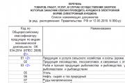
Procedure for filling out an income tax return
An example of a correct income tax return in 2017, download a new current form for free in Excel. What...
Horn fractures are common in cattle and less common in goats and sheep. In young bulls kept loose, horn injuries are accompanied by separation of the sheath from the horny process of the frontal bone or fracture of the process at its base.
Etiology. Occurs due to impacts, falls, damage from a mechanical harness or a moving conveyor, inappropriate fall, or pinching between hard objects. Massive injuries to the end of the horn are recorded in cows kept in stalls as a result of the gradual grinding of the horny sheath against the walls of the concrete feeder.
Clinical signs. When the horny process is fractured at its base, pain and swelling of the surrounding tissues are noted, the horn hangs down or is separated along with the horny sheath, while the sinus of the horny process is exposed, through it blood flows into the frontal sinus, and from it into nasal cavity(Fig. 5). The presence of blood in the frontal sinus is determined by percussion and radiography.
When the corneal process is fractured in the middle or near the apex with preservation of the horny sheath, mobility of the horn, pain and swelling of the tissues at the base of the horn are noted. The sinus of the corneal process is located below its middle, so bleeding may not be visible.
Tearing off the horny sheath from the process of the frontal bone leads to bleeding at the base of the horn, the tissue around the horn is swollen and painful. The horny cover is movable and can be easily removed from bone process. It is possible to completely separate the sheath from the horny process, and the connection between the leaves of the base of the skin of the horn and the leaves of the bony process is disrupted. Diagnosis. Cracks and fractures of the corneal process in the middle and upper parts established by palpation and radiography.
Forecast. In case of fractures of the corneal process and blood flow into the frontal sinus, the development of purulent frontal sinusitis, meningitis or phlegmon at the base of the horn, auricle and temporal fossa, the prognosis ranges from cautious to unfavorable.
Treatment. The horny cover, which has lost connection with the producing layer, is removed; A bandage impregnated with ichthyol or tar ointment, aminocaproic acid is applied to the exposed bone process.
When a bone process is fractured while the horny sheath is preserved, an improvised splint is applied to the damaged horn, which facilitates the formation of a strong callus.
If the horn fracture occurs at its base, then the soft tissue is prepared, the uneven edges of the bone process are smoothed with a rasp, saw or forceps, then a skin incision up to 4 cm long is made along the outer frontal ridge and a second one is made in the occipital direction. Both cuts at the base of the horn are connected by two semicircular cuts. The sinus of the corneal process is closed with displaced skin flaps, the latter are brought together with knotted sutures. In the presence of purulent inflammation, surgical treatment of the wound is performed; a bandage impregnated with Vishnevsky's liniment, dermato-tar or ichthyol ointments, irrigate with an aerosol of Chronicin, Kubatol, Libyan, Lifusol.
Rice. 5. Fracture of the horn at the base
Prevention. When keeping livestock loose, it is advisable to dehorn cows and prevent the growth of horns in calves. The zoohygienic conditions of keeping, tethering and grazing animals should not be violated.
Amputation of horns. In ruminant animals for agricultural purposes, especially in cattle, damage to the horns is observed in the form of fractures and cracks of the horny process of the frontal bone at the base, in the middle and approximately at the apex; detachment of the horny sheath from the corneal process; shedding of the horny sheath.
Etiology. Impacts, falls, damage from mechanical harnesses, unskillful fall of farm animals, pinching of horns between wooden or metal objects. Diseases such as osteodystrophy, osteomyelitis of the corneal process, etc., lead to damage to the horns.
Signs. At complete fracture The horny process of the frontal bone at the base of the horn, as a rule, hangs down, held partly by the soft tissues of the corolla of the horn. Intense bleeding appears, the sinus of the corneal process is exposed, blood flows into the frontal sinus, and from it into the nasal cavity of the side where the horn is damaged.
In case of fractures of the corneal process in the middle or in the area of the apex with preservation of the horny sheath, mobility of the horn when it sways and painful sensations are observed. The animal resists when held by the horns. There is likely hemorrhage in the sinus of the corneal process and in the frontal sinus.
Tearing off the horny sheath from the horny process of the frontal bone is accompanied by a disorder of integrity skin along the rim of the horn, exposing the bleeding base of the skin of the horn.
When the horny sheath is separated from the horny process, the connection between the leaves of the base of the skin of the horn and the horny leaves disappears. The horny sheath is held on the corneal process, but tissue rupture and bleeding are noted along the corolla.
On palpation, the horn is painful, an increase in local temperature indicators is observed, an inflammatory process is formed, and after 2-3 days a purulent exudate appears. The horny cover is made movable, and can be removed without much effort.
Diagnostics. The clinical picture of a complete fracture of the horny process of the frontal bone at the base is typical and no additional research is required.
Forecast. In case of fractures of the apex and middle of the horn, disruption or separation of the horny sheath, the prognosis is favorable; in case of a fracture of the corneal process at the base, the prognosis is cautious, since in these episodes the formation of purulent exudate is likely.
Treatment. At open fractures the tops and middle of the horn try to stop the bleeding, remove visible particles of dirt from the surface and apply an antiseptic bandage (with Vishnevsky's emulsion, tar), which must be fixed around the healthy horn, the bandages are laid in a figure eight. This treatment is carried out before the creation of a young scar horn.
If the corneal process is fractured at the base of the horn, the surgical field is prepared and the nerve of the horn is anesthetized. The outer ridge of the frontal bone is measured. Halfway between the orbit and the base of the horn, the skin is pierced with a short injection needle, pointing it slightly down and inward to a depth of 1-1.5 cm, and ten ml of a 2-3% novocaine solution is injected. Anesthesia sets in within 5-10 minutes.
After this, the entire horn is removed, the sharp ends of the fracture are leveled with bone forceps or a saw, the bleeding is stopped, the cavity frontal sinus tamponed with gauze soaked in a solution of furatsilin (1:5000). Several layers of gauze are applied to the stump, which is soaked in Vishnevsky’s emulsion and tar. Then the stump is covered with an adhesive bandage, which is replaced after 2-3 days.
When the horny sheath is shed, as well as when the latter loses its connection with the base of the skin of the horny process, the engraftment of the horn does not occur. For treatment, after carefully removing various contaminants from it with a warm solution of potassium permanganate, a bandage is applied to the exposed corneal process, which is soaked in tar or Vishnevsky ointment. The bandage is changed after 5-6 days. The corneal process is covered with a new horn.
Amputation of horns in cattle and rams it is also necessary to carry out in those episodes when growing horns injure soft tissues with their ends, giving rise to ulcerative pathologies, and often stick into tissue. For this purpose, the end part of the horn is eliminated with a bow saw (hacksaw) or using special scissors designed by V.K. Vasin.
2.7 Obstetrics, gynecology and biotechnology of agricultural animal reproduction
During my practice, I came across a case of serous mastitis (mastitis serosa) in a 5-year-old cow. When making a diagnosis, I took into account clinical data.
Mastitis is an inflammation of the mammary gland that occurs as a result of exposure to external and internal environmental factors with a decrease in the resistance of the animal’s body and complications with infection. There are 2 forms of mastitis - clinical, with obvious signs of inflammation of the mammary gland (redness, pain, swelling, temperature and impaired secretory activity) and subclinical, occurring latently, in which there are no signs of inflammation except for a decrease in milk production. Among the clinical forms of mastitis there are: serous, catarrhal, fibrinous, purulent, hemorrhagic, specific.
Serous mastitis is characterized by: effusion of serous exudate into the subcutaneous tissue and interlobular tissue of the udder. In animals, mild depression is sometimes noted, appetite decreases, and body temperature rises slightly (up to 39.80C). More often than not, one or two quarters of the udder are affected; they increase in volume, become painful, thickened, with reddened skin and increased local temperature. The nipples are enlarged, the suprauder lymph node on the side of the affected part of the udder is enlarged and painful. Milk secretion is reduced by 10-30%, and in the affected quarter by 50-70%. At the beginning of the disease, the milk does not change in appearance, but later it becomes watery, flakes and casein clots appear.
Differentiated from congestive edema, from which serous mastitis is distinguished by strong redness of the skin, increased local temperature with pain, in addition, with swelling of the mammary gland tissue, it is testy, which is easy to determine by palpation, and with serous mastitis, the consistency of the udder is rocky and dense.
Also differentiated from:
1) clinical mastitis(Mastitis catarrhalis) - Characterized by damage to the epithelium of the mucous membrane of the mammary cistern, milk ducts and canals, and glandular epithelium of the alveoli. The general condition of the animal remains satisfactory. Most often, only one quarter of the udder is affected; compactions are found in it, but the pain is mild. The nipple feels doughy to the touch. Milk is liquid with a bluish or yellowish tint and contains a lot of flakes and casein clots.
2) fibrinous(Mastitis fibrinosa) - Inflammation of the udder, in which fibrin is deposited in the thickness of its tissues, the lumen of the alveoli and milk ducts. The animal is depressed, often refuses food, body temperature is greatly elevated (40-41.0C), and lameness is noted. A quarter, half or all of the udder is affected. The affected quarters are greatly enlarged, red, hot, and very painful. Their tissues become very dense and the nipple is swollen. The suprauterine lymph node is enlarged, painful and inactive. The overall milk yield is reduced by 30-70%, the milk from the affected quarters is yellowish-gray, with fibrinous clots, films, often mixed with blood, and is difficult to milk.
3) purulent mastitis(Mastitis purulenta) - Inflammation of the milk ducts and alveoli of the udder with the formation of purulent or purulent-mucous exudate. The animal is depressed, appetite is sharply reduced, body temperature is increased to 40-41.0C. The affected quarters of the udder are enlarged, painful, hot, the skin is reddened and very dense. The suprauterine lymph node is greatly enlarged. Total milk yield reduced to 80%. A small amount of thick purulent or mucopurulent exudate with yellowish or white flakes is milked from the affected quarters.
4) hemorrhagic mastitis(Mastitis haemorrhagia) - acute inflammation udder with multiple hemorrhages and tissue soaking with hemorrhagic exudate. The disease occurs more often in the first days after childbirth. The cow is depressed, body temperature is increased to 40.0C. The affected quarters of the udder are enlarged, their skin is swollen, covered with burgundy spots, hot, and painful. The nipple is swollen and swollen. Overall milk yield is reduced by 25-40%, and of the affected quarters - by 60-95%. The milk is watery, reddish in color, with flakes.
If the animal is not helped in a timely manner, acute mastitis may develop into chronic form, and then a slow atrophy of the parenchyma occurs in the udder tissues, it is replaced by connective tissue. Milk yield is steadily declining, milk becomes mucopurulent. Complications are possible, including udder gangrene.
5) Subclinical mastitis visible signs are absent or weakly expressed, milk secretion and its quality are slightly changed.
The latent inflammatory process is accompanied by a sharp increase in the number of somatic cells in milk, which number over 500 thousand per 1 ml.
The following treatment was prescribed:
D.S. intercisternally, during the first 2 milkings after milking
3) Rp.: Solutionis Calсii chloridi
10%-100ml.
D.S. intravenously once
4) Rp.: Masticidum 150000 ED 5%-10.0
S.: intercisternally, inject 2 times. per day for 5 days.
5) Light massage from bottom to top for 10-15 minutes for 5 days.
2.8 Operative, general and private surgery
Due to the poor material and technical base and shortage of medicines, it does not have the ability to perform complex operations. The most common operation is castration of boars. Surgical assistance for injuries is also provided.
During the period of practice, surgical pathologies in animals were encountered. These are: loss of the left eyeball, disruption of the horny sheath, damage to the arch of the interclaw fissure, udder bruise, horn fracture, abscess.
| Disease | Cattle | pig |
| Abcess | 1 | |
| Destruction of the horny sheath | 1 | |
| Udder bruise | 1 | |
| Horn fracture | 1 | |
| Superficial wound of the udder | 1 | |
| Damage to the arch of the interclonal fissure | 1 |
|
| castration | 6 | |
| Total: | 6 | 6 |
Abscess, or abscess (abscess), is a limited pathological cavity filled with pus, resulting from spatially localized acute purulent, often infectious, inflammation of loose tissue, less often - other tissues and organs.
Symptoms: On the right side in the thigh area there is a spherical swelling, painful, dense, softening can be felt: in the center and in the lower part of the swelling.
Treatment: A wide dissection was made in the softening zone (in the lower part). The cavity was washed with a 3% solution of hydrogen peroxide. Intramuscularly "Nitox200" - 15 ml.
Destruction of the horny sheath.
Symptoms: The right horny sheath is torn off, moderate capillary bleeding, the wound is not contaminated.
Treatment: A bandage with 30% tar ointment is applied to the horny process of the frontal bone.
Udder bruise.
Symptoms: In the area of the posterior right lobe of the udder there was swelling on palpation - pain, a rounded hematoma with a diameter of 3 cm under the skin, the milk contains blood clots.
Treatment: Removing milk from a damaged udder, exposure to cold. The hematoma was opened and the contents were removed; the wound was powdered with streptocide powder.
Horn fracture.
Symptoms: The right horn is broken at the base heavy bleeding.
Treatment: The bleeding was stopped by destroying the intrahorn vessels with a scalpel and tamponing, then, against the background of novocoin blockade, blind amputation of the horn stump was performed according to Grigorescu. A bandage with 30% ointment is applied to the horny process of the frontal bone.
Superficial wound of the udder.
Symptoms: When trying to touch the nipple, the cow gets anxious and tries to hit with her pelvic limb. There is a superficial wound on the left anterior nipple.
Treatment: The wound was washed with furatsilin solution and sprinkled with antiseptic powder.
Damage to the arch of the interclofactual fissure.
Symptoms: The animal rests on 3 limbs; upon examination of the interhoof gap of the left anterior limb, damage to its arch was discovered, the cause of which was injury from a fixed chain.
Castration. It is held mainly in September - October, as well as in spring. This is due to the fact that during these periods the weather is most suitable for avoiding postoperative complications (infections, suppuration, etc.). As with any other surgical intervention, before castration it is necessary to prepare the surgical field and the surgeon’s hands (methods are presented below)
Castration of 3 boars one month old, owned by Akhpashev V.E. Castrated
in an open, bloody way.
Animals were fixed in the dorsal position. The skin of the scrotum was treated with a 5% alcohol solution of iodine. The skin of the scrotum is strained on the testis fixed with the left hand. We cut the layers of the scrotum layer by layer. The scrotal incision is made with a scalpel parallel to the suture (at a distance of 1-1.5 cm from it in the direction of the abdomen) along the entire length of the testis so that the common vaginal tunica is also opened. After cutting the vaginal ligament with scissors or a scalpel, the common tunica vaginalis is separated from the epididymis and spermatic cord. The thinned part of the spermatic cord was twisted to the point of breaking. Then the wound was sprinkled with streptocide mixed with iodoform.
Closed method castration was used for castration of an adult boar for 1.5 years. Using a scalpel, I carefully cut the scrotum to the length of the entire testis, without destroying the integrity of the common vaginal membrane. With an energetic movement of the fingers of her left hand, she squeezed the testis, covered with a common vaginal membrane, through the wound. Having pulled it out of the wound to the thinned part of the spermatic cord and pushing the edges of the scrotum towards the inguinal ring, he placed a ligature on the spermatic cord along with the common vaginal membrane. At a distance of 2 cm from the last, the spermatic cord was cut with scissors.
2.9 Veterinary and sanitary examination
During my practical training, unfortunately, I practically never encountered issues related to the examination of livestock products. My place of practice does not deal with issues of veterinary and sanitary examination. We only conduct a pre-mortem inspection, which included an examination of the animal as being clinically healthy, temperature, blood pressure, pulse, body condition, when vaccinations were carried out, a certificate form No. 4 was issued. After slaughter, the owners of the killed animals go to Tashtyp to the “Tashtyp Veterinary Station”.
| Veterinary work | Cattle | Pigs | Sheep |
| Ante-mortem inspection | 41 | 28 | 14 |
| Total | 41 | 28 | 14 |
Test
in operative surgery
on the topic: “Partial removal of cattle horns using a bloody method”
Introduction
1. Definition of the concept
Indications for surgery
Animal registration
Preparation surgical field
Preparing the surgeon's hands
Material support for the operation
Fixing the animal
Anesthesia
Selected method of operation
Online access
Operational reception
Conclusion
Application
Introduction
Literature review. Dehorning of adult cattle is recommended to be carried out at the age of no older than one and a half to two years. At this age, animals tolerate surgery more easily and complications are relatively rare. In animal husbandry practice, in order to prevent injuries, one should use those methods of dehorning that will be more economical and practically convenient in specific conditions. An integral condition for the transfer of livestock farming to an industrial basis is the creation of large complexes with high level mechanization of production processes, large concentration of animals in limited areas.
This technology of livestock farming, with all its positive features, caused the emergence of mass surgical diseases, one of them is injuries caused by sharp horns of animals, which causes considerable economic damage.
The pathogenic effect of trauma on the animal’s body has a number of features, the essence of which is as follows. Firstly, in acute cases injury may be accompanied by an immediate danger to the life of the animal, due to damage to vital tissues and organs, bleeding, etc.
Secondly, with extensive closed tissue damage and intensive absorption of tissue decay products, traumatic toxicosis of animals often occurs. Thirdly, in case of injuries caused by a strong impact of a mechanical factor, rupture of internal organs (liver, stomach, intestines, bladder and etc.).
Fourthly, when pathogenic microbes penetrate injured tissues, injuries are often complicated by abscesses, phlegmon, necrobacteriosis, actinomycosis, etc.
Fifthly, in some cases, injured animals develop neurotrophic disorders in the form of paresis, paralysis, and atrophy, which significantly worsen the general condition of the injured animal. A large number of injuries when animals are kept in large groups are caused by horns. Therefore, the task of farm veterinary specialists is to create polled herds.
1. Definition of the concept
DECORNUATION, decornuatio, onis, f (from the Latin de destruction, separation - cornu horn) - dehorning, surgical removal of horns or artificial prevention of their growth.
2. Indications for surgery
Produced to prevent horn injuries, as well as in case of abnormal growth, fractures and their diseases; Dehorning is carried out in cattle in order to prevent injuries due to diseases of the horns, their damage, neoplasms, rotting and abnormal growth. If surgery is not performed on a sick animal in a timely manner, this can lead to complications and even in some cases the death of the animal.
The operation was performed on a clinically healthy animal for educational purposes. But this operation can also be carried out with economic purpose. Since after the successful completion of this operation there is an increase in live weight and milk yield of cattle. This operation is mainly carried out to prevent injuries and to keep cattle loose.
Anatomical and topographical data of the site operational access to the organ
1. Horny capsule
Basal layer of epidermis
Papillary layer of horn dermis
Reticular layer of horn dermis
Subcutaneous layer
Horny process of the frontal bone
Frontal sinus
External structure of cattle horn
Base of the horn
Horn body
Top of the horn
Horn rings
The horns are located on the border of the aboral and external frontal crest. They belong to the frontal bones, but are still derivatives of the skin.
The base of the horn is formed by the horny process of the frontal bone, 7 to 20 cm long. Inside the process has a sinus covered with a mucous membrane, which communicates with the frontal sinus. The horny process is covered by the base of the horn skin, which fuses with its periosteum. The outer layer of the base skin of the horn forms the papillae, covered with the productive layer of the epidermis; the latter produces a dense stratum corneum that forms the horny sheath of the horn. The outer layer of the horn is represented by a horny sheath, protruding beyond the boundaries of the corneal process.
On the frontal bone, at the site of the future formation of the horny process above the periosteum, exostosis occurs, and a horny rudiment is laid in the thickness of the skin, which creates a horny tubercle. The exostosis and the horny rudiment are separated from each other by the periosteum, and then they grow together. At the same time, a small cavity appears in the horny tubercle, connected to the sinus of the frontal bone. During the process of growth, its cavity continues into an enlarging horny process.
In young animals, the horn cavity has a large number of partitions, varying in size, shape and direction. As the animal grows, the partitions become thicker, and their length, on the contrary, decreases, due to which the horn cavity becomes larger.
The horn is divided into root, body and apex.
The root of the horn - radix cornus - is the thinnest part of the horn, which is located at the junction of the horn with the skin of the forehead. The body of the horn - corpus cornus - continues from the root to the apex and is the most extensive and massive part. The apex of the horn is the pointed free end of the horn. At the root of the horns outer surface ring-shaped interceptions are noticeable, which in a cow are associated with the pregnancy period.
Innervation. The main nerve is the nerve of the horn - n. cornus - branch of the ophthalmic nerve. Coming out of the orbit, it runs along the external frontal crest, being covered by skin, fascia, fronto-scutellum muscle and a layer of fat. The branches of the frontal and subtrochlear nerves approach the base of the horn, which, connecting with their branches, form a kind of plexus. In addition, the branches of the dorsal trunks of the first cervical nerves.
The blood supply to the horn is provided by the horn artery - a. cornus, originating from the temporal superficial. It runs along the external frontal ridge, accompanied by the nerve of the same name, and divides at the base of the horn into lateral and medial branches.
trauma operating room animal surgeon
4. Animal registration
Gender - Bull
Breed - black and white
Color: black with white spots
Weight - 200 kg
Age - 2 years
Owner - Vivarium of the Department of Surgery.
The laboratory animal is clinically healthy, the operation is performed for educational purposes.
T - 38.4 ?S P - 72 D - 34
5. Preparation of the surgical field
Treatment of the surgical field takes place in several stages:
Mechanical cleaning - removal of foreign bodies contaminating the surface, as well as removal of grease and epidermal crusts. Mechanical cleaning was carried out using a fat-soluble substance (0.5% ammonia solution).
Field cultivation was carried out at 5% alcohol solution Yoda.
6. Preparing the surgeon's hands
Hand treatment consists of three stages: mechanical cleaning, disinfection, leather tanning.
Mechanical cleaning - the use of substances that dissolve grease and crusts of the epidermis.
Disinfection is a set of measures aimed at destroying pathogens infectious diseases and destruction of toxins at sites external environment.
Tanning - conclusion excessive amount water from the skin and closing skin pores.
The preparation of the surgeon's hands was carried out according to Olivkov's method:
Washing hands in 0.5% ammonia solution - 5 minutes.
They are thoroughly washed alternately in two basins for 2.5 minutes or under a running stream using a haze napkin.
Disinfection and tanning were carried out with a solution of iodized alcohol 1:1000 or 1:3000 until crepitation. This is done by wiping your hands with a swab - 2 times.
Treatment of the ends of the fingers, under the nail spaces and nail beds with 5% iodine.
7. Material support for the operation
Tools:
scalpel
rubber band
syringe with a capacity of 20 ml
luer lock syringe
injection needle
Tools special purpose
sheet saw
Dressing material used:
Sterilization of instruments.
Sterilization - destruction of all types of microorganisms, including bacteria<#"justify">8. Fixation of the animal
The animal being operated on is fixed in the machine in a standing position. For more reliable fixation, the animal’s head can be held by nasal septum and squeeze it with your fingers, you can also use nasal forceps.
9. Pain relief
Xylosine was used as a neuroleptic at a dose of 0.5 ml. per 100 kg. Animal weight. The weight of the operated bull was 200 kg, 1.0 ml was injected. xylazine. Palpation determines the outer crest of the frontal bone. Halfway between the orbit and the base of the horn, the skin is pierced with a needle and 20 ml of a 3% novocaine solution is injected. Then the needle is directed under the ridge to a depth of 1-1.5 cm and another 20 ml of 3% novocaine solution is injected. Anesthesia occurs within 5-10 minutes.
Neuroleptanesthesia and horn nerve block were used
10. Selected method of operation
To remove adult horns cattle 2 methods are used:
Bloodless
Bloody
Bloodless. A rubber ring made of white vacuum rubber with an outer diameter of 35 mm and an inner diameter of 10 mm is placed on the base of the horn. Before applying the ring, the hair at the base of the horn is cut off, the skin is cleaned of dirt and disinfected. The horn is anesthetized. To put on the ring, use a dilator or gauze straps. The ring is finally fixed with a metal spatula, using it to move the ring under the raised edges of the horny capsule. Constant pressure of the ring causes atrophy of the edge of the horny capsule, soft tissues and underlying bone tissue. The horns fall off within 28-47 days from the moment the rings are applied. The defects are covered with a small amount of fibrinous exudate, which then turns into a dense dry scab. Its formation prevents the development of infection in the frontal sinus. In the first days after the application of the rings, animal anxiety and loss of live weight (from 3 to 20%) are observed. 6-7 days before the horns completely fall off, the animals begin to worry again. After rejection of the horns, the general condition of the animal is normalized, milk yield and fatness are restored.
2. Bloody. The surgical site is prepared in the generally accepted manner. Then neuroleptanesthesia and horn nerve block are performed: the outer crest of the frontal bone is determined by palpation. Halfway between the orbit and the base of the horn, the skin is pierced with a needle and 20 ml of a 3% novocaine solution is injected. Then the needle is directed under the ridge to a depth of 1-1.5 cm and another 20 ml of 3% novocaine solution is injected. During the operation, a tourniquet is applied around the base of the horn to compress the right and left arteries of the horn and thereby prevent arterial bleeding. Amputation is carried out with a sheet saw. Bleeding areas are smeared with a scalpel to accelerate thrombus formation. The amputation wound can be treated with an antibiotic. Then apply cotton gauze bandage. Healing of wound defects after amputation of horns lasts 1.5-2 months.
The bloody method of operation was chosen. It was more expedient to use it, because the animal endures the operation not as painfully as with the bloodless method of operation, and the productivity of the animal is practically not reduced.
11. Online access
According to the existing nomenclature, the name of the operation is based on the name(s) of the organs being operated on and the main surgical technique used. In this case (in most cases in words of Greek origin), the name of the operation is combined into one word (enterotomy, cholecystectomy, pneumonectomy, omentocardiopexy, endarterectomy, biovarectomy). In some cases (mainly in words of Latin origin), the name of the operation consists of two words (resection of 7/8 of the stomach, amputation of the thigh in the middle third). Features of surgical access and anesthetic management introduce qualifying adjectives into the name of the operation (puncture liver biopsy, endoscopic papillosphincterotomy).
The surgical stage consisted of removing a third of the horn.
12. Surgical procedure
After treatment of the surgical field, neuroleptanesthesia and horn nerve block are performed: the outer crest of the frontal bone is determined by palpation. Halfway between the orbit and the base of the horn, the skin is pierced with a needle and 20 ml of a 3% novocaine solution is injected. Then a tourniquet is applied to the base of the horns. Amputation of the horn is carried out with a sheet saw, and the head is tilted towards the amputated horn in order to prevent the flow of blood into the frontal sinus. At the end of the amputation of the horns, the tourniquet is removed and the places where bleeding is observed are smeared with a scalpel to cause faster thrombosis of the damaged area.
The final stage.
The final stage This operation involves applying an eight-shaped gauze bandage to the horn.
The bandage is applied in rounds, with both hands, alternately rotating the bandage around the horn with one or the other hand.
Postoperative condition.
Postoperative wound healing in the animal proceeds favorably, without complications.
Complications.
None.
Conclusion
The expediency of this operation is not doubtful. Since cattle are kept en masse in livestock complexes, there is a very high risk of surgical trauma caused by the sharp horns of the animal, and considerable economic damage is caused.
Injuries caused to cattle can be very dangerous both to other animals and to workers of livestock farms, since after being hit by a horn they can occur serious injuries or even fatal outcome.
List of used literature
1.Olive general surgery 2010
.Operative surgery. Workshop: Proc. Benefit. I. I. Magda, V. M. Vlasenko, E. M. Ponomarenko.- K-: Higher. school, 2010
.Somatic systems of domestic animals. Textbook Benefit. I.V. Yatsenko, V.P. Gorbatenko, V.I. Simonenko and others.
.Operative Surgery with Basics topographic anatomy pets I. I. MAGDA, B. Z. ITKIN, I. Y. VORONIN MOSCOW “KOLOS” 2009
.General veterinary surgery. A. D. Belov, M. V. Plakhotin, B. A. Bashkirov and others; Ed. A. D. Belov, V. A. Lukyanovsky. - M. Agropromizdat, 2009
6.Anatomy of domestic animals. Akaevsky A.I. M.: “Kolos”, 2011
Application
Cattle before surgery
Treatment of the surgical field
Pain relief for cattle horns
Applying a tourniquet to the base of cattle horns
Amputation of part of a cattle horn
After amputation wound
Need help studying a topic?
Our specialists will advise or provide tutoring services on topics that interest you.
Submit your application indicating the topic right now to find out about the possibility of obtaining a consultation.
(TRAUMA CORNUUM)
In ruminants, especially in cattle, damage to the horns occurs in the form of fractures and cracks of the horny process of the frontal bone at the base, in the middle and near the apex; separation of the horny sheath from the corneal process; tearing off the horny cover.
Etiology. Impacts, falls, damage from mechanical tethering, unskillful felling of animals, pinching of horns between wooden or metal objects. Osteodystrophy, osteomyelitis of the corneal process, etc. predispose to damage to the horns.
^ Clinical signs . With a complete fracture of the horny process of the frontal bone at the base, the horn usually hangs down, partially held by the soft tissues of the corolla of the horn. Severe bleeding occurs, the sinus of the corneal process is exposed, blood flows into the frontal sinus, and from it into the nasal cavity of the side where the horn is damaged.
In case of fractures of the corneal process in the middle or near the apex with preservation of the horny sheath, mobility of the horn when it staggers and pain is noted. The animal resists when it is held by the horns or when the horn is percussed. In case of rupture of blood vessels, hemorrhage into the sinus of the corneal process and into the frontal sinus is possible.
Tearing off the horny sheath from the horny process of the frontal bone is accompanied by a violation of the integrity of the skin along the corolla of the horn, exposing the bleeding base of the horn skin, which in many places is torn down to the bone and is often contaminated with earth, dust or manure.
When the horny sheath is separated from the horny process, the connection between the leaves of the base of the skin of the horn and the horny leaves is lost. The horny sheath is held on the corneal process, but tissue rupture and bleeding are noted along the corolla. On palpation, the horn is painful, an increase in local temperature is noted, inflammation develops, and after 2...3 days a purulent exudate appears. The horny sheath becomes movable without special effort it can be removed.
Diagnosis. The clinical picture with a complete fracture of the horny process of the frontal bone at the base is characteristic and does not require additional research. When diagnosing fractures in the middle and upper parts of the horn or fissures of the corneal process, radiography is indicated. Forecast. In case of fractures of the apex and middle of the horn, disruption or separation of the horny sheath, the prognosis is favorable. If the corneal process is fractured at the base, the prognosis is cautious, since in these cases blood may flow into the frontal sinus and the development of purulent frontal sinusitis.
Treatment. In case of open fractures of the apex and middle of the horn, stop the bleeding, remove visible particles of dirt from the surface and apply an antiseptic bandage (Vishnevsky emulsion, tar), which is fixed around the healthy horn, placing the bandage in a figure eight. This treatment is carried out until a young scar horn forms.
In cases of fractures of the horny process while maintaining the integrity of the horny sheath, metal or wooden splints are used for fixation or a plaster cast is applied to the horn.
In case of a fracture of the corneal process at the base of the horn, the surgical field is prepared and anesthetized, after which the horn is completely removed, the sharp ends of the bone fracture are straightened with bone forceps or a saw, the bleeding is stopped, the cavity of the frontal sinus is tamponed with gauze soaked in a solution of furatsilin (1:5000). Several layers of gauze impregnated with Vishnevsky's emulsion, tar or molten paste of the following composition are applied to the stump: Cegae flayae (paraf-fini) - 10.0; Olei vaselini - 2.0; Picis liquidae (Ichthyoli) - 2.0. The stump is then covered with an adhesive bandage. The bandage is changed after 2...3 days. When changing the dressing for the first time, remove the gauze swab from the frontal sinus. If amputation is done in such a way that there is no growth of the horn, then the skin should be removed 1 cm from the base of the horn.
I. Ya. Tikhonin and M. A. Feldshtein recommend polymer glue-gikhlovul, melted in a water bath at a temperature of 100... 120 ° C, for closing the stump after amputation of the horn. A gauze cloth folded in four is soaked in it, and the wound is covered with it. The wound is hermetically sealed with a bandage for 30 days or more.
When the horny sheath is torn off, as well as when the latter loses its connection with the base of the skin of the corneal process, the engraftment of the horn does not occur. For treatment, after thoroughly removing contaminants from it with a warm solution of potassium permanganate, a bandage soaked in tar or Vishnevsky ointment is applied to the exposed corneal process. The bandage is changed after 5...6 days. The corneal process is covered with a new horn.
Amputation of horns in cattle and rams also has to be performed in cases where the growing horns, changing their direction, injure soft tissues with their ends, causing ulcerative lesions, and often stick into tissue. For this purpose, the end part of the horn is removed either with a bow saw (hacksaw), or using special scissors designed by V.K. Vasin. Prevention. To prevent damage to the horns, zoohygienic conditions for keeping, housing, tethering and grazing animals should be observed. Do not clutter the premises where animals, pastures, etc. are kept with foreign objects; take precautions when knocking down animals.
In recent years, in connection with the transfer of cattle to free-stall housing to prevent injuries to cows caused by horns to each other, there is a need to create polled herds by dehorning calves. (For the surgical technique, see the textbook on operative surgery.)
^ PURULAR INFLAMMATION OF THE BASIS OF THE SKIN OF THE HORNS
(PODODERMATITIS PURULENTA CORN US)
According to L.I. Tselishchev and V.L. Kupchinsky, purulent inflammation of the base of the skin in breeding rams occurs in 7...8% of cases of the total.
Etiology. Purulent inflammation the basis of the skin occurs as a result mechanical damage her when struck by horns at the moment of butting.
Pathogenesis. In case of injuries in the area of the base of the horn, the vessels of the base of the skin of the horn rupture, and hemorrhages occur between the base of the skin and the horny sheath. Inflammation develops at the base of the horn, as a result of which the base of the skin at the corolla separates from the horny sheath and exposes it. On the exposed base of the skin, ulcers with overgrown granulations appear, which are easily destroyed with the formation of a foul-smelling exudate. Inflammation can spread to the horny process of the frontal bone, causing osteomyelitis and bone abscess.
In places of lesions in the summer, the Wohlfarth fly lays larvae, which cause the breakdown of soft tissues with the release of ichorous exudate and the proliferation of granulation tissue.
^ Clinical signs . The animal stands with its head lowered towards the damaged horn. At the base of the horn, purulent inflammation, ulcers, overgrown foci of granulation tissue with the presence of liquid ichorous exudate are found, around which there is a proliferation of soft and. flabby horn. With wolfhartiosis, fly larvae can be found in the lesion.
^ Diagnosis. Purulent pododermatitis of the horn is diagnosed based on the clinical picture.
Forecast. With timely treatment, the prognosis is favorable.
Treatment. Surgical treatment of the ulcerative surface is performed and the horn is amputated 6 cm above its base, so as not to open the horny sinus. A paraffin-gauze application and a pressure bandage are used on the stump. For three days after the operation, streptomycin is administered intramuscularly once a day, 500 thousand units in a 0.5% solution of novocaine.
When complicated by wolfarthiosis, the ulcerative surface is treated with Estrosol, Dichlorvos, and Chlorophos aerosols in order to destroy fly larvae. Then the larvae are removed, and the affected areas are washed with a 1% solution of potassium permanganate, creolin or Lysol and a bandage with Vishnevsky’s emulsion is applied.
^ WOUNDS OF THE JAW JOINT
(VULNERA ARTICULATIO MANDIBULAR IS)
Etiology. Wounds jaw joint are inflicted on animals by random objects, spikes of a forged horse, or horns. There are gunshot wounds, complicated by intra-articular and periarticular bone fractures and disruption of the integrity of the articular cartilage.
^ Clinical signs . At puncture wounds on the first day, you can observe a small amount of clotted blood, which in the form of a crust is retained on the skin and closes the entrance to the wound canal. There is pain on palpation and opening oral cavity and slight swelling in the joint area.
With bruised and lacerated wounds, bleeding with an admixture of synovium is observed, and then on the 3rd...4th day, with the development of purulent inflammation, purulent exudate with an admixture of synovium begins to be released. The edges of the wound are swollen and uneven.
If the integrity of the bones is damaged, fragments are found in the lumen of the wound; eating and chewing food is painful, so the animal refuses food or has difficulty accepting it, although the appetite is preserved.
Diagnosis. The nature and type of wound is diagnosed based on the clinical picture and radiography.
Forecast. For wounds of the jaw joint, the prognosis is cautious. The development of purulent arthritis, osteomyelitis and deforming arthritis, joint ankylosis is possible, which leads to exhaustion of the animal.
Treatment. For puncture wounds, the hair around the wound opening is removed, the skin surface is treated with a 0.5% ammonia solution, wiped dry with a cotton swab, and then the skin and blood clots on the surface of the wound are twice lubricated with a 5% alcohol solution of iodine, and an alcohol bandage is applied. If on the 2...3rd day the inflammation does not intensify and the painful reaction decreases, then alcohol dressings continue to be used until 7...8 days. If inflammation intensifies, swelling increases, and local temperature rises, then novocaine-antibiotic solutions (penicillin, bicillin-3, bicillin-5) are injected into the joint cavity with a sterile needle.
For penetrating gaping wounds, primary surgical treatment of the wound is performed, the joint cavity is washed with a solution of furatsilin (1:5000) or ethacridine lactate (1:500), and then irrigated with a 0.5% solution of novocaine with penicillin. In the case of penetrating wounds with damage to the articular surfaces, the wound is inspected, loose bone fragments are carefully removed, and then the same treatment is carried out as in previous cases.
Animals with penetrating joint wounds are given crushed food to reduce chewing movements. After the wound heals, chewing function is restored.
^ INFLAMMATION OF THE JAW JOINT
(ARTHRITIS MANDIBULARIS)
Inflammation of the jaw joint can be acute and chronic, serous, aseptic, purulent and deforming.
Etiology. Acute serous aseptic inflammation of the jaw joint occurs as a result of blows and rough manipulations. Purulent inflammation develops with penetrating wounds, complicated fractures, with the transition of the inflammatory process to the joint from surrounding tissues, with penetration into the tissues surrounding the joint by the spines and leaves of feather grass, etc.
Chronic deforming arthritis develops as a result of bruises, purulent arthritis, dental disease, prolonged one-sided chewing, etc.
^ Clinical signs . In acute serous aseptic arthritis, a limited, fluctuating, painful swelling with an increase in local temperature appears in the joint area. Pain also appears when opening the mouth. Chewing function is impaired. In the chronic course, atrophy of the masticatory muscles occurs.
With purulent arthritis, the swelling in the joint area is larger, the pain is more severe, and it is possible to increase not only local, but also general body temperature. If arthritis has developed due to a penetrating wound, then from the wound opening on the 3rd...4th day synovial fluid with purulent exudate, and then begins copious discharge purulent exudate mixed with synovium. There is destruction of articular cartilage and caries of the articular surfaces of bones. With prolonged arthritis, joint stiffness develops and scissor teeth form. Sick animals lose weight quickly.
With deforming arthritis, an oval or spherical painless thickening is observed in the area of the affected joint, sick animals have difficulty opening their mouths, they develop scissor teeth, and atrophy of the masticatory muscles appears. Chewing occurs only on the unaffected side of the jaw. In horses, feed masses accumulate between the cheek and teeth of the affected side, which, if not removed in a timely manner, are compressed into dense formations. Animals progressively lose weight.
Diagnosis. The basis for diagnosis is characteristic clinical signs. Forecast. In case of acute aseptic inflammation of the jaw joint, the prognosis is favorable, in case of purulent and deforming arthritis - cautious or unfavorable.
Treatment. At the beginning of the development of acute serous inflammation On the first day, cold is used, and then alcohol or alcohol-ichthyol warming compresses. During the same period, a short novocaine blockade. Subsequently, irradiation with a Sollux lamp is used 2 times a day for 25...30 minutes, applications of paraffin, ozokerite, electrophoresis of iodine ions. After reducing inflammation and pain reaction, massage with iodine preparations is used.
For chronic arthritis, irritating ointments are rubbed in and diathermy is used.
For purulent arthritis, the joint cavity is washed with antiseptic solutions (furacilin, ethacridine lactate) and the wound is inspected, during which bone fragments are removed, pockets are opened, fistulas are expanded, dead tissue is removed with a sharp spoon and capillary drainage soaked in antibiotics or Vishnevsky's emulsion is introduced. In case of destruction of the articular surfaces, as well as in case of ankylosis of the joint in especially valuable animals, resection of the articular process can be performed lower jaw.
For deforming arthritis in cases where there is no stiffness of the joint, UHF, point cauterization is used, and acutely irritating ointments are rubbed in.
If the animal is not of particular value, then it is advisable to discard it in all cases of complicated purulent arthritis, deforming arthritis and ankylosis of the joint.
^ DISLOCATION OF THE LOWER JAW
(LUXATIO MANDIBULAE)
Mandibular luxation occurs primarily in dogs and cats; it is often bilateral, sometimes complicated by a fracture of the articular process.
Etiology. Impacts to the lower jaw, falling animals, excessive opening of the oral cavity.
^ Clinical signs . The animal's mouth is open, passive closure is impossible, drooling, prolapse of the tongue, displacement of the jaw to the side, and exophthalmia due to displacement of the coronoid process are observed. Dislocation is most often observed with a displacement of the lower jaw forward, as a result of which it appears longer than the upper one.
Diagnosis. Dislocation of the lower jaw is diagnosed based on clinical signs. This disease should be differentiated from paralysis of the lower jaw (rabies), in which the mouth is easily closed with the hands, and from foreign bodies getting stuck between the teeth, which are identified during examination of the oral cavity. Forecast. For dislocations not complicated by a fracture of the articular process, the prognosis is favorable.
Treatment. The mandibular nerves are anesthetized and antipsychotics (aminazine, rompun) are administered. When reducing a dislocation, the lower jaw must be pulled down and pushed back, using the following technique: a stick, 1...2 cm thick (for dogs) and 4...5 cm (for horses), is inserted into the mouth between the molars as far as possible back, then grab the ends of both jaws and bring them closer to each other, while simultaneously pushing the lower jaw back. If there is a displacement of the coronoid process to the side, then with your hand you press on the jaw from the side towards the midline.
U small dogs and in cats, reduction of the dislocation can be done manually by pushing the coronoidal processes down and back. As soon as articular surfaces will fall into place, the mouth will close freely.
^ FOREIGN BODIES IN THE ORAL AND PHYNAX
(CORPORA ALIENA IN CAVO ORALI ET PHARYNQEO)
Foreign bodies in the mouth and throat occur in all types of pets. They enter the oral cavity along with food, and in dogs, in addition, due to the habit of catching objects with their mouths. Foreign bodies in the oral cavity most often occur in dogs and cats (pins, needles, bones, wire, etc.), less often in cattle and horses (plant spines, pieces of wood and wire and other objects); In birds, foreign bodies in the oral cavity often contain threads and hairs that cover the tongue. Sharp foreign objects pierce the tongue, palate, gums and cheeks; blunt objects get stuck between the teeth and cheeks, ring-shaped ones - on the tongue, enveloping it in a ring.
In sheep with gingivitis and periodontitis, changing teeth, Wohlfarth fly larvae can be found in the oral cavity at the site of these lesions.
Foreign bodies in the pharynx are most often found in cattle and dogs, and less commonly in horses. Usually, large animals get stuck with roots and tubers, pieces of uncrushed cake, corn cobs, cabbage stalks, etc., and small animals get stuck with needles and bones. In horses, the larvae of the gastric gadfly - Gastrophyli - sometimes enter the pharyngeal cavity and attach to the mucous membrane of the posterior and lateral walls of the pharynx, around the larynx and on the soft palate.
^ Clinical signs . If there are foreign bodies in the oral cavity, salivation is noticed, sometimes mixed with blood and food particles. Often spreads from the oral cavity bad smell. Careful chewing of food is noted. Hard bulky objects stuck between the teeth interfere with the intake and chewing of food. Dogs and cats often experience agitation. If the tongue is pinched by a ring-shaped object, it increases in volume and takes on a reddish-bluish color.
With foreign bodies in the pharynx, salivation, coughing and vomiting, difficulty breathing and swallowing are noted. In cattle, there is no belching or chewing of cud, and rumen tympany develops.
Diagnosis. A thorough examination of the oral cavity and pharynx is required to make a diagnosis. For this purpose, a bilateral blockade of the mandibular nerves is performed or muscle relaxants and neuroplegics are used.
At differential diagnosis Rabies should be excluded. A foreign body in the pharynx is detected by examining its cavity with the hand. In some cases, especially in small animals, radiography is used.
Forecast. If the foreign body is easily removed and does not cause significant tissue damage or subsequent complications, the prognosis is usually favorable. If a foreign body causes a rupture of the great palatine artery - a. Palatinae major, inflammation of the tongue, gums, acute tympany and other complications, then the prognosis is cautious and even unfavorable.
Treatment. First of all, it is necessary to remove the foreign body. After blocking the mandibular nerves and using neuroplegics, open the mouth wide and, depending on the location and size of the foreign body, use tweezers, forceps or Musot forceps or remove the foreign body directly by hand. If there are deep wounds from foreign bodies, then the oral cavity is irrigated with a freshly prepared warm solution of potassium permanganate for 4-5 days after their removal.
If horses have larvae of the cavitary gadfly located on the mucous membrane of the pharynx, they are removed through the mouth with a forceps or hand, or using a catheter inserted through the lower nasal entrance, 10 ml of an emulsion of the following composition is injected into the pharyngeal cavity every other day: Creolini - 8.0 ; Trypani coerulei - 0.5; Glycerini, Aquae destillatae aa - 45.0. Under the influence of the emulsion, the larvae are separated from the mucous membrane.
If Wohlfarth fly larvae are present, they are removed with tweezers, the ulcers are treated with a solution of furatsilin (1:5000) or potassium permanganate (1:200) and lubricated with an emulsion of synthomycin or streptocide (E. T. Dyachenko).
Prevention. It is necessary to monitor the condition of the feed and clean it of any objects that could become a foreign body in the oral cavity or pharynx.
^ HYPERKINESIS OF THE TONGUE IN CATTLE
(HYPERKINESIS LINGUAE)
Tongue hyperkinesis (the term was proposed by I.I. Magda) is understood as a phenomenon in which an animal periodically raises its head, stretches its neck, removes its tongue from the oral cavity, or makes sudden movements with it in the mouth.
The phenomenon was first described in 1857 by Willer and was called by him Zungen spiele (“playing with the tongue”).
Etiology. There are several points of view regarding the causes of this vice: imitation (bad habit); lack of feed minerals(calcium, phosphorus, manganese, cobalt) and vitamins (B and D); transmission of this defect from mother to offspring (heredity).
^ Clinical signs . Tongue hyperkinesis occurs in adult animals and calves, affecting up to 26% of the Simmental and Black-and-White population - 3...9%. The disease clinically manifests itself in two ways (Fig. 6). In some cases, a sick animal opens its mouth and, sticking out its tongue, makes sudden, all kinds of movements with it for a long time at a speed of up to about one movement per minute. In other cases, the animal opens its mouth and makes movements in the oral cavity with its tongue, bending its free part towards the hard palate, while making clicking or slapping sounds. In both cases, the process involves the activity of the muscles of the neck, facial and chewing muscles, and the muscles of the pharynx. Sometimes you can observe periodic one-time, rapid withdrawal of the tongue from the oral cavity towards the auricle.
Rice. 6. Hyperkinesis of the tongue (according to O. B. Bondarenko); A - “play with language”; B - “tongue spanking”
In all cases, sick animals lose a large amount of saliva, which flows from the mouth onto the ground. With the loss of saliva, its flow into the gastrointestinal tract, and consequently, the supply of enzymes and proteins decreases. Periodic tympany of the scar is noted.
O. B. Bondarenko found that calves with the phenomenon of hyperkinesis of the tongue are stunted in growth, the content in the blood serum of such calves decreases total protein and globulins, especially gamma globulin, the content of organic phosphorus decreases compared to healthy peer calves. Cows show a decrease in milk production.
Diagnosis. All of the above clinical signs are the basis for making a diagnosis.
Treatment. No treatment has been developed. Shtrub recommends threading a ring with a diameter of 3...4 cm of soft wire with a cross-section of 3 mm into the frenulum of the tongue. The presence of a ring in the frenulum causes pain, and with hyperkinesis, animals stop abnormal movements of the tongue. The ring is removed after 6 months.
According to O. B. Bondarenko, inserting a ring into the frenulum of the tongue does not always achieve its goal.
Animals with tongue hyperkinesis should be culled and not allowed to reproduce. It is also necessary to pay attention to the completeness of feeding animals using balanced diets.
^ NEW FORMATIONS IN THE ORAL CAVITY
(NEOPLASMATA IN CAVO ORALI)
In the oral cavity of dogs, papillomas (fibroepitheliomas) ranging in size from a pinhead to a bean are very often found on the lips, tongue, cheeks, and palate. Warts can merge with each other. Papillomas are somewhat less common in the oral cavity of horses and cattle.
In cattle, pigs, horses, and less often in dogs, neoplasms are found in the oral cavity - epulis - “supragingival”, which is a growth of the periosteum alveolar process. In horses, sarcomatous, fibromatous and carcinomatous epulis occur; in cattle - actinomycotic; in dogs - sarcomatous; in pigs - iapillomatous.
^ Clinical signs . Single papillomas in dogs can go unnoticed. With multiple papillomas, some of them are injured while eating roughage, resulting in bleeding from the mouth and an unpleasant odor of decaying tissue.
Epulis are located under the gum and are difficult to recognize at the onset of the disease. As they grow, they destroy the mucous membrane of the gums and appear in the form of a spherical brown-red or bluish tumor of dense consistency. As the epulis increases in volume, it becomes difficult to eat food, loosening of the teeth and progressive weight loss are observed. Periodic bleeding from the oral cavity is noted. The large size of the tumor does not allow the mouth to be closed.
If the tumors are located at the base of the tongue, swallowing becomes difficult.
^ Diagnosis. To clarify the nature and type of neoplasm, a histological examination is carried out.
Forecast. For fibropapillomas, the prognosis is favorable. With epulis, especially of a malignant nature, the prognosis is unfavorable.
Treatment. Individual papillomas and other pedunculated tumors are cut off with scissors and the cut surface is cauterized with a 10% solution of iodine or silver nitrate. Ether is injected under the base of large papillomas, after which, after some time, they disappear on their own. Good healing effect for massive papillomatosis in dogs gives intravenous administration 0.5% solution of novocaine (B. M. Obukhov). Novocaine is administered 5 ml daily for three days. After 5 days, the course of treatment is repeated. Warts gradually decrease in size and disappear without a trace.
For papillomatosis in animals, the use of tissue preparations according to V. P. Filatov is also indicated. In addition, burnt magnesia is recommended.
For epulis, surgery is indicated at the onset of the disease. This involves radical excision of the tumor, extraction of the affected teeth and the edges of the alveolar process. Thermocautery is used to stop bleeding.
In advanced inoperable cases, as well as malignant tumors animals are culled. In cattle with epulis of actinomycosis origin, 50...80 ml of auto- or homoblood with antibiotics is injected into the thickness of the tumor and around it. Repeated injections are given after 7...10 days.
^ RETENTION CYSTS AND RANULA IN THE ORAL CAVITY
(CISTAE RETENZIONIS ET RANULA IN CAVO ORALI)
Retention cysts and ranulae are more common in dogs and cattle, less common in horses. They are located on the floor of the oral cavity under the tongue on the side of the frenulum of the tongue in the form of oval and cylindrical elastic, fluctuating swellings, the size of egg and more.
The name ranula (“frog tumor”) was given for its resemblance to the pharyngeal bladder of a frog. They develop from the mucous glands or excretory ducts of the sublingual and submandibular salivary glands due to their blockage.
In horses, retention cysts the size of a pea are also found on the mucous membrane of the lips. Etiology. The causes of cyst formation are not well understood. It is assumed that when the glands and their excretory ducts are bruised with a bit, or they are injured by the thorny spines of plants or the ends of bones, the integrity of the glands and excretory ducts is disrupted, their inflammation occurs, as a result of which the latter are blocked.
^ Clinical signs . When examining the oral cavity, a sharply limited spherical swelling the size of a chicken or duck egg is found under the tongue, hyperemia of the mucous membrane, and abundant salivation (Fig. 7). Palpation establishes that the swelling is painless, soft, elastic, and fluctuating. When the cysts are opened, a thick yellowish color liquid. Eating food is difficult and sometimes impossible.

Rice. 7. Ranula (cyst) of the sublingual salivary gland (according to I. I. Magda)
In horses, retention cysts, located on the mucous membrane of the lips, also interfere with food intake.
When the excretory duct of the submandibular salivary gland is blocked, a fluctuating, painless, varying size swelling is located in the intermaxillary space.
In dogs, cases of calcification of the wall of the submandibular gland cyst have been reported.
^ Forecast. In general, the prognosis for retention cysts and ranulae is favorable.
Treatment. Gives the greatest effect surgical treatment: the wall of the cyst is cut with a scalpel, the contents of the cyst cavity are removed, the wall of the cyst is lubricated with a 10% alcohol solution of iodine. If possible, the wall of the cyst should be removed, otherwise after a certain period of time, as soon as the hole made in the wall closes, the cyst will recur.
In cases of frequent relapses, it is advisable to remove the submandibular salivary gland. For this purpose, an operation is performed from the intermaxillary space.
^ WOUNDS AND ULCERS OF THE TONGUE
(VULNERA ET ULCERA LINGUAE)
Tongue sores occur in all types of domestic animals, but are more common in horses. They can be superficial and deep, punctured, torn, bitten with tearing and tearing the end of the tongue. Ulcers are more common in cattle. Etiology. Tongue wounds occur when sharp teeth, anomalies of teeth and their improper abrasion on the lower jaw, tearing of the tongue and rupture of the frenulum with a bit, inept use of a dental rasp when filing sharp teeth, as well as rough manipulation in the oral cavity or inept insertion of an esophageal probe. Part of the tongue can be torn off due to animal bites and gunshot wounds.
The causes of tongue ulcers can be: prolonged course of the wound process, implantation significant amount in one place foreign bodies (plant spines), specific infection(necrobacteriosis, actinomycosis).
^ Clinical signs . Clinical signs wounds of the tongue are bleeding, salivation, difficulty eating food. When examining the oral cavity, abrasions, cuts, punctures, lacerations or biting wounds, a tear in the frenulum of the tongue or its tip are found. If the outflow of blood and lymph is disrupted, the tongue becomes swollen with a dark blue tint and falls out of the oral cavity.
Ulcers come in various shapes and sizes, with thickened, often calloused edges. In cattle, ulcers are located mainly in the transverse groove in front of the tongue roll (Fig. 8). Eating and swallowing are difficult, and drooling is observed.

Rice. 8. Cattle tongue ulcers
Forecast. For wounds and avulsions of the apex of the tongue, the prognosis is favorable. In this case, we mean that the tissues of the tongue and its mucous membrane quickly regenerate and their damage in most cases heals without complications. In cases of wound complications with nercosis or actinomycosis, the prognosis is cautious. For simple tongue ulcers, the prognosis is favorable; necrotic ulcers- careful.
Treatment. The cause is eliminated, the oral cavity is sanitized with a warm, freshly prepared solution of potassium permanganate (1: 500), the wound surface is lubricated with iodine-glycerin (1: 3). For more significant manipulations on the tongue, it is necessary to anesthetize it (blockade of the nerves of the tongue according to I.I. Magda, blockade of the mandibular nerves), and if necessary, neuroplegics are used: rompun, aminazine, kombelen, stresnil.
Large wounds with a tear in the tongue after surgical treatment and excision of wound edges with a sharp razor is recommended to be closed with loop-shaped sutures (I. E. Povazhenko). If the free part of the tongue is completely torn off, sutures are placed on the stump and the frenulum is trimmed in order to ensure free mobility of the stump after the wound heals.
For the first 5...6 days after tongue surgery, animals are fed liquid food; at the end of feeding, the oral cavity is irrigated with a solution of potassium permanganate.
For simple tongue ulcers, antiseptic sanitation of the oral cavity and lubrication of the ulcers with iodine-glycerin are used.
For ulcers that arise due to the penetration of plant awns, surgical excision of the ulcerative surface along with the awns is performed, and loop-shaped sutures are placed on the edges of the resulting wound. IN postoperative period animals are given liquid food and the oral cavity is sanitized after feeding.

An example of a correct income tax return in 2017, download a new current form for free in Excel. What...

P. S. Pallas (1741 - 1811) - naturalist and traveler-encyclopedist, who glorified his name with major contributions to...
Today, all issues related to the placement of government orders are regulated by the Law on the Contract System -...
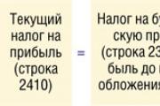
Accounting Regulations Accounting for calculations of income tax of organizations PBU 18/02 (as amended by Orders of the Ministry of Finance of the Russian Federation...
A trainee salesperson is usually called those salespeople who are not yet ready to work completely independently. Process...
Educational institution "Gomel State Medical University" Department of Neurology and Neurosurgery...

One of the most controversial and controversial methods for the early development of children was developed in the 80s by a sociologist...

Contents Dietary supplement based on an extract obtained from the fly beetle (or...

Ekaterina MirimanovaSystem minus 60. RevolutionSystem minus 60 with Ekaterina Mirimanova“System minus 60....
Heaviness and bloating are the causes of both ordinary overeating and more serious digestive problems...

The second blood group, Rh-negative, appeared many years ago, when a person ceased to be...

PSYCHOLOGICAL ASPECTS OF ANOREXIA PHENOMENON (EXPERIMENTAL STUDY) T. V. Tarasova, E. V. Arsentieva...
Contents Since the skin in this area is thin, it is more prone to the appearance of various types of spots....

vseslav Sat, 10/17/2015 - 20:50 Vasileostrovskaya station is one of the oldest stations...

P. S. Pallas (1741 - 1811) - naturalist and traveler-encyclopedist, who glorified his name with major contributions...
Today, all issues related to the placement of government orders are regulated by the Law on Contract...