Act on damage, damage, scrap of inventory items
Form TORG-15 is drawn up in the case when during transportation, movement between and within a warehouse, during storage...
Gall bubble- This is a small sac located in the lower surface of the liver and is a reservoir for bile. If liver function is disrupted or due to poor nutrition, the balance of acids and cholesterol is disturbed, which leads to the formation of stones. In severe cases it is required emergency removal gallbladder. In order to make an accurate diagnosis and determine a subsequent treatment strategy, you should undergo the necessary examination.
You will need
1. If you are worried about pain in the right hypochondrium, digestive disorders, nausea, heartburn, bad mood, fluctuations in body temperature, only after diet violations, the skin has acquired a yellowish tint, immediately consult a gastroenterologist-hepatologist. You will be prescribed an examination.
2. Particularly true results of the examination can be obtained by performing an ultrasound of the abdominal cavity. If you have been prescribed this examination, then you need to prepare for it. Follow a diet for several days, conduct a course of cleansing enemas. Maximum emptying of the intestines and stomach allows you to get real results. More often than not, each ultrasound diagnosis is enough to make an accurate diagnosis and prescribe subsequent treatment.
3. In addition to ultrasound, you may be prescribed cholecystography. To carry it out, you will be given a special substance intravenously or orally in tablets. After this, the doctor will conduct an X-ray diagnosis, which is done on an empty stomach and after meals. This allows you to determine how much bile bubble copes with its function. If bile does not accumulate and is not supplied to process food, then this means that the bile bubble does not work.
4. You may also be prescribed dynamic scintography. To carry it out, you will be injected with a radioisotope, the one that penetrates the bile, after which, with the support of the unit, the doctor will determine the degree of damage to the gallbladder. The last two research methods are used in extreme cases, if ultrasound diagnostics show that there are no stones in the gall bladder, and all signs of the disease indicate a malfunction of the gallbladder.
5. In addition, you will be prescribed to undergo a general and biochemical blood test, on the basis of which the doctor will be able to make a more accurate diagnosis and prescribe appropriate treatment.
6. Occasionally, based on the results of the examination, it is necessary to carry out emergency surgical intervention, because the presence of stones can lead to the formation of dangerous complications incompatible with life due to the development of peritonitis and sepsis.
Checking the intestines is suitable even for healthy people who do not experience painful sensations or difficulties with digestion. With a simple examination, you can make sure that everything is in order, or notice the first signs of the disease and stop it in the bud. Modern methods make the procedures fairly painless and the results accurate.

1. First you need to contact a gastroenterologist or proctologist. The doctor will listen to the patient’s complaints, conduct a consultation and prescribe examinations. At the first appointment, the doctor will perform palpation. At this stage it is possible to obtain or confirm doubts about a particular disease.
2. The core type of diagnosis, the one that is especially accurate and recommended for use by doctors, is colonoscopy. This procedure is carried out using a special device - a probe. It is injected into the patient’s intestines, and the doctor sees on the monitor the condition of the mucous membranes, cavities, inner surface this organ. The manipulation is carried out in case of various complaints from patients, the main of which are: abnormal stool, flatulence, the presence of continuous or periodic pain, discharge of blood or mucus during the bowel movement. With the help of colonoscopy, it is possible to identify or exclude: colitis, polyps, oncological tumors. Colonoscopy will not be performed if a person has poor clotting blood, there are abnormalities in the functioning of the lungs or heart, and there are also acute diseases and infections.
3. The colonoscopy procedure is not painful. Many patients experience slight discomfort during the procedure, but tolerate the manipulation extremely well. During the examination with a probe, you should follow all the recommendations of the doctor and his assistant. They will rigorously try to reduce discomfort to a minimum or completely eliminate them. With modern equipment, it is virtually impossible to get an infection during the examination process. In addition, doctors strictly follow the disinfection procedure. It is necessary to carry out a little preparation for a colonoscopy. It consists of taking a painkiller in case discomfort occurs and an antispasmodic to relax the muscles. A colonoscopy will take no more than 20 minutes. The patient needs to remove clothing below the waist. Some hospitals provide disposable underwear for the duration of the diagnosis. As soon as the patient is ready for manipulation, a probe is inserted into the lumen of the colon, gradually moving it deeper. At the same time, air is supplied, which allows the colonoscope to move in the intestine. There may be some slight bloating, but this feeling will pass soon. During a colonoscopy, doctors usually ask patients to turn on their side, back, or lie on their stomach. After the procedure, the person is advised to follow a diet for a couple of weeks. It is necessary to relieve bloating. To do this, exclude foods from the diet causing flatulence. Among them: cabbage of all kinds, legumes (lentils, beans, peas), black bread, bakery products based yeast dough, apples. The doctor will recommend medications to remove gases as quickly as possible.

4. Another equally famous and effective method of checking the intestines is sigmoidoscopy. This type The procedure allows diagnosing cancer, colitis, enterocolitis. Using this method, it is possible to examine not only colon, but also the rectum. The probe is buried an average of 35 cm, which allows for particularly accurate surveys.
5. Diagnose different important diseases intestines is permissible and with the support of x-ray equipment. This method is called irrigoscopy. A barium suspension is injected into the patient through the anus. When taking pictures, the patient is fixed with the side part, and then in a direct projection. This type of diagnosis makes it possible to detect the presence of polyps, various growths, fistulas, inflammatory phenomena, neoplasms, including malignant ones, in the intestines. Through an X-ray examination of the intestine, the doctor evaluates the elasticity of its walls. A contraindication to irrigoscopy is the period of complications of the disease.
6. Ultrasound examination is also widely used for diagnosis. various ailments gastrointestinal tract. To check the intestines on this unit it will take no more than 15 minutes. But you need to prepare for the procedure a couple of weeks in advance. You must follow a special diet. You need to avoid foods that cause increased gas formation. It is worth excluding fat foods, alcohol, soda, smoked foods, sweets, excessively salty foods, and dishes with a huge number of spices from the diet. A couple of days before an ultrasound of the intestines, activated charcoal is prescribed. Before the examination, an enema is given at night. Only if all the above conditions are met will the result of the survey be optimal and accurate. Before the procedure, liquid is injected into the intestines at ease and the organ is immediately scanned. Ultrasound of the intestine can be performed rectally, which does not require the introduction of a special liquid.
7. In order to evaluate the functioning of the intestines, it is also possible to conduct laboratory blood tests. This will not help diagnose the type of tumor or the presence of polyps, but through surveys it is possible to establish existing abnormalities in the functioning of the body and correlate them with other previously conducted examinations. When looking at the results of a blood review, the number of red blood cells and hemoglobin level are assessed. A review of the blood is done for tumor markers. These are substances that are released into the blood if the body has malignancy. In laboratory conditions, it is recommended to evaluate the condition of stool. It can be checked for the presence of mucus or hidden traces of blood. This is typical for tumors and polyps. The doctor's laboratory research also includes performing a biopsy. A piece of tissue removed from the intestine is examined under a microscope.

The condition of each organism depends on the condition of the gallbladder. The functions of the gallbladder are: “launching” the gastrointestinal tract, regulating metabolic processes, exclusively water-salt, participation in production synovial fluid, which is a lubricant for joints. In addition, gall bubble controls the body's pain threshold, the development of neuroses, insomnia, increased nervous and muscle excitability, etc. There are so many functions of the gallbladder in the body that if it malfunctions, every organism fails.

You will need
1. Gall bubble It is charmingly cleansed using the probeless liver dubage method. It is impossible to eat on the day of gallbladder cleansing. During the day, drink a glass of antispasmodic tea three times. To prepare it, take a teaspoon of lemon balm, lavender and rose petals, pour a glass of boiling water and leave for 30-40 minutes.
2. In the evening, drink a sorbitol solution - 10 grams per half glass of hot water. Lie on your right side, applying a hot heating pad to the liver area.
3. After 40-50 minutes, drink a glass mineral water weak mineralization with half a teaspoon of salt diluted in it. Lie down for another 30 minutes without removing the heating pad from the liver.
4. After this, pour 1/3 cup olive oil, add 2/3 cup of lemon juice, stir, drink and lie down for 30 minutes.
5. After 30 minutes, drink one a raw egg from domestic chickens and drink half a glass of antispasmodic tea. Lie on the heating pad for another 2 hours, and after that, removing the heating pad from the liver, sleep until the morning.
Note!
You should know that removing a gallbladder clogged with sand and stones does not solve problems with their formation in the future, only in the bile ducts. Therefore, it is better to maintain the typical condition of your gallbladder and not bring its condition to the point of forced removal.
Helpful advice
If pain occurs in the liver, take 1-2 tablets of no-shpa. Follow this gentle cleansing of the gallbladder for 5 days vegetarian diet, and another 5 days - a diet including fermented milk products and lean boiled fish. It is recommended to carry out one-day fasting - once a week. This is an excellent preventive measure for removing excess bile from the body.
Ultrasound examination today is the most accessible and informatively accurate way to diagnose the condition of the human abdominal organs. These organs are of sufficient size and density, as a result of which they perfectly reflect ultrasound and are amenable to scanning. Ultrasound is painless and harmless at any age.

2. Survey liver with ultrasound support, it is usually performed in the morning, on an empty stomach. But in in case of emergency The survey is carried out at any time of the day. Special preparation for ultrasound liver traditionally not required, but in patients with enormous weight and patients with increased flatulence conducting the examination may be difficult.
3. To prepare for an ultrasound liver, it is recommended that a few days before the scheduled examination you exclude from food foods that cause increased gas formation in the intestines. These are cabbage, legumes, carbonated drinks, raw vegetables, rich in fiber, black bread, whole milk.
4. For patients suffering from constipation, in order to prepare for an ultrasound liver, for several days before the procedure it is recommended to take 1 tablet 3 times a day enzyme preparations and enterosorbents – Activated carbon, espumizan, mezim-forte, festal, which reduce flatulence in the intestines.
5. What diseases can ultrasound detect? liver? These are: cirrhosis, acute or chronic hepatitis, potassium hypertension, metastases, fatty infiltration liver, hemangioma, cyst and cystic formations, hepatoma, calcification liver, Budd-Chiari syndrome.
6. Results of ultrasound examination liver in most cases they are decisive in establishing a diagnosis and prescribing treatment. In some cases, to establish a 100% accurate diagnosis, a combination of ultrasound and fine-needle cytobiopsy is necessary.
Video on the topic
Gall bubble is a hollow organ. Its core function is the accumulation and saturation of bile, which periodically enters the duodenum.

Gall bubble is a reservoir for storing bile, the volume of which is approximately 60 ml. Bile is produced by liver cells that work non-stop. After this, it flows naturally into the gall bubble, where its saturation increases significantly. This is due to the fact that part of the liquid is absorbed. The composition of bile is quite rich, it includes bile pigments, bile acids, bilirubin and cholesterol. Bilirubin is formed during the breakdown of hemoglobin. One part of it is absorbed into the blood, the other is excreted in the urine. The main part of the pigments is excreted in the feces (in addition, it gives color to the secretions). Consequently, when the function of the gallbladder is impaired (say, an inflammatory process or stones), when the path of bile is blocked, the feces become virtually colorless. Metabolic disorders cause the formation of stones in the bladder and bile ducts, because cholesterol falls out in undissolved form. With the participation of bile, certain enzymes are activated, fat is broken down into small particles, the process of absorption of fats and vitamin K is improved, and motor function intestines, prevents the formation of putrefactive processes. Core function biliary tract is the excretion of bile into the intestines. The more food enters the stomach, the more bile is released. The intake of bile is enhanced by the consumption of egg yolks and food with a high content of vegetable and animal fats. With irregular nutrition and large intervals between meals, bile stagnates in the bladder, which is a direct path to the occurrence of inflammatory processes. In turn, inflammation thins the walls of the gallbladder, and when a large amount of bile accumulates, a breakage may occur. Infected bile flows into the abdominal cavity and negatively affects the patient’s condition. To prevent such consequences, it is necessary to systematically carry out an ultrasound examination in order to identify disruptions in the functioning of the gallbladder in the early stages.
Video on the topic
Stomach- a very important organ, through which nutrients enter the human blood and tissues to ensure its functioning. Failures in the functioning of the stomach are fraught with dysfunction of many organs. Consequently, it is very significant when the slightest violations Work to check your stomach by contacting an expert. There are many ways to help diagnose the problem and treat the disease at an early stage.

1. An experienced and competent expert gastroenterologist is able to make an early diagnosis during the first conversation with the patient. Based on the nature of the complaints and having studied the signs, the doctor determines what disease caused them. Additional information it can be obtained during examination of the patient by palpating the stomach. By pressing and feeling it through the abdominal wall, the doctor will determine whether the patient has tumor-like formations or hernial canals.
2. For more detailed examinations, doctors use a probe located at the end of a thin, elastic tube. Once swallowed, the probe enters the esophagus and slowly moves along it towards the stomach. A nutrient can be introduced through a tube, which, once in the stomach, forces it to work. Gastric juice samples are taken through a tube.
3. The level of acidity in the stomach is determined not only by probing; it can also be checked by another method. To do this, the patient swallows a special substance containing a harmless chemical dye. Later processing in the stomach, the dye is excreted from the body along with urine. A review of the urine will determine the acidity - the more dye left in the urine, the higher the level of acidity.
4. There is a special electronic sensor, which can also be used to determine the acidity level. It needs to be swallowed and, once in the stomach, it will emit signals, the frequency of which depends on the level of acidity. The sensor leaves the body naturally.
5. One of the most authentic ways that allows a doctor to literally “examine” the condition of the stomach from the inside is endoscopy. With its support, it is possible to diagnose tumors and ulcers. The process of movement of the endoscope through the esophagus and stomach can be monitored on the monitor screen.
6. Famous diagnostic method stomach diseases is ultrasonic survey. It allows you to examine this organ in detail and diagnose it in a timely and painless manner.
Video on the topic
In order to check your intestines, it is best for everyone to see a doctor. There are several research methods, and the expert will select the best one. In addition, you can use folk remedies.

1. Only an expert will be able to positively evaluate and analyze the functioning of any organ. There are several methods for examining the intestines, one of them is ultrasound. With the help of ultrasound, it is possible to find some pathologies, but not all. This is due to the accessibility of only parts of the intestine located naturally next to abdominal wall. You should prepare for ultrasonic testing in advance. For 3 days you need to follow a diet and also take medications that improve digestion and reduce gas formation. Before the procedure, the intestines must be emptied.
2. Checking the intestines can be carried out using different hardware methods, such as anoscopy, colonoscopy or sigmoidoscopy. They are all similar and involve the entry into the rectum through the anus of special devices equipped with cameras. Such a survey allows us to find polyps, fissures, tumors, and stagnation of feces. All hardware diagnostic procedures require certain preparation, in particular diet and bowel movements. And because these manipulations are quite unpleasant and even painful, anesthesia is often used.
3. Another method of checking the intestines is an x-ray examination called irrigoscopy. The essence this method consists in the fact that first a contrast agent, the role of which is played by a barium solution, is introduced into the intestines with the help of an enema. After this, a photo is taken. X-rays are absorbed by barium, which makes it possible to detect some pathologies. Most often, each survey is carried out twice. The 1st photo is taken immediately after the introduction contrast agent, another - after the intestines are freed from the solution. This way, the doctor will be able to see the difference and assess the condition of the intestine.
4. If you want to check yourself, then try using folk remedy. Rub raw beets grated, and then squeeze the juice out of the pulp and let it brew for two hours. Drink half a glass and watch your bladder or bowel movements. If the urine becomes beet-colored and the bowels do not empty, this may indicate serious problems.
Note!
If you have problems with bowel movements, consult your doctor immediately. Constipation is very dangerous and threatens the formation of fecal stones and intestinal perforation.
Helpful advice
Always monitor your digestion to avoid serious snags.
It is known that most diseases appear unexpectedly. They develop gradually, becoming more complex and progressing every day. This also applies to diseases associated with the gallbladder, which have become especially common in the last couple of decades. In general, you may not have problems with your gallbladder, but, nevertheless, there are some areas of the body where lesions may indicate a malfunctioning gallbladder.
Symptoms that may appear with gallbladder diseases.
Signs indicating problems with the gallbladder may include the following:
ear problems;
presence of tinnitus;
headache in the temporal region;
pain in the eyes;
neuralgia trigeminal nerve;
pain in the right hypochondrium;
tension in the chest before the cycle;
discomfort in the hip joint.
Speaking about the gallbladder, it must be said that it is made in the form of a small sac, which is a special reservoir for a substance such as bile.
How to check gallbladder for the presence of stones and other diseases.
Everyone has probably heard about the formation of gallstones. They appear as a result of an imbalance of cholesterol and acids due to poor nutrition, as well as malfunctions of the liver. If you do not undergo a timely examination, you may be left without a gallbladder (in case of complications, it is removed surgically). If you do not know how to check the gallbladder, it is recommended to contact a specialist in this field - a gastroenterologist, who will guide you necessary examinations, will find out the reasons for your poor health and prescribe necessary treatment.
If you suffer from pain in the right hypochondrium, feel nauseous all the time, suffer from heartburn, feel unwell, or experience changes in body temperature and your skin becomes yellowish, this may indicate that you have a disease such as hepatitis. To know for sure, it is best to visit a gastroenterologist - a hepatologist who will make sure you undergo necessary diagnostics to check if you have gallbladder disease.
When prescribing and performing an ultrasound of the abdominal cavity, you have the opportunity to obtain accurate results. This method is used to make the correct diagnosis of a particular disease and prescribe the appropriate course of treatment. Before doing an ultrasound, it is necessary to carry out certain preparations for your body. It is necessary to sit for at least a week special diet and cleanse your body. Enemas will also help you with this. Only with a cleansed intestine and stomach can we talk about obtaining accurate and truthful examination results during an ultrasound scan.
In addition to ultrasound, it is also quite common and effective method cholecystography is considered. The essence of this method is the introduction of a special substance orally in the form of tablets or orally. To find out about the functioning of the gallbladder, the doctor performs an x-ray, which is done on an empty stomach or after eating food. If there is no accumulation of bile and it does not participate in the process of processing food, then this indicates that the function of the gallbladder is disabled.
In addition to the methods described above, there is dynamic scintography, which is also used to check the gallbladder. It is carried out using a radioisotope, which determines the degree of damage to the gallbladder.
To verify the correctness of the diagnosis, the doctor may prescribe blood tests, based on the results of which, taking into account the examination using the above methods, the necessary course of treatment can be prescribed.
Gallbladder problems occur in 300 people per 100 thousand population. Timely detection of the disease is very important. Not the least place in the diagnosis of this group of diseases is occupied by tests, since they are the most informative and allow one to fairly accurately determine the presence of disturbances in the functioning of this organ.
Diagnosis of diseases is carried out using laboratory and instrumental research methods. Laboratory research methods are various tests. Instrumental - using special equipment. To check the condition of the gallbladder and the entire biliary system, it is necessary to undergo both types of examination.
The most informative and common types of examination of the biliary system are: duodenal intubation, Ultrasound, general blood test, biochemical blood test, general urinalysis, coprogram.

Duodenal sounding preparation
Main instrumental methods research:
In addition to these two methods, X-ray examination of the gallbladder, cholangiopancreatography and computed tomography are sometimes used.
X-rays are used to find stones in the gallbladder and to evaluate its function. Cholangiopancreatography is necessary to examine the exit site of the bile duct into the duodenum and is used if blockage is suspected. Computed tomography is used when other types of instrumental examination cannot be applied to the patient due to contraindications.


Gallbladder size is normal in adults
The most necessary tests to determine problems with the biliary system:


In addition to the above analyses, diagnostic purposes Other, less well-known tests are also considered: tests for alkaline phosphatase, C-reactive protein, AST and ALT. An increase in alkaline phosphatase indicates not only gallbladder pathology, but also liver problems. Level C-reactive protein increases during the inflammatory process, in particular, may indicate inflammation in the gallbladder. ASAT and AlAT- important indicators liver function.
When experiencing abdominal pain, many people ask the question: how to check the gallbladder? They arise for a reason: according to statistics, they are registered annually in 0.3% of the world's population. If you are experiencing pain in the right side of your abdomen, nausea, occasional vomiting, bad taste in the mouth - it is advisable to visit a doctor. Remember, only a highly qualified doctor will conduct a high-quality examination, correctly diagnose and prescribe correct treatment.
The gallbladder is a hollow, pear-shaped organ located on the lower surface of the liver. Its main function is the temporary storage of bile. Bile is a waste product of liver cells and performs a digestive function - it ensures the breakdown of fats and activates motility. small intestine. U healthy person After food enters the stomach, bile with pancreatic juice is released in certain portions into the duodenum, preparing it for work.
Contraction of the smooth muscles of the gallbladder ensures the timely removal of bile into the lumen of the duodenum. When the ducts are blocked or the walls are insufficiently contracted, bile stagnates and precipitates. Gradually, the sediment thickens and forms stones, which have a local damaging effect on the mucous membrane and can also cause blockage of the cystic duct.
As a result of these disorders, the following may develop:
The clinical picture in some cases is asymptomatic. The severity of symptoms depends on the type of pathology.
General symptoms All diseases are characterized by:

If you are experiencing severe pain in the abdomen and increased body temperature - urgently seek qualified medical advice medical care.
Factors contributing to the occurrence of local disturbances are:

If you experience complaints indicating gallbladder disease, do not try to treat yourself. It is necessary to urgently visit a gastroenterologist.
It is this specialist who will collect the necessary information about the disease, conduct visual inspection, will direct you to take tests, undergo the appropriate examination and make a competent diagnosis. The doctor will also prescribe treatment, select a diet, and monitor the rehabilitation period.
If you decide to have your gallbladder checked, the examination begins with a visit to a specialist. The doctor will ask you in detail about the nature of the symptoms and their first appearance. If your relatives have ever had diseases of the liver, gallbladder or pancreas, you should definitely inform your doctor about this.
 Then the doctor will conduct a clinical examination skin and visible mucous membranes, will perform palpation (tactile examination) of the abdomen to determine the nature of the pain, as well as the spread of pain during its implementation.
After the examination, the doctor will order an examination, which will provide further information for making a diagnosis.

So, diseases of the gallbladder are indicated by an increase in the concentration of C-reactive protein, a shift in the leukocyte formula to the left and an increase in ESR, an increase in the level of bilirubin, cholesterol and alkaline phosphatase, darkening of the urine and lightening of the stool, a decrease or absence of urobilin in the urine.
It is an informative method for confirming the diagnosis. The essence of the method is that the doctor inserts a duodenal tube into the cavity of the duodenum.
Before the procedure, several conditions must be met:
 This method allows you to examine the contents of the duodenum. Immediately before the procedure, the doctor gives a brief instruction. During the study, the time of appearance of bile and the duration of its secretion are recorded. Several samples are taken at intervals of 5-10 minutes - the consistency of bile, volume and color are examined. If necessary, bile is sent for microbiological analysis.
The “favor” of cholecystitis is evidenced by cloudy bile with an admixture of flakes and reduced acidity.
Ultrasound examination is one of the most informative methods. The use of sound waves causes minimal discomfort during the procedure and gives the clearest possible picture of the structure of the biliary system.
Allows you to identify:
 Gallbladder scintigraphy is a study based on the intravenous injection of a weak radioactive isotope into the body, which is accumulated by liver cells and then transported with bile to the gallbladder. The scan is carried out with a gamma tomograph, which detects weak radioactive radiation and displays it on the monitor. The procedure lasts about two hours, scanning is carried out at intervals of 10-15 minutes. Irradiation is absolutely safe and does not cause any harm to the patient. Scintigraphy allows you to identify features of the structure and motility of the gallbladder, obstruction of the bile ducts.
In addition to the above methods, the gallbladder is examined using magnetic resonance imaging (MRI), which allows visualization.
If you have symptoms indicating any problems in the gallbladder area, it is strongly recommended to consult a specialist. It is he who will indicate what measures and diagnostic examinations may be necessary in this particular case. It is very important that all of them are carried out exclusively by a qualified specialist - only in this case will it be possible to maintain 100% confidence in the correctness of the results obtained.
Diagnostics and remedial measures for the non-aggravated form gallstone disease is handled by a gastroenterologist. The diagnosis can be identified based on a person's complaints, as well as a number of additional studies.
Talking about clinical analysis blood, it is strongly recommended to pay attention to the identification of leukocytosis. In addition, we should not forget about the importance of identifying a shift in the leukocyte formula to the left, as well as accelerating ESR indicators - these are values that indicate the addition of an inflammatory algorithm. They are most characteristic precisely for the exacerbation phase pathological condition associated with the gallbladder area.
The next type of blood test, which will help determine the current state of this organ, is biochemical. With the addition of cholestasis, namely stagnant bile syndrome, there may be an increase in direct bilirubin, as well as y-GT, alpha2- and beta-globulins.
 Duodenal sounding allows you to identify indirect clinical manifestations cholecystitis. First of all, you should pay attention to cloudy bile with the presence of flakes, a decrease in bile pH, and the addition of sand. It is strongly recommended not to forget that:
Ultrasound examination of the gallbladder area makes it possible to determine the exact size of the organ. In addition, this is how thickening in the area of the gallbladder walls (more than three mm) and deformation are identified. We should not forget that ultrasound is informative when it is necessary to identify infiltration of peri-vesical tissues, the addition of constrictions, stagnant bile, as well as cholesterosis, stones and neoplasms. In addition, we should not forget about the presence of gas in the gallbladder area. A test with the use of a choleretic breakfast determines the specific type associated with gallbladder dyskinesia. In general, according to experts, CT scan(CT) does not have any advantages over ultrasound.
The presented technique involves the introduction of a radioisotope into the patient’s body.
After this, with the help of certain devices, the presence of the presented radioisotope is captured and identified.
In the same way, accordingly, bile is identified. If dynamic scintigraphy failed to find the gallbladder, this will be direct evidence of the cessation of its functioning. We should not forget about the need to carry out some other diagnostic checks for all those who are wondering how to check the gallbladder.
 First of all, it is strongly recommended to pay attention to the X-ray examination. Speaking about this, they mean taking a survey image of the peritoneum, which is carried out if there is a suspicion of perforation in the area of the gallbladder. This is what makes it possible to exclude the presence of calculous cholecystitis, as well as calcification of the gallbladder.
Further, endoscopic retrograde chmay well be used. It is carried out to identify obstructive (impaired patency) lesions in the area of not only the bile ducts, but also the pancreatic ducts. Next diagnostic examinations worthy of attention are those that represent radioisotope research:
It should, however, be taken into account that the last two methods are currently in medical practice are used quite rarely. The formulation of the so-called differential diagnosis deserves no less attention.
A similar diagnosis is made in connection with chronic form calculous cholecystitis, functional abnormalities in the biliary tract. In addition, one should not forget about peptic ulcer disease duodenum. It is also strongly recommended to pay attention to what exactly the nutrition process should be.
 Patients are strongly advised to consume foods and dishes such as bread, namely black and white, but it is advisable that it be yesterday’s baked goods. We should not forget about first courses, for example, dairy or vegetable soups, borscht, cabbage soup, as well as vegetarian beetroot soup. Speaking about main courses, we should not forget about lean meat of rabbit, chicken, beef, fish, and various steamed cutlets.
It is permissible to consume vegetable side dishes and broths made from vegetables of all varieties, in addition to fried ones. It is also allowed to eat such dishes that belong to pasta and cereals, for example, porridge, casseroles using dried apricots, raisins or even cottage cheese. Another acceptable category of products for consumption are dairy products, for example, kefir, milk, cottage cheese, yogurt.
Sour cream and cream can also be used, but only in limited quantities.
Once the diagnosis is made, the patient's diet may contain some fats, in particular vegetable oil. Limited consumption of butter is also acceptable. It is allowed to prepare certain dishes from eggs, namely no more than one egg per 24 hours, an omelet based on proteins. The diet may also contain some sweet foods and dishes, for example, honey, jelly, as well as compotes and fresh fruits. We should not forget that it is acceptable to use some snacks, for example, soaked herring, boiled fish or mild cheese. Speaking of drinks, it is strongly recommended to pay attention to the use of weak coffee with the addition of milk, weak tea, currant and rosehip infusions.
Thus, checking the functioning of the gallbladder is prerequisite, allowing you to determine its current state. It is after a full examination that it will be possible to talk about deciding on a specific treatment, as well as prescribing certain preventive measures.
Important!
HOW TO SIGNIFICANTLY REDUCE THE RISK OF CANCER?
Time limit: 0
Navigation (job numbers only)
0 out of 9 tasks completed
Information
TAKE THE FREE TEST! Thanks to detailed answers to all questions at the end of the test, you can REDUCE the likelihood of disease by several times!
You have already taken the test before. You can't start it again.
Test loading...
You must log in or register in order to begin the test.
You must complete the following tests to start this one:
results
Time is over
1.Can cancer be prevented?
The occurrence of a disease such as cancer depends on many factors. No person can ensure complete safety for himself. But significantly reduce the chances of occurrence malignant tumor everyone can.
2.How does smoking affect the development of cancer?
Absolutely, categorically forbid yourself from smoking. Everyone is already tired of this truth. But quitting smoking reduces the risk of developing all types of cancer. Smoking is associated with 30% of deaths from oncological diseases. In Russia, lung tumors kill more people than tumors of all other organs.
Eliminating tobacco from your life - best prevention. Even if you smoke not a pack a day, but only half a day, the risk of lung cancer is already reduced by 27%, as the American Medical Association found.
3.Does excess weight affect the development of cancer?
Look at the scales more often! Overweight will affect not only the waist. The American Institute for Cancer Research has found that obesity promotes the development of tumors of the esophagus, kidneys and gallbladder. The fact is that adipose tissue serves not only to preserve energy reserves, it also has secretory function: Fat produces proteins that influence the development of chronic inflammation in the body. And oncological diseases appear against the background of inflammation. In Russia, WHO associates 26% of all cancer cases with obesity.
4.Do exercise help reduce the risk of cancer?
Spend at least half an hour a week training. Sport is on the same level as proper nutrition when it comes to cancer prevention. In the United States, a third of all deaths are attributed to the fact that patients did not follow any diet or pay attention to physical exercise. The American Cancer Society recommends exercising 150 minutes a week at a moderate pace or half as much but at a vigorous pace. However, a study published in the journal Nutrition and Cancer in 2010 shows that even 30 minutes can reduce the risk of breast cancer (which affects one in eight women worldwide) by 35%.
5.How does alcohol affect cancer cells?
Less alcohol! Alcohol has been blamed for causing tumors of the mouth, larynx, liver, rectum and mammary glands. Ethanol decomposes in the body to acetaldehyde, which then, under the action of enzymes, turns into acetic acid. Acetaldehyde is a strong carcinogen. Alcohol is especially harmful for women, as it stimulates the production of estrogens - hormones that affect the growth of breast tissue. Excess estrogen leads to the formation of breast tumors, which means that every extra sip of alcohol increases the risk of getting sick.
6.Which cabbage helps fight cancer?
Love broccoli. Vegetables not only contribute to a healthy diet, but they also help fight cancer. This is why recommendations for healthy eating contain the rule: half of the daily diet should be vegetables and fruits. Particularly useful are cruciferous vegetables, which contain glucosinolates - substances that, when processed, acquire anti-cancer properties. These vegetables include cabbage: regular cabbage, Brussels sprouts and broccoli.
7. Red meat affects which organ cancer?
The more vegetables you eat, the less red meat you put on your plate. Research has confirmed that people who eat more than 500g of red meat per week have a higher risk of developing colorectal cancer.
8.Which of the proposed remedies protect against skin cancer?
Stock up on sunscreen! Women aged 18–36 are especially susceptible to melanoma, the most dangerous form of skin cancer. In Russia, in just 10 years, the incidence of melanoma has increased by 26%, world statistics show an even greater increase. Equipment for this is also blamed fake tan, And Sun rays. The danger can be minimized with a simple tube of sunscreen. A 2010 study in the Journal of Clinical Oncology confirmed that people who regularly apply a special cream have half the incidence of melanoma than those who neglect such cosmetics.
You need to choose a cream with a protection factor of SPF 15, apply it even in winter and even in cloudy weather (the procedure should turn into the same habit as brushing your teeth), and also not expose it to the sun's rays from 10 a.m. to 4 p.m.
9. Do you think stress affects the development of cancer?
Stress itself does not cause cancer, but it weakens the entire body and creates conditions for the development of this disease. Research has shown that constant worry alters the activity of immune cells responsible for triggering the fight-and-flight mechanism. As a result, a large amount of cortisol, monocytes and neutrophils constantly circulate in the blood, which are responsible for inflammatory processes. And as already mentioned, chronic inflammatory processes can lead to the formation of cancer cells.
THANK YOU FOR YOUR TIME! IF THE INFORMATION WAS NECESSARY, YOU CAN LEAVE A FEEDBACK IN THE COMMENTS AT THE END OF THE ARTICLE! WE WILL BE GRATEFUL TO YOU!
Task 1 of 9
Can cancer be prevented?
Task 2 of 9
How does smoking affect the development of cancer?
Task 3 of 9
Does excess weight affect the development of cancer?
Task 4 of 9
Does exercise help reduce the risk of cancer?
Task 5 of 9
How does alcohol affect cancer cells?

Form TORG-15 is drawn up in the case when during transportation, movement between and within a warehouse, during storage...

Nutritionists say that for good health and a slim figure, you must include snacks in your diet....
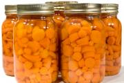
Delicious pickled carrots for the winter can be prepared in a variety of containers, it can be a wooden container,...

When I have a few minutes left to prepare breakfast, the simplest and fastest recipes are used. This option...

An elegant table, a decorated Christmas tree, tangerine spirit spilled throughout all the rooms, soon the most magical holiday - New...

Each of us has repeatedly faced financial difficulties and difficult periods in life when Fortune mocked...

If you are new to magic, then it will be useful for you to get acquainted with the signs by which you can accurately...

SECRETS OF DREAMS Why does day follow night? What is life? What is death and what is sleep? These questions...

Main meaning: Whatever version of Madame Lenormand’s deck we take, we can definitely say that this is one of...

An example of a correct income tax return in 2017, download for free in excel the new current...

P. S. Pallas (1741 - 1811) - naturalist and traveler-encyclopedist, who glorified his name with major contributions...

Today, all issues related to the placement of government orders are regulated by the Law on Contract...

Accounting Regulations Accounting for income tax calculations of organizations PBU 18/02 (as amended by Order...

A trainee salesperson is usually called those salespeople who are not yet ready to work completely independently....

Nutritionists say that for good health and a slim figure, you must include snacks in your...
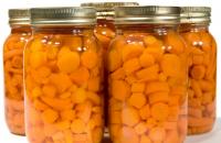
Delicious pickled carrots for the winter can be prepared in a variety of containers, it can be a wooden...