What is possible and not possible during the Nativity Fast?
In 2018, the Nativity Fast will begin on November 28. During this period, Orthodox believers prepare to celebrate Christmas...
In the process of evolution, different animal species have developed different abilities to absorb food of a certain quality. Depending on the nature of nutrition and living conditions, the digestive system of animals also developed. Let us consider the structure of the gastrointestinal tract of ruminant mammals using the example of the structure of the stomach of a cow.
Plant foods have a number of features. On the one hand, they are easily available for consumption. However, on the other hand, they are not as beneficial for absorption as animal feed - plant feeds are significantly inferior to them in terms of nutritional value . Moreover, such a basic structural component Plants like cellulose (or fiber) are not broken down in most animals due to the absence of the cellulase enzyme in their digestive juices. This enzyme is synthesized only by bacteria and unicellular organisms, as well as some invertebrates.
Mammals are incapable of this. Therefore, in order for them to be able to use plants as food, animals need the help of symbiont microorganisms.
 The use of rough plant feed contributed to some changes in the digestive system. Thus, in herbivorous mammals, there was a change in the dental system, an increase and complexity of the digestive system, and the formation of forestomach and cecum.
This can be observed in such representatives of the animal world as horses and rabbits. In their long intestines there is a set of bacteria that partially digest cellulose fibers. But representatives of the suborder of artiodactyl mammals, the ruminants, have learned to use the energy stored by plants most effectively.
Ruminants include such representatives of the animal world as:
Herbivorous mammals evolved a stomach adapted for digesting plant fibers, and in parallel there was an evolution of bacteria and microorganisms that live in the digestive tract. This complex of microorganisms forms an entire ecosystem of bacteria and protozoa, which form a symbiosis with the host animal.
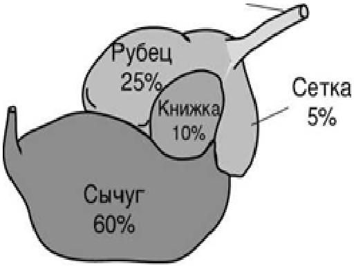
 The structure of the stomach in all ruminant animals (goats, sheep, cows and other cattle) is quite different from the stomachs of other representatives of the class of mammals. But the cow’s stomach has the most complex structure. A cow has one stomach, but it has 4 sections or 4 chambers:
The first three sections are parts of the esophagus; in fact, we can say that the esophagus is three-chambered. Let's look at the structure of the cow's digestive system and the sections of its four-chamber stomach.
Lips, tongue and teeth serve for capturing, tearing and chopping plant foods. The main food-feeding organ of a cow is the tongue. It is designed in such a way that with its help the cow effectively grabs grass, leaves and other herbaceous feed.
 The rumen is the largest section of the stomach of ruminants. Happening here primary processing digestive mass by enzymes and cellulose is broken down by microorganisms. As a result of processes occurring in the rumen, organic acids, carbon dioxide, methane and water. Acids, carbon dioxide and water are absorbed through the walls of the rumen, and methane is removed from the body during respiration. The scar has a complex structure and 3 separate parts: dorsal, ventral and cranial.
The rumen is connected to the mesh - the second section of the cow's stomach. Fermentation and digestion processes continue in this section. The walls of the rumen and mesh have highly developed muscles. This promotes efficient nutrient fermentation process. After the accumulation of a certain amount of cellulose fibers in the rumen, its contraction occurs. Hard-to-digest fibers are regurgitated back into oral cavity cows, where they are repeatedly chewed and ground.
Secondary chewed food enters the book - the third section of the cow's stomach. Absorption of water, as well as fatty acids and other nutrients occurs here. Book connects to the mesh with a groove and has thin partitions, which look like the pages of a book. That's why this department has this name. Here the crushed plant mass is exposed to bacteria, and the fermentation process occurs. This allows the cow’s body to absorb the maximum amount of fiber from rough plant foods. Next, the food moves into the abomasum.
Abomasum is the name given to the fourth section of the stomach of ruminants, which is not much different from the stomachs of other animals. Digestion here occurs due to the action of acid, as well as the animal’s own enzymes.
The stomach of the cow and all ruminants ends with abomasum, but digestive processes continue in other parts of the digestive system. In the duodenum, the processes of absorption of nutrients supplied by microorganisms continue. The part of the food that is not digested enters the large intestine. After that, in a blind and colon, what bacteria could not break down in the stomach is exposed to the following groups microorganisms. What remains after exposure to these bacteria is the toughest part of the food and is excreted from digestive tract.
Thus, the cow's stomach has 4 sections, its structure is complex. Each camera has its own specific function. The process of digesting food into the cow's stomach takes from 8 hours. The stomach is designed in such a way that it allows the most efficient extraction and absorption of nutrients from rough plant food.
Attention, TODAY only!
The cow's stomach is designed in a special way - it has four sections or chambers, each of which performs its own function. Malfunction of at least one part of the digestive system entails various pathologies of the animal’s health.
Cows have an interesting digestive system - this animal swallows food whole, almost without processing it with its teeth, and then, when it rests, it regurgitates it in parts and chews it thoroughly. This is why the cow is often seen chewing. The mechanism of regurgitation and chewing food from the stomach is called cud. If this process stops for a cow, it means that something is wrong with her.
The digestive system of a cow has the following structure:
The structure of the stomach of a cow is also of interest - this organ consists of 4 chambers:
The real stomach in the full sense of the word is the abomasum; the remaining chambers serve for preliminary processing of food, they are called forestomach. The rumen, book and mesh do not have glands that produce gastric juice; only the abomasum is equipped with them. But in the forestomach, fermentation, sorting and mechanical processing of food occurs. Let's look at the sections of the cow's stomach in detail.

The first section of a cow's stomach is called the rumen. It has the largest volume compared to other chambers - about 200 liters! It is located in abdominal cavity From the left side. Ingested food enters this proventriculus. The rumen is filled with microorganisms that ensure the primary processing of food.
Reference. The rumen contains a huge number of microorganisms, their total mass is about 3 kilograms. They promote the synthesis of B vitamins and protein in the animal’s body.
The scar consists of a double muscle layer and is divided into 2 parts by a small groove. The mucous membrane of the proventriculus is equipped with ten-centimeter papillae. It is in the rumen that starchy compounds and cellulose are broken down into simple sugars. Thanks to this process, the animal receives the necessary energy.
This section of the stomach is much smaller in volume than the previous one. Its capacity is no more than 10 liters. The grid is located in the area chest, one section of it is adjacent to the diaphragm. The main function of the net is to sort the feed. Small fractions of food from here move to the next section of the stomach, and larger fractions are regurgitated and enter the cow's mouth, where they are chewed. The mesh, as it were, filters the food, passing food that has already undergone primary processing further through the digestive system.

Small pieces of food move into the book - the third section of the stomach. Here the food is thoroughly crushed mechanically, thanks to the special structure of the mucous membrane. It consists of folds resembling leaves. In the book, further processing of coarse fibers and absorption of water and acids takes place.
The abomasum is the only part of the cow's stomach that is equipped with glands for secreting gastric secretions. It is located in the area between the 9th and 12th ribs with right side. Its volume in adults reaches 15 liters.
In calves, the abomasum is actively working, while the remaining parts of the stomach remain unused until almost three weeks of age. Their rumen is in a folded position, and the milk immediately enters the abomasum through a gutter, bypassing the mesh and book.
Cows often suffer from pathologies of the digestive system. They pose a serious danger to the life of the ruminant animal. Common digestive problems in cows:
Tympany or bloating - very dangerous condition, arising due to a sharp change in the cow’s diet, the animal’s consumption of large quantities foods that contribute to increased gas formation. Tympany may occur due to a blockage in the esophagus. Symptoms:

Attention! This condition It is dangerous for the life of the cow, since the increased size of the scar strongly compresses the diaphragm, preventing the animal from breathing normally. If help is not provided, the cow will die from lack of oxygen.
Methods to help with bloating include:
You can open up your stomach with a massage. It is performed on the left side of the abdominal cavity, in the area hungry hole fist. Pouring this area often helps. cold water. A cow needs to run to get her stomach working.
The digestion process often stops in cows due to improper feeding, for example, if concentrates predominate in the diet or the animal has eaten rotten hay. Also, gastric arrest occurs when the esophagus is blocked. Symptoms of the pathology: loss of chewing gum and appetite, general depression. If the cow's stomach has stopped, this can be checked. You need to lean your fist into the area of the hungry pit and listen to whether contractions occur.
Treatment of this pathology begins immediately. The first thing to do is to keep the animal on a starvation diet for 24 hours. In the future, digestible feed is gradually introduced - silage, a small amount of root vegetables, high-quality hay.

To start the stomach use:
Sometimes the stomach stops due to a blockage of the book. This happens when the animal’s diet is dominated by dry food, bran or grain waste. The cause of the pathology may be sand or dirt in the feed. The symptoms of a blocked book are similar to those observed when the stomach stops. Quite difficult to identify the real reason cessation of digestion. For diagnosis, a puncture of the stomach with a needle is used. If it enters with difficulty, it means that we are talking about a blockage.
If the diagnosis is confirmed, it makes sense to rinse the stomach. To do this, use a solution of sodium sulfate or chloride at a concentration of 10%. The procedure will require about a liter of this solution. To start the digestion process, use the same means as discussed above - vegetable oil, hellebore tincture, vodka.
Since the cow swallows unprocessed food, dangerous objects - wire, nails, wood chips, sharp stones - often get inside along with the food. Such foreign bodies can cause serious injuries animal - to pierce the stomach or pierce its walls. Injuries to the mesh are often through and through; sharp objects can touch nearby organs– heart, spleen, lung.
Symptoms of traumatic reticulitis:
Treatment is aimed at removing foreign object from the stomach. Metallic foreign bodies are removed with a magnetic probe. If it is not possible to remove the object, they resort to surgery or the animal is slaughtered.
All parts of the stomach of ruminants perform their functions. If at least one of them stops working, the entire digestive system suffers. It is important to diagnose the development of pathology in time and begin treatment.
The digestive tract is one of the important parts in the human body. It processes food. It breaks down into small components - vitamins, microelements, fiber and others. Also he has beneficial microflora. It protects the body from invasion harmful bacteria and supports metabolic processes fine. One of the abdominal organs is the stomach. Where is it located, what is it responsible for and what is its structure? We'll tell you.
It does not depend on us how to arrange the digestive organs in the body. If we talk about the structure of the stomach, it is quite complex. This is the first organ where food is processed by enzymes. gastric juice, hydrochloric acid and bile.
The stomach is usually understood as a hollow, elastic instrument that has a sac-like shape. It connects the esophagus and the intestinal canal. There the collection, digestion, and transformation of food from solid to liquid occurs.
Some parts of the stomach are distinguished as:
In this way, you can learn about the complete structure of the gastric organ.
Anatomy is one of the branches of medicine that allows you to better understand the human structure. This group also includes the functions of the stomach.
This body allows:
If your stomach starts to hurt, it means one of the sections is affected. This adversely affects the functionality of the organ and general condition person.
Almost every patient knows where the stomach is. It is located in the upper left, presumably behind the ribs. The empty hollow organ does not touch the peritoneum, but there is a space between them. But it is difficult to describe the exact shape, since all organisms are individual. A huge role in changing the structure is played by:
The stomach may look different. It can be pear-shaped, retort-shaped, crescent-shaped, or bag-shaped. The volume of an empty hollow organ is 500 milliliters. When it is filled, it increases to 1 liter. There are people who eat a lot. This also affects. Usually its walls stretch. In such cases, the organ can accommodate up to 3-4 liters of food.
If we talk about the structure of a child’s stomach, it will differ significantly from that of an adult. First of all, the question arises, where can the stomach be located? In newborns, the organ is relatively small in size. In appearance it resembles a ball. It holds no more than 35 milliliters breast milk or mixtures.
As the child grows, the stomach gradually stretches. By the age of one year it acquires an oblong shape. In addition to all this, its volume also increases. If it holds up to 350 milliliters of food per year, then by the age of seven the amount increases 2.5 times.
If we consider gastric organ in the section, you can determine the thickness of the walls and the length of the sides. If a person is completely healthy, then the thickness of the walls will not exceed 5-6 millimeters. The length of the emptied hollow organ ranges from 18 to 20 centimeters. When filled, it drops to 22-26 centimeters.

The stomach consists of several sections. But him anatomical structure turns out to be even more difficult.
The walls of the stomach consist of several layers.
The layers of the stomach have a complex structure. Moreover, each of them is responsible for certain functions.
When considering the lining of the stomach and its parts, it is worth learning about the glands. They play one of important roles during the digestion process. They are divided into two types. The first type of glands form chemical components. The second type helps to bring these connections through the output paths.
The glands are located in different sections, each of which performs its own function.
This group also includes endocrine glands. They affect the functioning of the stomach.

The digestive process is one of the main activities. It connects two types of state in the form of internal and external. The first of them is responsible for digestion, the second for the feeling of hunger, touch, and vision.
As you know, the digestion process begins in the oral cavity. Food is exposed to saliva and then chewed. With the help of swallowing movements, it enters the esophagus. The sphincter is activated, the valve opens and the consumed products enter the stomach through the opening.
The processing mechanism begins. It is divided into several stages.
Food can remain in the stomach for 1-2 hours. If a lot of food has been eaten, then some will remain undigested and simply get stuck between the esophagus and stomach. This process leads to the suspension of the organ's work. In order for it to begin performing its functions again, you will have to perform special exercises, use traditional methods or rinse the stomach.
The most common complaint of patients is pain on the left side in the area of the stomach. It doesn’t matter what the organ consists of. With any adverse effect, the anterior surface and mucous membrane begin to suffer first. This process subsequently negatively affects functions.
If you don't take care of your health, various diseases can develop. The causes of pathologies can be anything. Most often, the stomach suffers from regular stressful situations, poor nutrition and overeating.
This is one of the organs designed in such a way that all symptoms affect the external state of the body.
The main signs of stomach damage include:
In medicine they distinguish big list stomach diseases in the form of:
There are others no less dangerous diseases in the form of erosion, bulbitis, pneumatosis, disorder.
You can prevent their occurrence if you follow a few recommendations.
The stomach has a complex structure. But despite this, he is primarily exposed to an unfavorable environment. Therefore, you should be careful about your health.
The process of raising animals on a farm or backyard is often called fattening. And this is no coincidence: the final result – timely weight gain and achievement of standard indicators – depends on the quality of feed, its absorption and quantity. In order for the result of the work to be good, before starting the project it is necessary to become familiar with the structural features digestive organs pets and their physiology. Especially a complex system- stomach of ruminants.
From the mouth, food enters one of the stomach sections through the esophagus.
The stomach of this group of inhabitants of a farmstead or farm has a special structure. It consists of 4 departments:

Each of the parts has its own functions, and physiology is aimed at assimilation of feed as completely as possible - obtaining energy and “ building material" for body.
This is not a true stomach, but rather one of its 3 vestibules, which are called proventriculi. The rumen is the largest part of the gastric system. It is a bag of a curved configuration, occupying a significant part of the abdominal cavity - almost the entire left half and the posterior part of the right. The volume of the scar increases as it grows and by the age of six months reaches:

The walls of the rumen do not have a mucous membrane and do not secrete enzymes for digestion. They are lined with many mastoid formations, which make the inner surface of the section rough and increase its area.

A small round sac, the mucous membrane of which forms transverse folds resembling a network with holes of different diameters. Digestive enzymes are not produced here, as in the rumen, but the size of the cells allows you to sort the contents and allow only pieces of feed of a certain caliber to pass through.

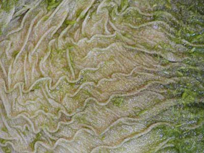
The border organ between the forestomach and the true stomach. The mucous membrane of the department is grouped into unidirectional folds of different sizes adjacent to each other. At the top of each “leaf” there are rough short papillae. The structure of the book provides for further mechanical processing of the incoming feed and transit to the next department.
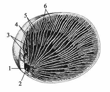
Scheme of the structure of the book: 1- bottom; 2- entrance; 3-6 - leaves
This is a real stomach with all the functions inherent in this organ. The shape of the abomasum is pear-shaped, curved. The expanded section is connected to the exit from the book, and the narrowed end is smoothly connected to the intestinal cavity. Internal cavity lined with mucous membranes and has digestive secretion glands.

For the full development of the animal, the process of processing and assimilation of feed in ruminants must be constant. This does not mean that you need to constantly fill the feeder. Nature provides a long period processing of each portion of food in adult ruminants.
The absorption process begins in the oral cavity. Here the food is moistened with saliva, partially crushed, and the fermentation process begins.

Solid and dry food ends up in the rumen. A favorable environment for the development of microorganisms has been created here:

The food fragments entering the rumen are no longer as coarse as in the feeder. Due to primary chewing and exposure to saliva, they become susceptible to grinding on the rough surface of the rumen epithelium and processing by microbes.
Undergoing these processes, the feed remains in the rumen for 30 to 70 minutes. During this period, a small portion of it reaches the desired condition and enters the book through the mesh, but the main part undergoes the chewing process.
Chewing is the process of repeatedly regurgitating food from the rumen into the oral cavity in order to increase its digestibility.
The reflex mechanism involves a process that occurs periodically and continuously. Not all the food received is burped, but individual portions of it. Each portion moves back into the oral cavity, where it is again moistened with saliva and chewed for about a minute, then again enters the first pregastric region. Consecutive contraction of the mesh fibers and rumen muscles moves the chewed part of the food deeper into the first section.

The chewing period lasts about an hour (about 50 minutes), then stops for a while. During this period, contractile and relaxing movements (peristalsis) continue in the digestive system, but belching does not occur.
Important! The entry of chewed feed into the rumen activates microorganisms, which, feeding on their juices, increase the availability of food for absorption by the animal.
The complex digestion of plant proteins is facilitated by the activity of bacteria that constantly live in the gastric digestion sections of ruminants. These microorganisms reproduce several generations of their own kind per day.
In addition to participating in the breakdown of cellulose, rumen microorganisms are also the most important suppliers in the ruminant menu:
Such “mutually beneficial cooperation” - the use of the host organism for the life of bacteria and assistance to this macroorganism in carrying out physiological processes is called symbiosis - a widespread phenomenon in nature.

The digestion of ruminants is multifaceted: many processes occur simultaneously. Individual portions of food are constantly moving into a mesh, which allows pieces of a suitable size to pass through, and pushes large ones back with contractile movements.
After a period of rest, which lasts for ruminants different time(depending on the conditions, type of food and type of animal), a new period of chewing begins.
Important! The chewing process does not stop at night, but, on the contrary, is activated.

The rumen is called the fermentation chamber of the ruminant body, and for good reason. It is in the rumen that 70–75% of the feed, including cellulose, undergoes breakdown, which is accompanied by the release of large volumes of gases (methane, carbon dioxide) and fatty (so-called volatile) acids - sources of lipids (acetic, propionic, butyric). The food becomes suitable for digestion.
Only food particles that are already sufficiently fermented (by saliva, plant sap and bacteria) pass through the mesh.
Between the leaves of the book they are:
Active absorption of volatile fatty acids (up to 90%) - a source of glucose and fats - occurs here. By the time it comes out of the book, the lump of food is a uniform (homogeneous) mass.

Unlike other animals, the stomach of ruminants (abomasum) produces juice containing digestive enzymes continuously, and not in response to food intake. During the day, rennet juice containing pepsin, lipase, chymosin and hydrochloric acid is produced from 4 - 11 liters in sheep to 40 - 80 liters in adult cows. The continuity of rennet secretion is explained by the constant supply of a sufficiently prepared mass of food from the proventriculus.
The quantity and quality of rennet juice directly depends on the composition of the feed. The largest volume and most significant activity of the secretory fluid is observed after the intake of fresh grass or hay from legumes, grains, and cakes.
In the process of digesting food, hormones from the liver, pancreas, thyroid, gonads and adrenal glands take part in the abomasum.
The walls of the abomasum, and later the intestines, complete the digestion process, absorbing previously undigested substances. Undigested residues are excreted as manure. Thanks to deep bacterial processing, it is a very valuable agricultural product, always in demand on the market and widely used in crop production.
Functions of the gastric sections
| Department | Functions |
|---|---|
| Scar | Fermentation, fermentation, creation and maintenance of an environment for symbiotic bacteria, food enrichment, chewing gum, cellulose breakdown, absorption of substances available for absorption |
| Net | Sorting pieces of food |
| Book | Transit + additional grinding of individual particles; Absorption of water and fatty acids |
| Abomasum | Final digestion with the participation of internal digestive organs and partial absorption, transport of food residues to the intestines |
The harmonious development of livestock directly depends on the correct composition of the feed according to age.
In young ruminants, the phenomenon of rumination, as well as the chambers of the gastric system, are not formed from birth. The abomasum at this time is the largest chamber of the gastric system. The milk that newborns are fed at the beginning of life goes directly into the abomasum, bypassing the undeveloped proventriculus. Digestion of this type of food occurs with the help of gastric secretions and partly enzymes from the mother’s body present in the product.

To enable the chewing process and the start of the rumen, plant foods and their inherent microorganisms are necessary. Usually, young animals are switched to plant foods from 3 weeks of age.
However modern technologies cultivation allows some acceleration of the process of establishing the typical digestion of ruminants:
Young animals fed on milk should be gradually transferred to plant foods. If the cubs are born during the grazing period, then the mixing of feed in the diet occurs naturally - along with mother's milk, newborns very soon try grass.

But most of the calving occurs in autumn - winter, so the transition to mixed, and then plant based diet depends entirely on the owner of the herd.
It is during the period of mixed nutrition that:
The bacterial component of the diet and the species composition of microorganisms changes with a change in food (even plant food). Therefore, the transfer, for example, from dry food to succulent food should also not occur at once, but be extended over time with a gradual replacement of components. A sudden change in diet is fraught with dysbacteriosis, and therefore worsening digestion.
And of course, with any type of feeding, the food should be varied. Only if this condition is met will it ensure the supply of sufficient amounts of proteins, fats, carbohydrates, vitamins and microelements to the ruminant’s body.

The predominance of one type of feed can unbalance the harmonious processes in the body, shifting them towards increased fermentation, gas formation or peristalsis. And any strengthening of one of the aspects of digestion certainly weakens the others. As a result, the animal may become ill.
Important! In addition to feed great importance has a sufficient supply of livestock drinking water even when kept on pasture. Its deficiency slows down digestion, reduces chewing activity and digestibility of feed.
So it's good organized meals taking into account the peculiarities of digestion in ruminants - a guarantee proper development farm animals and excellent results of their rearing.
The normal residence time of the contents (digested food) in the stomach is about 1 hour.
The figure on the right shows: 1. Body of the stomach. 2. Fundus of the stomach. 3. Anterior wall of the stomach. 4. Greater curvature. 5. Small curvature. 6. Lower esophageal sphincter (cardia). 9. Pyloric sphincter. 10. Antrum. 11. Pyloric canal. 12. Corner cut. 13. A groove formed during digestion between the longitudinal folds of the mucosa along the lesser curvature. 14. Folds of the mucous membrane.
The following anatomical structures are also distinguished in the stomach:
The shape of the stomach depends on the position of the body, the fullness of food, functional state person. With average filling, the length of the stomach is 14–30 cm, width 10–16 cm, length of the lesser curvature 10.5 cm, greater curvature 32–64 cm, wall thickness in the cardiac region 2–3 mm (up to 6 mm), in antrum 3–4 mm (up to 8 mm). The stomach capacity is from 1.5 to 2.5 liters (the male stomach is larger than the female). The normal weight of the stomach of a “conditional person” (with a body weight of 70 kg) is 150 g.

The stomach wall is made up of four main layers (listed from inner surface walls to outer):
 The mucous membrane of the stomach is formed by a single layer of columnar epithelium, a layer of its own and a muscular plate that forms folds (relief of the mucous membrane), gastric fields and gastric pits, where the excretory ducts of the gastric glands are localized. In the proper layer of the mucous membrane there are tubular gastric glands, consisting of parietal cells that produce hydrochloric acid; main cells producing the proenzyme pepsin pepsinogen, and accessory (mucosal) cells secreting mucus. In addition, mucus is synthesized by mucous cells located in the layer of the surface (integumentary) epithelium of the stomach.
 The surface of the gastric mucosa is covered with a continuous thin layer of mucous gel consisting of glycoproteins, and underneath is a layer of bicarbonates adjacent to the superficial epithelium of the mucosa. Together they form the mucobicarbonate barrier of the stomach, which protects epithelial cells from the aggression of the acid-peptic factor (Y.S. Zimmerman). The mucus contains antimicrobial activity immunoglobulin A (IgA), lysozyme, lactoferrin and other components.
The surface of the mucous membrane of the body of the stomach has a pitted structure, which creates conditions for minimal contact of the epithelium with the aggressive intracavitary environment of the stomach, which is also facilitated by a thick layer of mucous gel. Therefore, the acidity on the surface of the epithelium is close to neutral.  The mucous membrane of the body of the stomach is characterized by a relatively short path for the movement of hydrochloric acid from the parietal cells into the lumen of the stomach, since they are located mainly in the upper half of the glands, and the main cells are in the basal part. An important contribution to the mechanism of protecting the gastric mucosa from the aggression of gastric juice is made by the extremely rapid nature of gland secretion, caused by the work of the muscle fibers of the gastric mucosa. On the contrary, the mucous membrane of the antral region of the stomach (see the figure on the right) is characterized by a “villous” structure of the surface of the mucous membrane, which is formed by short villi or convoluted ridges 125–350 µm high (Lysikov Yu.A. et al.).
By the birth of a child, the fundus and cardiac part of the stomach are not sufficiently developed, and the pyloric part is much better, which explains frequent regurgitation. Regurgitation is also promoted by swallowing air during sucking (aerophagia), with improper feeding technique, short frenulum of the tongue, greedy sucking, and too rapid release of milk from the mother's breast.
The gastric juice of a healthy person is practically colorless, odorless and contains a small amount of mucus.
Basal secretion, not stimulated by food or otherwise, in men is: gastric juice 80-100 ml/h, hydrochloric acid - 2.5-5.0 mmol/h, pepsin - 20-35 mg/h. Women have 25–30% less. About 2 liters of gastric juice are produced in the stomach of an adult per day.
The gastric juice of an infant contains the same components as the gastric juice of an adult: rennet, hydrochloric acid, pepsin, lipase, but their content is reduced, especially in newborns, and increases gradually. Pepsin breaks down proteins into albumins and peptones. Lipase breaks down neutral fats into fatty acid and glycerin. Rennet (the most active enzyme in infants) curdles milk (Bokonbaeva S.D. et al.).
 The main contribution to the total acidity of gastric juice is made by hydrochloric acid produced by the parietal cells of the fundic glands of the stomach, located mainly in the area of the fundus and body of the stomach. The concentration of hydrochloric acid secreted by parietal cells is the same and equal to 160 mmol/l, but the acidity of the secreted gastric juice varies due to changes in the number of functioning parietal cells and neutralization of hydrochloric acid by alkaline components of gastric juice.
 Normal acidity in the lumen of the body of the stomach on an empty stomach is 1.5–2.0 pH. The acidity on the surface of the epithelial layer facing the lumen of the stomach is 1.5–2.0 pH. The acidity in the depths of the epithelial layer of the stomach is about 7.0 pH. Normal acidity in the antrum of the stomach is 1.3–7.4 pH.
Currently, the only reliable method for measuring gastric acidity is intragastric pH-metry, performed using special devices - acidogastrometers, equipped with pH probes with several pH sensors, which allows you to measure acidity simultaneously in different areas of the gastrointestinal tract.
Stomach acidity in conditionally healthy people(without any subjective sensations in gastroenterological terms) changes cyclically during the day. Daily fluctuations in acidity are greater in the antrum than in the body of the stomach. The main reason for such changes in acidity is the longer duration of nocturnal duodenogastric reflux (DGR) compared to daytime, which throws duodenal contents into the stomach and, thereby, reduces the acidity in the lumen of the stomach (increases pH). The table below shows the average acidity values in the antrum and body of the stomach in apparently healthy patients (Kolesnikova I.Yu., 2009):
The general acidity of gastric juice in children of the first year of life is 2.5–3 times lower than in adults. Free hydrochloric acid is determined at breastfeeding after 1–1.5 hours, and with artificial feeding – 2.5–3 hours after feeding. The acidity of gastric juice is subject to significant fluctuations depending on the nature and diet, and the state of the gastrointestinal tract.
 |
|
Motor activity of various parts of the stomach and duodenum (Gorban V.V. et al.)
The figure on the right shows a diagram of the fundic gland (Dubinskaya T.K.): 1 - mucus-bicarbonate layer Microflora of the stomachUntil recently, it was believed that due to the bactericidal effect of gastric juice, microflora that penetrated the stomach died within 30 minutes. However modern methods microbiological research this has been proven not to be the case. The amount of various mucosal microflora in the stomach of healthy people is 10 3 –10 4 / ml (3 lg CFU / g), including those detected in 44.4% of cases Helicobacter pylori(5.3 lg CFU/g), 55.5% - streptococci (4 lg CFU/g), 61.1% - staphylococci (3.7 lg CFU/g), 50% - lactobacilli (3. 2 lg CFU/g), in 22.2% - fungi of the genus Candida(3.5 lg CFU/g). In addition, bacteroides, corynebacteria, micrococci, etc. were sown in an amount of 2.7–3.7 lg CFU/g. It should be noted that Helicobacter pylori were determined only in association with other bacteria. The environment in the stomach turned out to be sterile in healthy people only in 10% of cases. Based on their origin, the microflora of the stomach is conventionally divided into oral-respiratory and fecal. In 2005, strains of lactobacilli that adapted (similar to Helicobacter pylori) to exist in the sharply acidic environment of the stomach: Lactobacillus gastricus, Lactobacillus antri, Lactobacillus kalixensis, Lactobacillus ultunensis. At various diseases(chronic gastritis, peptic ulcer, stomach cancer) the number and diversity of bacterial species colonizing the stomach increases significantly. For chronic gastritis greatest number mucosal microflora was found in the antrum, with peptic ulcer- in the periulcerous zone (in the inflammatory ridge). Moreover, the dominant position is often occupied by non- Helicobacter pylori, and streptococci, staphylococci, |

In 2018, the Nativity Fast will begin on November 28. During this period, Orthodox believers prepare to celebrate Christmas...

Starting a family is the dream of most women. They want to have a loving husband and a bunch of kids. But it's not always a relationship...

This article contains: the most powerful prayer for divorce - information taken from all over the world, the electronic network and...

Information site about icons, prayers, Orthodox traditions. Prayer for scandals and quarrels in the family, with husband, with children...

What would New Year be without champagne, tangerines, Olivier, aspic and everyone’s favorite “Herring under a fur coat”. With the last one...

Let's prepare the necessary ingredients for the cookies. The first thing to do is put the water to boil. We need...

Is it possible to register an employee for the position of financial director - chief accountant? The chief accountant claims...

The head of a small business can easily manage the budget independently. CHECKED! If you manage...

The creation of new projects involves a preliminary economic justification for their feasibility, subsequent...

Reporting is generated by the RM, is agreed upon (approved) by the Risk Committee under the Management Board and transmitted to...

At the edge of a large, very large meadow, on a long emerald blade of grass lived a tiny Ladybug. God's little...

Nowadays, it is quite common for people to turn to the stars. With the help of a horoscope a person can find out...

Business or friendly. If you were pursuing a stranger, it means your level of trust in the female sex...

according to Freud's dream book If you dreamed about how you were fishing, it means that in real life you can hardly switch off...

Starting a family is the dream of most women. They want to have a loving husband and a bunch of kids. But it's not always...

This article contains: the most powerful prayer for divorce - information taken from all over the world, electronic...