Procedure for filling out an income tax return
An example of a correct income tax return in 2017, download a new current form for free in Excel. What...
To quickly cure cough, bronchitis, pneumonia and strengthen the immune system, you just need...
“Pulmonary sarcoidosis: what is it and how to treat it?” – this question can be found on many medical forums.
Today, sarcoidosis is a rare disease that can affect different organs and systems human body. Most often, pathology chooses the lungs as its target.
Diseases in which accumulations of inflammatory cells are observed in organs are called granulomatosis, and the accumulations themselves are called granulomas (nodules). This is exactly what a disease is.
It is a systemic pathology that can affect not only respiratory system(bronchi, lungs, intrathoracic lymph nodes), but any organs.

The course of the disease is unpredictable: nodules can resolve or become the cause of fibrosis - proliferation connective tissue. Many people confuse sarcoidosis with lung cancer, but doctors say that these pathologies are completely different. To the question “Is Beck’s sarcoidosis contagious or not?” doctors answer in the negative.
As a rule, pulmonary sarcoidosis (in Ukrainian – sarcoidosis legen) is observed in patients aged 20 to 40 years. Moreover, representatives of the fairer sex suffer from the disease more often than men. The peak incidence occurs in winter and early spring.
Today there are five stages of the disease:
On initial stages Sarcoidosis can occur without symptoms, but as the disease develops, characteristic symptoms arise.
Typically, sarcoidosis manifests itself:
The exact causes of the disease remain unknown. Some scientists suggest that pulmonary sarcoidosis occurs under the influence of negative external factors: fungi, viruses, bacteria, pine pollen, clay, talc, as well as zirconium, beryllium and aluminum compounds.
According to this hypothesis, granulomas in the lungs can appear in firefighters, sailors, smokers, and employees of agricultural and chemical enterprises.
Given the fact that previous version is not proven, doctors tend to believe that the cause of the development of sarcoidosis is the simultaneous influence of genetic, autoimmune and environmental factors.
Because some people have an innate vulnerability to the disease, scientists believe that sarcoidosis is inherited.

Many patients wonder whether the disease “pulmonary sarcoidosis” is terrible and, if so, how bad it is.
The answer is simple: if you do not consult a doctor when the first symptoms of the disease appear, this can lead to unpleasant consequences.
Sarcoidosis of the lung is dangerous because... against this background it is possible to develop:
Such complications develop in cases where sarcoidosis does not go into remission.

To make a diagnosis, a patient with suspected sarcoidosis is admitted to the hospital.
Standard diagnostics is a combination of laboratory and instrumental studies:
The essence of the Kveim test is the subcutaneous injection of sarcoid antigen. After about 20 days, the injection site is excised and the tissue is sent for histological analysis. Tissue samples obtained during biopsy are examined in the same way.
The Kveim test detects granulomas in the lungs and is performed in all patients with suspected sarcoidosis.
As for a biopsy, the basis for its implementation is the need to examine the cells under a microscope. In this procedure, tiny pieces of lung tissue are removed using a bronchoscope or needle.
The presence of sarcoidosis can be judged by enlarged lymph nodes, dilated bronchial vessels, elements of granulomas without symptoms of inflammation and necrosis, plaques or warty growths of the mucous membranes.
Sometimes a patient with sarcoidosis feels great, but x-rays reveal a pathological process in the lungs.
What the radiologist sees in the image depends on the stage of the disease:

Many patients wonder who treats sarcoidosis and how this disease is treated. Typically, patients suffering from this pathology are treated by a sarcoidologist. If the disease does not go away on its own within six to eight months (as often happens), the doctor begins to treat the patient.
The use of medications for pulmonary sarcoidosis is the basis of symptomatic therapy.
Typically the patient is prescribed the following medications:
Many drugs used for sarcoidosis have serious side effects. However, people have to take them for several months.
Prednisolone is most often prescribed to patients with sarcoidosis.
Therapy begins with high dosages, after which the doses are gradually reduced over four to six months. In case of poor tolerability of the drug or exacerbation of concomitant diseases, the medication is taken at intervals of 1-2 days.

If the patient’s body responds better to combination therapy, it is recommended to alternate Prednisolone with taking drugs such as Indomethacin and Voltaren.
A number of people (about 10%) show resistance to doses of glucocorticoids, such as Chlorambucil, Pentoxifylline, Azathioprine, Cyclophosphamide, Infliximab, etc. Such patients are prescribed a course of Methotrexate, an antitumor drug.
The disadvantage of this drug is that it takes a long time to develop the therapeutic effect (from 6 to 12 months). Once a stable dose of medication has been achieved, the patient's liver enzymes and blood composition should be monitored.
Pulmonary sarcoidosis can often be cured using inhalations. Inhalations are carried out using Fluticasone, Budesonide and others similar drugs. These drugs are prescribed in the initial stages of the disease and help get rid of a debilitating cough when the bronchi are damaged.
Sarcoidosis is often treated with glucocorticoids taken by inhalation and orally (by mouth).
If you have sarcoidosis, you need to eat little and often. The basis of the diet should be protein food stewed and boiled. It is advisable that the menu include seaweed, sea buckthorn, apricot kernels, black currants, nuts, pomegranates, honey, fresh basil leaves and legumes.

As for the following foods, their consumption in sarcoidosis should be minimized:
For pulmonary sarcoidosis, diet can reduce pathological changes in the lymph nodes and prevent worsening inflammatory process and the formation of kidney stones.
Treatment for pulmonary sarcoidosis folk remedies It makes sense only in the initial stages of the disease.
The most popular are the following recipes:
Note:
In parallel with the listed treatment methods, you can practice consuming bear or badger fat, because it contributes speedy recovery from sarcoidosis.
Preventing the development of pulmonary sarcoidosis is much easier than treating the disease and its complications. The first thing you need to do is completely reconsider your lifestyle.
It is important to give preference healthy eating, refuse bad habits Take walks in the fresh air as often as possible.
Patients suffering from pulmonary sarcoidosis wonder how long they can live with it.
Doctors say that the pathology can end:
Thanks to modern medicine, the prognosis for life with pulmonary sarcoidosis is usually favorable. The active period proceeds without severe symptoms, with no visible deterioration. In 1/3 of patients, the disease degenerates into a state of remission with periodic exacerbations.
In 10-27% of cases, a chronic type of disease develops and, as a result, pulmonary fibrosis. This is fraught with development respiratory failure, not threatening the patient's life.
In most cases, pulmonary sarcoidosis is curable.
The development of a lethal outcome is possible only with a progressive form of the disease, if the patient does not ask the question “How to treat sarcoidosis?” and does not bother to see a doctor.
This is a very rare disease. The disease can affect many organs, but in 90% of cases it targets the respiratory system. For a long time, the pathology was called by the names of the doctors who studied it: Beck-Besnier-Schaumann disease. Then a short formulation took root: Beck’s sarcoidosis.
When inflammatory cells are concentrated in organs, the formation of such accumulations is called granulomas (nodules), and the diseases are called granulomatosis. The disease sarcoidosis is one of them. What is its nature, what is pulmonary sarcoidosis? The disease is systemic and can affect not only the lungs, bronchi, intrathoracic lymph nodes, but also any organ. Granulomas either resolve or lead to fibrosis - an increase in proliferating connective tissue.
Medical statistics record this disease and its relapses, as a rule, in people of young and mature age - 20-40 years. Among them, women are more common than men. Another feature of the disease is the rise in incidence in early spring and in winter. There is no reason to be afraid of a person with this disease, since Beck's sarcoidosis is not contagious.
So, what is pulmonary sarcoidosis, in terms of the development of the disease? According to the accepted classification, there are 5 stages:
What is pulmonary sarcoidosis? This can be judged by the manifestations of the pathology. In the initial stages, the disease often occurs without symptoms. Later, as a rule, the very first sign of the disease is chronic fatigue syndrome. Patients often complain of the following symptoms of sarcoidosis:
What is pulmonary sarcoidosis and how the inflammatory process proceeds can be imagined if we take into account the phases of the disease. There are three of them:
According to the speed of occurrence pathological lesions Various variants of the development of the disease may occur:
The disease sarcoidosis begins with mild degree– local damage to the alveoli. Then granulomas form in the tissues of the bronchi and pleura. In severe disease, the inflammatory process affects the heart, kidneys, liver, eyes, and brain. Chronic illness may lead to respiratory failure. If eye pathology is not treated, there is a high risk of vision loss. In the vast majority of cases, the prognosis is favorable.

Doctors continue to struggle with this mystery. However, the exact causes of pulmonary sarcoidosis remain unknown. There are only hypotheses. Scientists believe that Beck's sarcoidosis is a reaction to negative external factors:
The result of this reaction is the development of granulomas, the main symptom of sarcoidosis. Nodules most often form in the lung tissues and lymph nodes, but are found in eyeballs, sinuses, heart, liver, kidneys, skin. However, this version has not been proven. Most scientists are inclined to conclude that the disease is a consequence of the simultaneous influence of environmental, autoimmune and genetic factors.
Diagnosis of sarcoidosis is carried out only in a hospital setting. Only a complex of instrumental and laboratory studies is informative, including methods such as:
The doctor notes a positive result from the Kveim test, indicating the presence of granulomas and dangerous diagnosis. Sarcoid antigen is injected under the patient's skin, then after about 3 weeks the injection site is excised and the tissue is analyzed histologically. Her biopsy sample is examined in the same way.

Miniature fragments of lung tissue are removed with a needle or bronchoscope. A lung biopsy for sarcoidosis is performed when it is necessary to examine the cells under a microscope. Direct and indirect signs of the presence of the disease:
The patient may feel well, but the presence of an inflammatory process is immediately revealed by the R-image. What is pulmonary sarcoidosis from the point of view of a radiologist? At the first stage of the disease, chest x-ray shows an increase in intrathoracic lymph nodes. In the second stage, the image shows new enlarged lymph nodes in the roots of the lungs and the mediastinum (the space between the sternum and the spine). Later stages are characterized by fibrous lesions of the lung tissue.
Often the disease goes away on its own. Taking this into account, the patient is monitored to determine the need for treatment. Observation is carried out for 6-8 months. If recovery does not occur, it is clear to the TB specialist that the patient needs to be treated. Drug treatment of pulmonary sarcoidosis is only symptomatic. The following groups of drugs are used:
Hormone therapy helps protect the patient from severe complications of the disease. Many medications, despite serious side effects, have to be prescribed in long, 2-6 month courses. To strengthen the immune system, the patient is prescribed high doses of vitamin E in combination with vitamins C and D. In addition to medications, physiotherapy methods are also effective.

Treatment with it for 4-6 months begins with large dosages, gradually reducing them. If the patient does not tolerate Prednisolone well or if the drug causes an exacerbation of concomitant diseases, use an intermittent regimen of taking the medication every 1-2 days. Often, a combined treatment strategy is more acceptable, in which Prednisolone is alternated with Voltaren and Indomethacin.
Approximately 10% of patients have resistance to doses of glucocorticoids (Azathioprine, Infliximab, Pentoxifylline, Cyclophosphamide, Chlorambucil and others), and they are prescribed a course antitumor drug Methotrexate. However, the therapeutic effect of this medicine often appears after six months or even a year. When a stable dose is achieved, regular monitoring of blood composition and liver enzymes is necessary.

Such procedures using drugs such as Budesonide, Fluticasone are prescribed for primary stages pulmonary sarcoidosis. They help treat debilitating cough in patients with bronchial lesions. These drugs are also effective in a number of cases of eye and skin pathologies. A combination of glucocorticoids taken orally and inhaled is often effective.
You should exclude fatty fish, dairy products, and cheeses, which increase the inflammatory process and provoke the formation of kidney stones. It is necessary to forget alcohol, limit consumption flour products, sugar, salt. A diet with a predominance of protein dishes in boiled and stewed form is required. Nutrition for pulmonary sarcoidosis should be frequent, small portions. It is advisable to include in the menu:

Pulmonary sarcoidosis- This systemic disease, accompanied by the formation of granulomas consisting of Piragov-Langhans cells and epithelial cells. Granulomas are also diagnostic sign, which is detected by microscopic examination, but sarcoid nodules are not accompanied by caseous necrosis and tuberculous mycobacteria are absent. Also, the nodules merge as they grow and form lesions of different sizes.
Not only the lungs, but also many organs are affected by sarcoidosis. Most often these are lymphatic, intrathoracic, tracheobronchial, bronchopulmonary nodes, spleen and liver. Possible damage to the organs of vision, bones, joints, nervous system, heart, parotid salivary glands, skin. However, pulmonary sarcoidosis can occur long time without clinical manifestations. It is also not transmitted from patient to patient and is not infectious.
The etiology is currently unknown. Any people age category susceptible to the disease, but pulmonary sarcoidosis in children is quite rare. What is known is that pulmonary sarcoidosis has racial and geographic characteristics. For example, per 100,000 African Americans there are 36-64 people who have sarcoidosis, in the United States there are 10-14 cases per 100,000 fair-skinned people. In European countries, there are 40 cases per 100,000 people, however, the incidence is much higher in the Nordic countries.
In sarcoidosis, granulomas of two types form on the bronchial walls and in the lungs:
The first type is sclerosing or stamped. Granulomas are small in size, having a border from the surrounding tissues, as well as connective tissue cells - fibroblasts - surround the granulomas;
The second type is large granulomas that do not have clear boundaries.
Quite often, sarcoid granulomas are confused with tuberculous granulomas. For precise definition Diagnosis requires laboratory testing of the tissue.
Depending on the location, the disease is divided into sarcoidosis of the intrathoracic glands and lungs, lymph nodes, respiratory system with damage to other organs and generalized sarcoidosis.
According to the course of the disease, it is divided into:
— Regression phase ( reverse development, process fading). The reverse development is accompanied by resorption, compaction and, quite rarely, calcification of formed sarcoid granulomas in the lymph nodes and lung tissue;
— Stabilization phase;
— Exacerbation phase or active phase.
Directly depending on the speed with which changes increase, pulmonary sarcoidosis is divided into:
— Chronic sarcoidosis;
— Delayed sarcoidosis;
— Progressive sarcoidosis;
— Abortive sarcoidosis.
Oddly enough, but real reasons Pulmonary sarcoidosis is still unknown. Some scientists believe the disease is genetic, others that pulmonary sarcoidosis occurs due to impaired functioning immune system person. There are also suggestions that the cause of the development of pulmonary sarcoidosis is a biochemical disorder in the body. But at the moment, most scientists are of the opinion that the combination of the above factors is the cause of the development of pulmonary sarcoidosis, although not a single theory put forward confirms the nature of the origin of the disease.
Scientists studying infectious diseases suggest that protozoa, Histoplasma, spirochetes, fungi, mycobacteria and other microorganisms are the causative agents of pulmonary sarcoidosis. Endogenous and exogenous factors can also cause the development of the disease. Thus, today it is generally accepted that pulmonary sarcoidosis of polyetiological origin is associated with a biochemical, morphological, immune disorder and genetic aspect.
The incidence is observed in persons of certain professions: firefighters (due to increased toxic or infectious exposure), mechanics, sailors, millers, agricultural workers, postal workers, chemical industry and healthcare workers. Pulmonary sarcoidosis is also observed in people with tobacco addiction. The presence of an allergic reaction to certain substances perceived by the body as foreign due to impaired immunoreactivity does not exclude the development of pulmonary sarcoidosis.
A cascade of cytokines is responsible for the formation of sarcoid granuloma. They can form in various organs, and also consist of a large number of T-lymphocytes.
Several decades ago, there was an assumption that pulmonary sarcoidosis was a form of tuberculosis that was caused by weakened mycobacteria. However, according to the latest data, it has been established that these are different diseases.
Pulmonary sarcoidosis begins with the involvement of alveolar tissue in the pathological process and the development of interstitial pneumonitis or alveolitis.
Pulmonary sarcoidosis does not have a clear clinical picture, since it is often asymptomatic. For example, in most patients, the intrathoracic lymphoglandular form of the disease does not manifest itself clinically. Most often, pulmonary sarcoidosis is suspected when lymphadenopathy of the roots of the lungs is detected. Signs of pulmonary sarcoidosis are as follows: joint pain, fever, shortness of breath, cough with sputum, chest pain, restless sleep, insomnia, night sweats. Fever, weight loss, loss of appetite, increased fatigue, weakness, anxiety, severe malaise.
Pulmonary sarcoidosis is divided into three stages: initial, mediastinal-pulmonary and pulmonary.
Symptoms of early stage pulmonary sarcoidosis are similar to the symptoms of many other diseases: causeless anxiety, weakness, sleep disturbances, etc. A common sign of pulmonary sarcoidosis is fatigue, which is felt in the morning (a person feels it without even getting out of bed), and in the afternoon . At this stage, as a rule, there is an asymmetric and bilateral enlargement of the lymph nodes: tracheobronchial, paratracheal, bifurcation, bronchopulmonary.
The second stage of pulmonary sarcoidosis is manifested by symptoms characteristic of respiratory diseases: pain in the chest, in the joints, cough, wheezing, shortness of breath, weakness. It is possible that an inflammatory process may develop in the subcutaneous fat. skin vessels. This stage of pulmonary sarcoidosis is accompanied by bilateral dissemination (miliary, focal), infiltration of lung tissue.
The third stage includes a combination of symptoms of the first and second stages of pulmonary sarcoidosis. However, increased wet and dry wheezing, pain in the affected area of the lungs, crunching and wheezing sounds, and arthralgia are observed. Also, the third stage is manifested by damage to the lymph nodes, parotid glands (Herford syndrome), eyes and other organs that are not associated with respiratory system. Damage to the brain nerves, formation of cysts in the bones, and enlargement of the liver are possible.
The last stage of pulmonary sarcoidosis can be manifested by severe fibrosis or pneumosclerosis of the lung tissue, while enlargement of the intrathoracic lymph nodes is not observed. The increase in emphysema and pneumosclerosis occurs due to the formation of drainage conglomerates as the disease progresses. The disease also manifests itself as cardiopulmonary failure.
As pulmonary sarcoidosis progresses, it manifests itself with extrapulmonary symptoms as adjacent tissues are affected.
Sarcoidosis extends beyond the lungs, affecting the spleen and liver, and does not manifest itself clinically. Ultrasound examination may show slight enlargements internal organs. If the liver is significantly enlarged, the patient feels heaviness in the right hypochondrium. The patient will complain of loss of appetite, but the functions of the spleen and liver will not be impaired. Occasionally, choleostasis also develops.
The differences between granulomatous and sarcoid hepatitis are unclear. Gastric granulomas are quite rare. Mesenteric lymphadenopathy causes pain in the abdominal area.
Affecting joints and bones, the disease does not manifest itself clinically, but enzymes may be elevated in patients. Sometimes acute or silent myopathy develops, accompanied by muscle weakness. There may be pain when moving. However, the bone damage in pulmonary sarcoidosis differs from arthritis in that it is less damaging to the joints and bones. The development of lymphadenopathy of the roots of the lungs, erythema nodosum, acute polyarthritis, and osteopenia cannot be ruled out.
If myocardial damage occurs, the main symptom of the disease will be episodic, and the heart rhythm will also be disturbed. An attack is possible sudden death in case of severe compaction of cardiac muscle granulomas. Pulmonary or contribute to the development of heart failure. Quite rarely develops.
Pulmonary sarcoidosis has a significant impact on the nervous system. Sensory loss may occur, unilateral facial paralysis, swallowing is more difficult, paralysis of limbs, dizziness. Eighth cranial nerve neuropathy causes hearing loss. The development of neuropathy is possible optic nerve And peripheral neuropathy, polyphagia.
If the kidneys are damaged due to sarcoidosis of the lungs, hypercalciuria most often occurs. Nephrocalcinosis also develops, requiring a kidney transplant, nephrolithiasis, caused by chronic renal failure and interstitial nephritis.
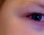
When the visual organs are damaged, a burning sensation occurs, the mucous membranes turn red, sensitivity to light is increased, and lacrimation is present. The disease is also accompanied high blood pressure(intraocular). Developing secondary glaucoma, optic neuritis, dacryocystitis, chorioretinitis, iridocyclitis, etc. If left untreated, progression leads to blindness, but most often resolves spontaneously.
At skin lesions, medium-sized reddish nodules form on the body. Severe damage is quite rare skin. Erythema nodosum develops: hard red nodules appear on the anterior surface of the lower limb. Nonspecific lesions include subcutaneous nodules, papules, macules, spots, hyperpigmentation, and hypopigmentation. The development of lupus pernio is possible: protruding spots appear on the ears, lips, cheeks and nose.
In sarcoidosis, the lymph nodes are usually not enlarged, with only occasionally visible enlarged lymph nodes in the groin or neck. In some cases, cervical or mild peripheral lymphadenopathy occurs.
According to its development, pulmonary sarcoidosis is divided into four stages:
Stage 0 is asymptomatic. In patients undergoing preventive medical examinations, the disease will not be detected even on x-rays;
At stage 1, the lung tissue remains unchanged, but slight enlargements of the intrathoracic lymph nodes are observed;
At stage 2, a pathological process is observed in the lung tissue, the intrathoracic lymph nodes are significantly enlarged;
Stage 3 is accompanied by significant changes in the lung tissue, however, the lymph nodes do not enlarge;
Stage 4 is accompanied by the formation of fibrosis - this is an irreversible process of compaction of lung tissue with the formation of scars on it (lung tissue is replaced by connective tissue).
The first three stages are not clinically apparent. Patients can learn about the presence of pulmonary sarcoidosis only from the results of a preventive X-ray examination during examination. Changes in lung tissue will be visible on the images. Quite rarely, there are patients with early stages of pulmonary sarcoidosis, in whom the body temperature rises, the joints of the limbs swell, and the lymph nodes are enlarged.
Diagnosing pulmonary sarcoidosis is not easy, but it is possible, regardless of stage. An accurate medical history of the patient, all clinical manifestations, and laboratory blood tests (accelerated ESR, eosinophilia, leukocytosis, increased globulins) are required. It is also necessary to conduct X-ray, ultrasound, computed tomography and magnetic resonance imaging, biopsy with bronchoscopy and further histological examination, radionuclide methods. The need for an ultrasound examination with a fine-needle biopsy of the lymph nodes is decided by a specialist. The patient is always prescribed a general urine test and a functional test of the kidneys and liver. Additional research will be ordered if complications are detected.
The acute course of pulmonary sarcoidosis is characterized by changes in laboratory blood values, which indicate an inflammatory process: a significant or moderate increase in ESR, lympho- and monocytosis, eosophilia. However, blood counts may be normal in pulmonary sarcoidosis. Leukocytosis will manifest itself if the bone marrow, spleen and liver are affected. To exclude kidney damage, urine tests are performed and functional tests (blood urea nitrogen, creatine) are determined.
More characteristic changes can be detected during x-ray examination. MRI and CT scans of the lungs make it possible to identify tumor-like enlargements of the lymph nodes, especially at the root, focal disseminations: fibrosis, emphysema, cirrhosis of the lung tissue.
Most patients have a positive Kveim reaction - after intradermal injection of a specific antigen (substrate of the patient's sarcoid tissue) 0.2 ml, a purplish-red nodule is formed.
During a biopsy with bronchoscopy, it is possible to detect direct and indirect signs of pulmonary sarcoidosis: dilated vessels at the mouths of the lobar bronchi, as well as sarcoid lesions of their mucous membranes (presence of warty growths, tubercles, plaques), signs of enlarged lymph nodes at the site of bifurcation, atrophic or deforming.
A more reliable method for diagnosing pulmonary sarcoidosis is a histological examination of biological material taken during bronchoscopy, open lung biopsy, transthoracic puncture, prescale biopsy, mediastinoscopy. In biological material, specialists identify elements of granuloma (epithelioid) without signs of perifocal inflammation and necrosis.
Angiotensin-converting enzyme (ACE) is a marker of the activity of the process and in pulmonary sarcoidosis its content in the blood is significantly increased. Also, increased levels of calcium in the urine and blood are evidence of complications in the body.
In order to exclude, it is necessary to conduct a Mantoux tuberculin test. If the body has an active form of pulmonary sarcoidosis, the Mantoux test is usually negative, however, there are exceptions.
Despite the fact that a lot of medical manipulations are required to make a diagnosis, it is the correct diagnosis that allows you to choose the right treatment.
Pulmonary sarcoidosis in most patients is accompanied by spontaneous remission and for this reason, the patient will be under observation for 8 months. This allows you to determine the prognosis and the need for specific treatment.
As a rule, for mild forms of the disease that occur without deterioration, treatment is not prescribed. Even in the case of minor changes in the lung tissue and the patient’s satisfactory condition, only observation is carried out. This is due to the fact that granulomas that have formed in the lungs dissolve and pulmonary sarcoidosis goes away on its own.
Severe forms of pulmonary sarcoidosis require treatment, as there is a risk of complications, including death. The development of tuberculosis and serious diseases of other organs is possible.
If pulmonary sarcoidosis is detected, a long course of antioxidants (Acetate, Tocopherol, Retinol and others), immunosuppressants (Azathioprine, Rezoquin, Delagil), anti-inflammatory drugs (Indomethacin), steroids (Prednisolone) is prescribed. If the patient is intolerant to Prednisolone, then non-steroidal anti-inflammatory drugs (Nimesulide, Diclofenac) are prescribed. On average, the course of treatment lasts 8 months, however, in severe cases of the disease, this period may be longer. In rare cases, specialists prescribe anti-tuberculosis drugs.
As a rule, during the first 4 months, Prednisolone should be taken 30–40 mg per day, after which the dosage is reduced to 5–10 mg. Accept this drug needed for several months. After 24–48 hours, the doctor prescribes glucocorticosteroid drugs in case of side effects from Prednisolone. The course of treatment also includes anabolic steroids and potassium preparations (Nerobol, Retabolil).
Treatment always depends on the activity, progression and severity of pulmonary sarcoidosis. When combination therapy, including Dexamethasone or Prednisolone, the drugs alternate with non-steroidal anti-inflammatory drugs (Indomethacin, Voltaren).
In rare cases, they are prescribed for severe cough inhaled glucocorticoids. They help reduce cough in patients with endobronchial lesions. Also, in rare cases of eye and skin lesions, topical glucocorticoids will be prescribed.
Dispensary observation of patients is carried out by a phthisiatrician. Patients with pulmonary sarcoidosis are divided into two dispensary groups:
♦ The first group includes patients with an active form of the disease;
Group IA includes persons in whom the disease is first diagnosed;
Group IB includes people whose disease has worsened or relapsed after the prescribed course of treatment;
♦ The second group includes people who have an inactive form of the disease.
Patients also need to pay attention Special attention diet. Table salt should be limited and consumed as much as possible more products, enriched with proteins. In order to restore immunity, therapy must include medicinal and food plants that concentrate certain biologically active substances (BAS) - zinc, manganese, silica and other minerals.
It is necessary to consume food plants that have immunocorrective properties - chokeberry, raw seeds sunflower, decoction of young shoots of sea buckthorn, walnuts, seaweed, noble bay, pomegranates, basil, legumes, blackcurrant leaves and fruits. The following products should be excluded from the daily diet: dairy products, cheese, sugar, flour.
Pulmonary sarcoidosis in children is also treated by a phthisiatrician. The medication course is selected individually, depending on the child’s condition. For the purpose of prevention, it is necessary to harden the child, accustom him to daily physical education, monitor his social circle to prevent pulmonary diseases. It is also necessary to include vegetables and fruits in it daily diet. Children who have had pulmonary sarcoidosis need to be explained that they should not start smoking in the future. Parents should protect their child from various contacts with chemicals. Many cleaning products contain a large number of chemical substances, which the child should not breathe.
Also, many patients include folk remedies in their treatment. For example, from medicinal herbs(calendula, goralthea, sage, oregano) a decoction is prepared at home, which must be taken 3 times a day for 1.5 months before meals, 50 ml. A tincture made from vodka and vegetable oil is also popular. Mix 50 ml and take 3 times a day for a year. Cases have been reported full recovery thanks to this tincture. You can also dilute 20% propolis tincture in warm water and 10–15 grams of the product per glass of water will be enough. Should be taken for 15 days 40 minutes before meals.
Most patients in the early stages of the disease prefer treatment with folk remedies. If the disease progresses similar methods become ineffective. Every patient should understand that most herbs have side effects. It is for this reason that treatment of pulmonary sarcoidosis with folk remedies usually causes a deterioration in the general condition.
Since pulmonary sarcoidosis is rarely diagnosed, a special diet has not yet been developed, however, it should be healthy image life. Sleep and nutrition should be adequate. It is recommended to stay in the fresh air as much time as possible and do physical exercise. However, direct contact with the sun's rays must be avoided (sunbathing is strictly prohibited). You should also avoid contact with vapors of chemical liquids, dust, and gases.
Typically, symptoms of pulmonary sarcoidosis go away without treatment. In 60% of cases, 9 years after diagnosis, patients have no symptoms. After a few months, extensive pneumonia and swollen lymph nodes may disappear. About 75% of patients who experience only enlarged lymph nodes and damage only to the lungs recover completely within 5 years.
The most beneficial effects of pulmonary sarcoidosis are in patients whose disease has not spread beyond the chest, especially if it began with erythema nodosum. In 50% of cases, relapses are observed.
Although patients often recover spontaneously, the manifestations and severity of pulmonary sarcoidosis are quite variable. In most cases, a repeat course of glucocorticoids is necessary. It is for this reason that regular monitoring is necessary to detect relapses. In 90% of cases where spontaneous recovery occurs, relapses develop in the first two years after diagnosis. In 10% of cases, relapses are detected after two years. Patients who do not experience remission within two years will have chronic pulmonary sarcoidosis.
Pulmonary sarcoidosis is generally considered chronic in 30% of patients and is permanent in 10–20%. The disease is considered fatal in 5% of cases. Most common cause the fatal outcome is pulmonary fibrosis with respiratory failure, after which pulmonary hemorrhage follows due to aspergilloma.
More unpleasant consequences Pulmonary sarcoidosis occurs in patients with the extrapulmonary form of the disease and in African-Americans. In 89% of cases, recovery occurs in European countries. Signs of a favorable outcome are the presence of acute arthritis and erythema nodosum. However, unfavorable signs of pulmonary sarcoidosis are: extensive pulmonary damage, myocardial diseases, nephrocalcinosis, neurosarcoidosis, chronic hypercalcemia, uveitis. In 10% of cases, damage to the respiratory organs and eyes develops.
Sarcoidosis is a disease that can affect many organs and systems and is characterized by the formation of granulomas in the affected organs, which can be seen at the microscopic level. The lymph nodes, lungs and spleen are most often affected. It is not an infectious disease and is not transmitted to others. Previously, sarcoidosis was considered rare disease, but with modern improvement diagnostic capabilities, with a fresher look at this pathology of doctors, it is no longer a rarity.
More often, sarcoidosis is detected in a younger group of the population from 20 to 40 years old. There is a slight predominance of women. This disease has been known for more than 100 years, but its causes are still unknown. There are suggestions that sarcoidosis is caused by various viruses, such as herpesvirus. There is also the possibility that this disease is hereditary, or is caused by various mycobacterioses (this is why phthisiatricians used to deal with sarcoidosis, sarcoidosis was considered a type of tuberculosis). Perhaps there are a number of factors that together lead to changes occurring in the body, leading to the development of clinical manifestations of sarcoidosis. On modern stage With the development of medicine, we can say that sarcoidosis is an independent disease, with an as yet unclear origin.
In accordance with the international classification of diseases, there are:
1. Pulmonary sarcoidosis
2. Sarcoidosis of the lymph nodes
3. Sarcoidosis of the lungs with sarcoidosis of the lymph nodes
4. Sarcoidosis of other specified and combined localizations
5. Sarcoidosis, unspecified
The onset of the disease can be either acute or gradual, and asymptomatic. Asymptomatic disease is more common when sarcoidosis of intrathoracic lymph nodes, without damage to other organs. In such cases, the disease is detected during a preventive fluorographic examination.
More often, the disease occurs gradually with such general symptoms as lethargy and fatigue, which are characteristic of many diseases. When the lungs are involved in the process, patients complain of difficulty breathing, coughing, and back pain. After 2-3 weeks, these symptoms may completely disappear. With a prolonged course, these manifestations can persist for a long time. Characterized by a dry cough with difficult to clear sputum. Subsequently, with the development of fibrosis (thickening of the lung tissue), respiratory and pulmonary-heart failure develops.
There is an acute course of sarcoidosis - Löfgren's syndrome. It is observed rarely, in 10% of cases. It is characterized by an increase in temperature to 38-39°C, the appearance of erythema nodosum (inflammatory nodes rising above the skin level, most often occurring in the area of the legs, forearm, less often the face), swelling and pain of the joints, as well as bilateral damage to the intrathoracic lymph nodes. These signs may not appear fully and the disease will progress more slowly in this case.
Erythema nodosum in sarcoidosis
In addition to this, there is also Heerfordt syndrome characterized by damage to the salivary glands and eyes.
Among the extrapulmonary localizations of the process, most often there are damage to peripheral lymph nodes, skin and subcutaneous tissue. The cervical and subclavian nodes, less often the axillary and inguinal nodes, are enlarged, densely elastic, mobile, the skin over them is not changed, and is not complicated by fistulas. Lymph nodes may also be affected. abdominal cavity. In 5% of cases the spleen and liver are enlarged. Bilirubin and aminotransferase activity are increased in the blood. Damage to skeletal bones It is rare and is characterized by a typical x-ray picture. Typically, the development of multiple foci of bone loss in the phalanges of the fingers and toes is associated with the leaching of calcium and is accompanied by an increased content in blood and urine tests. Damage to the central nervous system It is very rare and has a difficult course.
Occurs in 20-30% of cases sarcoidosis of the heart, which is asymptomatic. Defects can be detected using an ECG and ultrasound of the heart. The volume and size of the left chambers of the heart decrease, the right ventricle increases, and arrhythmias occur.
If the above symptoms occur, the patient must first contact the general medical network, where the therapist will prescribe necessary tests and other research methods. And if sarcoidosis is suspected, he will refer you to a specialist - a pulmonologist. Early detection and treatment of the disease ensures a favorable prognosis and recovery. Otherwise, gross changes in the lung tissue, respiratory and pulmonary-heart failure, blindness and other disorders occur, leading to the patient’s disability.
More often, sarcoidosis in its symptoms is very similar to tuberculosis, but the causes of occurrence, and, accordingly, the treatment of these diseases are enormously different. And treatment with anti-tuberculosis drugs, and possibly self-medication of the patient, can lead to a worsening of the condition, and in special cases and death. Also, in its course, sarcoidosis is similar to diseases such as lymphogranulomatosis, central lung cancer, toxoplasmosis, brucellosis and a whole group of diseases collectively called “granulomatous”. Therefore, timely consultation with a doctor, diagnosis and treatment by a specialist can prevent unwanted consequences.
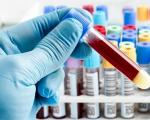
The tests that will need to be taken include: general blood test, blood biochemistry, Mantoux test, diaskintest, general sputum analysis, sputum analysis for mycobacteria, fibrobronchoscopy with biopsy, spirography, radiography (CT, MRI).
Anemia, leukopenia, lymphopenia, monocytosis, and an increase in erythrocyte sedimentation rate (more often in acute cases) may be detected in the blood. At the same time, in many patients, especially with sarcoidosis of the intrathoracic lymph nodes, changes in the blood are insignificant or absent. At biochemical research it is possible to detect an increase in fibrinogen, lipoproteins, C-reactive protein, some patients experience dysproteinemia. In 15%-20% of patients, there is an increase in calcium levels in the blood and urine. Both of these analyzes tell us the degree of organ damage in sarcoidosis, the severity of the process.
Most patients with sarcoidosis experience tuberculin anergy, reflecting impaired cellular immunity. Negative tuberculin test and diaskintest are more common in sarcoidosis with lung involvement.
A general sputum analysis and sputum analysis for mycobacteria will help us carry out differential diagnosis with other lung diseases, for example, such as: aspergillosis, tuberculosis. In rare cases, sputum analysis reveals Mycobacterium tuberculosis (1% of cases of combined pathology of sarcoidosis and tuberculosis).
During fiberoptic bronchoscopy of patients with sarcoidosis, various changes. Due to compression of the bronchi by enlarged lymph nodes, a narrowing of the bronchial lumen occurs. Deformation of their walls, bulging of the bronchial wall into the lumen, “sarcoid ectasia” - expansion, tortuosity, thickening of vessels in the form of a network or individual plexuses: “spiders”. Most patients experience nonspecific endobronchitis, usually bilateral. Rarely are multiple tubercles identified. Plaques, granulations.
When biopsying such areas using cytological and histological methods elements of a sarcoid granuloma can be detected. A morphological examination of the bronchi reveals various granulomas: typical sarcoid and numerous lymphoid cell accumulations. The most commonly used in our time are: transbronchial intrapulmonary biopsy, mediastinoscopy, transbronchial biopsy of intrathoracic lymph nodes, video thoracoscopy. In addition, a biopsy of the bronchi, peripheral lymph nodes, skin, liver, spleen and other organs involved in the process is also performed. The elements of sarcoidosis found in the biopsy material allow us to make a correct and timely diagnosis.
Examination of external respiratory functions (spirography, spirometry) helps to identify disorders varying degrees in patients with respiratory sarcoidosis. Degree of violation bronchial obstruction, an increase in the resistance of the bronchi to air flow, a change in the elastic properties of the lung tissue indicates the progression of respiratory failure. Obstructive disorders are more common in patients with an acute course of the disease, and restrictive in patients with a chronic course.
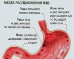
One of the main methods for diagnosing sarcoidosis of the intrathoracic lymph nodes and lungs is radiography. Lymph nodes are almost always enlarged on both sides. As a rule, bronchopulmonary lymph nodes are more affected. Shadows of lymph nodes appear on an x-ray as large conglomerates, but they can also appear in the form of separate groups of lymph nodes. The greatest changes in combined lesions of the intrathoracic lymph nodes and lungs in the acute phase occur in the middle and lower parts lungs, foci of dissemination are noted. They can be of different sizes. In some cases, the changes on the right are more pronounced than on the left. In the chronic course of the process, the following are noted: increased pulmonary pattern, emphysematous pulmonary fields, formations of the “bull” type. Against this background, fresh lesions may also arise.

Sarcoidosis of intrathoracic lymph nodes on x-ray
Isolated lung damage is not often observed. After sarcoidosis has been cured, x-rays may show residual changes in the form of pneumofibrosis of varying severity, cavity formations according to the bullous type.
The classification of stages of sarcoidosis associated with radiological signs, it distinguishes: stage 1 - enlargement of the intrathoracic lymph nodes, stage 2 - development of combined lesions of the intrathoracic lymph nodes and lungs. Stage 3 - combined damage to the intrathoracic lymph nodes and lungs with the development of fibrosis and large formations.
The whole set diagnostic methods, history of the disease, history of the patient’s life leads to an accurate diagnosis.
In the treatment of sarcoidosis, the following are used: corticosteroids, non-steroidal anti-inflammatory drugs, pentoxifylline, cytostatics (methotrexate, azathioprine), immunosuppressants (cyclosporine), immunomodulatory agents, physiotherapeutic methods, plasmapheresis. Currently, a number of hormone therapy regimens are used that reduce inflammation and improve the clinical picture of the disease. As a rule, prednisolone or its analogs (metipred) are prescribed. Inhaled corticosteroids - budesonide - are also used. Becotide, beclomethasone dipropionate. They selectively affect the affected bronchial mucosa.
The complex treatment regimen for sarcoidosis includes delagil or plaquenil; they reduce tissue oxygen demand, inhibit the development of granulation tissue, and are immunomodulators. During the entire treatment period, antioxidants are prescribed: vitamin E, sodium thiosulfate, vitamin C.
Patients with acute onset of the disease and microcirculation disorders are prescribed angioprotectors. It is necessary to avoid stress, avoid prolonged exposure to the sun and hypothermia. In most patients with mild symptoms of the disease, with a moderate degree of enlargement of the intrathoracic lymph nodes, in the absence of pulmonary lesions, observation is often prescribed for 3-6 months with the prescription of non-steroidal anti-inflammatory drugs and antioxidants: indomethacin, vitamin E, sodium thiosulfate, pentoxifylline, antimalarials (chloroquine ). More often, after such treatment, patients experience complete or almost complete remission. There is rarely a deterioration of the process during treatment, which forces one to prescribe hormonal treatment. For sarcoidosis with limited damage to other organs, treatment with an antimalarial drug with minimal doses of the hormone is prescribed. For generalized forms, methotrexate, azathioprim or cyclosporine are also used.
At the first stage of treatment, especially in acute stage diseases, recommended hospital treatment to select an individual treatment regimen and select doses of hormone therapy. Treatment will last about two months, with transition to outpatient monitoring. With unsystematized treatment, the patient's condition may worsen and progress. The duration of treatment depends on the improvement of the patient’s condition, tests, X-ray examination methods and depends on the extent of the process. Treatment for advanced cases can last up to two years.
The diet for sarcoidosis is not specific, but there are a number of restrictions. Since the process is inflammatory in nature, and carbohydrates beneficially increase inflammation, it is necessary to exclude from your diet: sugar, sweets, cakes, products made from yeast dough, sweet carbonated drinks. Any inflammatory process also intensifies when consuming: spicy, salty and fried foods. It is necessary to exclude foods containing calcium, as its content increases in the blood and urine (milk and fermented milk products, cottage cheese, sour cream). Food should be complete and easily digestible: steamed, boiled or stewed. It is useful to eat the fruits of sea buckthorn, black currant, gooseberries, cherries, pomegranates, onions, garlic, seaweed, buckwheat and oatmeal, beans, peas. At hormone therapy You should limit your salt intake, as fluid retention occurs in the body. Protein breaks down rapidly, so you need to use a protein diet.
Traditional methods of treatment include herbal treatment (marshmallow root, calendula, plantain, oregano, sage), if there is no allergy to them. In principle, such treatment is allowed; it does not lead to damage to organs and systems. But, for example, treatment with vodka and butter can affect liver function, which can worsen in sarcoidosis. Badger fat contributes to the deterioration of the process in the lungs, intensification of the inflammatory process, its use in sarcoidosis will only worsen the situation. Aloe and honey are natural immunomodulators, so they can be used in the treatment of sarcoidosis.
Non-drug rehabilitation: interstitial electrophoresis, magnetic therapy, heparin or lidase electrophoresis, ultrasound, laser treatment, physical therapy, manual therapy. Duration general treatment depends on the course of the disease and the patient’s condition.
The most frequent complications sarcoidosis are: severe emphysema, respiratory failure, pulmonary heart failure, broncho-obstructive syndrome, cor pulmonale. That is, the occurrence of irreversible processes, which subsequently become chronic and accompany the patient throughout his life, require treatment. Therefore, timely consultation with a doctor and annual fluorographic examination contribute to early detection diseases and eliminates the occurrence of complications.
Approximately 60% of patients experience spontaneous remission after two years. 25% of patients recover completely after treatment. In 10% of cases, remission cannot be achieved. Treatment of extrapulmonary sarcoidosis and the central nervous system is most often ineffective. Sarcoidosis is rarely a cause of permanent disability, especially when modern level medicine. More often, disability is associated with the development of pulmonary heart failure, pulmonary heart disease, as a result of a long-term sarcoidosis process. Death occurs rarely - in 0.5-7% of cases.
Since the possible causes of sarcoidosis are not fully known, it is difficult to talk about prevention of this disease. We can talk about maintaining a healthy lifestyle: avoid smoking and alcohol, prolonged exposure to the sun, avoid contact with chemicals, vapors, and substances that affect liver and lung function. Undergo an annual fluorographic examination.
Phthisiatrician L.A. Kuleshova
>For hypertensive patients >> READ → Shishkina Olga" url="https://feedmed.ru/bolezni/organov-dyhaniya/sarkoidoz-legkikh.html">Pulmonary sarcoidosis is a disease in which inflammatory nodules (granulomas) form in the affected tissues. Damage to the liver, lung and lymph nodes is more common. In another way, Beck's sarcoidosis has a benign course.
 The disease has a racial predisposition. It is more common among African Americans, Asians, Germans, residents of Ireland, and Scandinavia.
The reasons for the development of pathology have not been fully established.
The main ones are:
In case of infectious lesions of the lungs, pathogens can be:
There are also many studies that confirm the genetic nature of the disease, that is, when there were manifestations of pathology in families.

Currently, research shows that the disease is associated with immune deficiency in the body.
The development of pathology in individuals of specific specialties is observed.
These are people working:
Tobacco smokers and people who have allergic reactions for some substances are also at risk.
 The onset of sarcoidosis is characterized by the development of a pathological process in the alveolar tissue, resulting in pneumonia or alveolitis.
Then sarcoid granulomas begin to form in the subpleural and bronchial tissues.
The disease has three stages:
The disease is classified according to the speed of development of the inflammatory process:
Phases of the course of pulmonary sarcoidosis:
Sarcoidosis does not have a pronounced clinical picture and may even be asymptomatic.
The first symptoms of pulmonary sarcoidosis appear:
As the disease progresses, other symptoms appear:
With a strong cough, sputum mixed with blood may be discharged. The functioning of other organs is disrupted, which can lead to dysfunction of the heart and lungs. The spleen and liver may be affected. If the liver is significantly enlarged, the patient is bothered by heaviness in the right hypochondrium.
Sarcoidosis stage 2 is a pathology of the respiratory system.
At this stage, the intrathoracic lymph nodes are enlarged and granulomas develop in the lung tissue.
The first signs of pathology appear. The patient complains of fatigue, dry cough, discomfort in the chest and chest pain.
Such complaints are a reason to consult a doctor and full examination sick. It is difficult to make a diagnosis, since sarcoidosis has a similar clinical picture to other pulmonary pathologies.
 The disease is diagnosed based on clinical manifestations, medical history and hereditary predisposition.
A general blood test is prescribed, which, if this pathology is present, will include:
The most effective diagnostic method is histological analysis.
It is carried out over the material that is taken during bronchoscopy or biopsy. The Quain test is also reliable. A specific antigen is administered.
At positive result sample, a purplish-red nodule is formed.
At asymptomatic the disease is detected during preventive x-ray examination.
Be sure to perform a Mantoux test. In cases of sarcoidosis, it is negative, which indicates weak immunity.
 The disease has a long development, so the patient is under the supervision of a specialist throughout this period. Drug treatment pulmonary sarcoidosis is carried out depending on the periods of the disease.
The patient is being monitored at the dispensary.
There are several accounting groups:

The patient has been registered for two years with a favorable prognosis. In more severe cases up to five years. Then the patient is removed from the dispensary register.
Be sure to use for treatment:
There is no specific treatment at the moment, since the exact causes of the disease have not been established.
During treatment, the patient follows a protein diet, with limited use salt.
Most often, complications affect the respiratory system and cardiovascular system. These include cor pulmonale syndrome.
In this condition:
This leads to heart failure.
Pulmonary emphysema, tuberculosis, and bronchial obstruction often develop.
 Often the disease is benign. Since the course occurs without clinical manifestations, the condition does not cause discomfort to the patient.
In 35% of patients, the disease becomes chronic. Such patients are under the supervision of a doctor.
They are given prophylaxis against respiratory failure, which often develops in this condition.
In a small percentage of patients, the healing period begins immediately after the first treatment course.
In other cases, the patient experiences an exacerbation of the disease over many years.
Pathology is much easier to cure if it was detected in the initial stages. Therefore, you should not neglect preventive examination.
 First of all, it is recommended to lead a healthy lifestyle and not smoke.
Eat foods that contain non-natural ingredients as little as possible.
Limit the use of chemicals.
The likelihood of developing sarcoidosis occurs in those patients who have pathologies in the functioning of the immune system.
If there are minor suspicions, they should seek advice from a specialist and take care of their health.
Those who are already sick should take care of their health and not allow the disease to worsen. They are advised to limit their calcium intake. Sarcoidosis leads to the formation of stones in bladder, and calcium accelerates this process. Exposure to the sun is also limited.
Vitamin D, which is produced by exposure to sunlight, promotes calcium production.
It is necessary to reduce exposure to harmful chemicals and increase the body's immune reactivity.
If you suffer from shortness of breath and persistent cough, you need to consult a specialist and check your health.
The examination is necessary for people:
Those who have already been diagnosed with this disease need to be under the supervision of a doctor at all times.
A number of tests have been conducted that have shown that such a disease can be inherited by close relatives.
Some scientists believe that such a disease may appear as a result of a weakening of the body's protective functions.
He is not viral disease, therefore, if you come into contact with a patient with pulmonary sarcoidosis, you cannot become infected and get sick, that is, pulmonary sarcoidosis is not contagious.
Such a disease can be treated at home with folk remedies, but only when the disease does not progress and the patient does not need treatment. urgent hospitalization. Traditional methods can treat this disease very effectively, but do not neglect the help of a qualified specialist.
Used as home treatment herbal teas and tinctures:
There is no special diet to treat this disease. But, there are several recommendations that must be followed. Since this disease is considered inflammatory, it may worsen if you eat foods that contain large amounts of carbohydrates.
Therefore, you should not eat:
You should not exclude onions and garlic, they are very useful and have a beneficial effect on the body’s condition, while improving the name system.
During pulmonary sarcoidosis, the amount of calcium in the body increases, which leads to the formation of calcium stones in the urinary tract(kidneys, ureters, bladder).
Therefore, you should refrain from using:
With pulmonary sarcoidosis, you need to ensure that food is quickly digested and complete. It is better to stew, boil or steam the product. Also, meals should be taken regularly, in small portions 4-5 times a day.
If you have this disease, you can eat the following foods:
The following products are considered very useful:
It is recommended to consume as many freshly squeezed juices as possible. Carrot, apple, and pomegranate are especially useful. They contain many vitamins and microelements that help restore normal lung function.
Sarcoidosis can manifest itself in damage to the lymph nodes in the groin, armpits, as well as in the cervical and subclavian regions. It can also affect the lymph nodes that are located in the abdominal cavity. The pathological process is expressed in significant enlargement and swelling of the lymph nodes. But, upon palpation pain no, you can only visually notice and feel small moving seals. Skin color also does not change.
Very often there is a lesion in the chest area. This creates some problems with establishing an accurate diagnosis, especially in the early stages of the disease. This is due to the fact that enlarged lymph nodes in the chest area can be found with tuberculosis. A biopsy, a study of a tissue sample, helps identify the disease.
If a person catches sarcoidosis of the lymph nodes, then the first symptoms are sharp pains and heaviness in the abdomen, frequent loose stools. Sometimes, along with this disease, damage to the spleen can be observed.
This disease has a second name - Beck's sarcoidosis. The symptoms are very broad and can lead to damage to many internal organs and systems.
It affects the condition:
Most often, older women are affected by this disease. age group. Diagnosis is carried out through laboratory and x-ray studies. It is quite difficult to establish an accurate diagnosis, so they often resort to additional research, for example the Kveim reaction.

An example of a correct income tax return in 2017, download a new current form for free in Excel. What...
P. S. Pallas (1741 - 1811) - naturalist and traveler-encyclopedist, who glorified his name with major contributions to...
Today, all issues related to the placement of government orders are regulated by the Law on the Contract System -...
Accounting Regulations Accounting for calculations of income tax of organizations PBU 18/02 (as amended by Orders of the Ministry of Finance of the Russian Federation...
A trainee salesperson is usually called those salespeople who are not yet ready to work completely independently. Process...
Educational institution "Gomel State Medical University" Department of Neurology and Neurosurgery...

One of the most controversial and controversial methods for the early development of children was developed in the 80s by a sociologist...

Contents Dietary supplement based on an extract obtained from the fly beetle (or...

Ekaterina MirimanovaSystem minus 60. RevolutionSystem minus 60 with Ekaterina Mirimanova“System minus 60....
Heaviness and bloating are the causes of both ordinary overeating and more serious digestive problems...
The second blood group, Rh-negative, appeared many years ago, when a person ceased to be...

PSYCHOLOGICAL ASPECTS OF ANOREXIA PHENOMENON (EXPERIMENTAL STUDY) T. V. Tarasova, E. V. Arsentieva...
Contents Since the skin in this area is thin, it is more prone to the appearance of various types of spots....

vseslav Sat, 10/17/2015 - 20:50 Vasileostrovskaya station is one of the oldest stations...
P. S. Pallas (1741 - 1811) - naturalist and traveler-encyclopedist, who glorified his name with major contributions...
Today, all issues related to the placement of government orders are regulated by the Law on Contract...