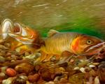
Infection of the body with Trichinella causes the development of a disease such as trichinosis
What is trichinosis
The term "trichinosis" refers to a helminthic disease accompanied by damage to the striated muscles and small intestine. Particularly vulnerable to this form helminthic infestation People. Human infection is associated with the appearance of predominantly expressed acute form symptoms and local damage to muscle tissue. Absence on time measures taken treatment most often results in death.
Trichinosis in beavers, wolves, wild boars, small rodents, and humans is caused by a pathogen called Trichinella. This roundworm relatively small in size - no more than half a centimeter, the body of which is twisted into a spiral. There is no particular specificity in the choice of carrier by Trichinella, that is, almost all types of animals, as well as humans, can become infected.
Trichinella development cycle
After birth, Trichinella larvae spread throughout the body through the blood and lymph flow, penetrating muscle tissue, where they grow and actively develop. A few weeks after birth, the larvae acquire the spiral shape characteristic of Trichinella and are covered with a protective shell - a kind of capsule.
Important! Infection of animals and humans with trichinosis occurs through consumption of infected meat. Other routes of helminthic infestation are almost completely excluded.
Prevalence of trichinosis
According to modern statistics, currently in some regions the incidence of trichinosis in wild animals is more than 90%. This creates a more than direct threat of helminthiasis for both domestic animals and humans. So high level The prevalence of trichinosis is due to the following aspects:
- The main route of infection is eating the meat of a sick animal. In the body of the host, the larvae can remain viable for several months even after the death of the host.
Thus, it is almost impossible to protect a person from infection using conventional methods, for example, boiling or frying contaminated meat. The only way protect yourself - do not eat meat that has not passed proper control.
Symptoms of the disease
Characteristic signs of infection appear on average five days after infection. Most often in animals, such as pigs, the disease occurs against the background of the following symptoms:
- Digestive disorders, expressed in the appearance of severe diarrhea and vomiting.

The disease may manifest itself as apathy in the pet.
- Severe exhaustion.
- Swelling of the eyelids and limbs.
- The animal becomes apathetic and weak, often lying motionless for a long time.
The disease can last from one month to a year, after which it progresses to chronic form and is characterized as a period of recovery. However, recovery can only occur in animals that have strong immunity. Most die on the tenth to fourteenth day from the moment of invasion.
Infection of a person with trichinosis is also accompanied by severe digestive disorders, swelling and muscle soreness, high temperature, as well as severe puffiness of the face. If the human body is weakened, death from trichinosis usually occurs within three to five weeks from the moment of infection. Timely and correct diagnosis is important for adequate treatment.

In humans, as in animals, infection causes swelling
Important! Penetration of more than five larvae into the human body at the same time is a kind of lethal dose.
To detect trichinosis in humans, the following methods are most often used:
- Intravenous allergy test.
- Biopsy of muscle tissue.
- Serological research methods.

Trichinosis can be detected in humans by serological studies
In addition, when trichinosis is detected in a person, it is necessary to examine the meat products that the infected person consumed before signs of infection appeared. This measure is necessary, among other things, to prevent mass infection of the population.
Treatment and prevention
Currently, methods for treating trichinosis in sick animals are relatively poorly developed, especially in those regions where the risk of contracting this type of helminthic infestation is relatively low. Most often, a foreign-made drug called Thiabendazole is used to treat domestic pigs. The effectiveness of its use depends on many factors, including the duration and degree of invasion.
Treatment of infected people is carried out, as a rule, in a hospital setting, due to the need for round-the-clock monitoring of the patient. Can be used during treatment A complex approach, including the use of anthelmintic drugs, as well as a number of drugs that restore and normalize the activity of the entire body as a whole.

If trichinosis is detected in a person, hospital treatment is required
The mortality rate among the population when infected with trichinosis is at least 20%. In this regard, it is necessary to pay attention to the prevention of invasion. Due to the fact that the source of human infection is most often infected pig meat, it is necessary:
- Before cooking pork, it is recommended to pre-freeze the meat at a temperature of at least 15 degrees. In order to completely destroy the larvae, it is necessary to keep the meat at a low temperature for about three weeks.
- In the early stages of animal infection effective method The destruction of larvae is by boiling or frying. But if the larva has already become covered with a layer of lime, this technique is completely ineffective.
In addition, owners of farms or farmsteads that keep pigs should pay attention to the conditions in which the animals are kept. It is necessary to completely exclude contact of pigs with small rodents - mice and rats. Also necessary measure is sanitary and epidemiological control over the quality of meat. Purchase products that have not passed this mandatory procedure, absolutely should not.
You will learn more about trichinosis in the video:
An anthropozoonotic acute and chronic disease of many mammal species of a pronounced allergic nature, caused by larvae and mature nematodes of the genus Trichinella.
Pathogen
Sustainability muscular trichinella to various external influences quite high. To destroy Trichinella in meat, especially in thick pieces, long-term heat treatment is necessary: the temperature in the thickness of the pieces should not be lower than 80 ° C. In meat stored at a temperature of -17...-27 °C, Trichinella remains viable for 6 weeks. Salting and smoking meat products does not neutralize Trichinella. Muscular trichinella are capable of secreting toxic substances that are highly resistant.
Epizootological data
Trichinosis affects pigs, wild boars, bears, badgers, dogs, cats, wolves, foxes, rodents, nutria, marine mammals of the far north, as well as humans.
Infection of animals and humans with trichinosis occurs through meat containing invasive Trichinella larvae. The meat is digested, and the released muscle trichinella are transformed into intestinal ones.
Pre-mortem diagnostics
There are no characteristic clinical signs of trichinosis. Intravital diagnosis of trichinosis in animals on pig farms consists of enzyme-linked immunosorbent assay (ELISA).
Post-mortem diagnostics
A reliable method for detecting trichinosis is trichinoscopy of pig meat, wild boars, bears and other animals. From the meat samples, use curved scissors along the muscle fibers to cut 12 pieces the size of oat grain. The slices are placed on a compressorium and crushed until newspaper print can be read through them. The prepared sections are carefully examined under a trichinelloscope, with a low magnification microscope (40-100 times) and a projection camera or screen trichinelloscope.
By doing differential diagnosis more accurate digestion method minced meat in artificial gastric juice followed by microscopy of the sediment.
At meat processing plants, a method of group testing of pork for trichinosis is used. It is based on digestion in special liquid samples of muscle tissue taken from the legs of the diaphragm of several pork carcasses, and the detection of Trichinella larvae in the sediment (digested mass). The study is performed using an apparatus for isolating Trichinella larvae (AT).
Differential diagnosis
Trichinella is differentiated from air bubbles, cysticerci, sarcocysts and calculi.
Veterinary and sanitary assessment
The following are subject to testing for trichinosis: carcasses, half-carcasses, quarters and pieces of carcasses of pigs (except for piglets up to 3 weeks of age), wild boars, badgers, bears, other omnivores and carnivores, as well as nutria.
If at least one Trichinella larva is detected (regardless of its viability), the carcass and offal having muscle tissue, esophagus, rectum, as well as depersonalized meat products are sent for disposal.
External fat(lard) is removed and reheated. Internal fat released without restrictions.
Intestines (except rectum) after usual processing are released without restrictions.
The skins are released after removing the muscle tissue. The latter is sent for disposal.
Prevention and control measures
In order to prevent the disease of animals with trichinosis in pig farms, it is necessary to promptly carry out deratization and disinfection, destroy stray animals, and dispose of animal corpses in compliance with veterinary and sanitary rules.
On a farm where trichinosis is detected in pigs, restrictive measures (quarantine) are imposed, and a set of liquidation and preventive measures are carried out.
A farm is declared free from trichinosis if, during a repeat serological examination after 1 year of the entire livestock, no positive animals are found, and when slaughtered for meat and trichinelloscopic examination of the carcasses, no Trichinella larvae are detected in them.
Trichinosis is an acute or chronic zooanthroponotic disease of many species of mammals and humans, which has a pronounced allergic nature and is caused by larvae and mature nematodes (Trichinella spiralis, Trichinella natuva and Trichinella pseudospiralis) parasitizing in the intestines and striated muscles.
The disease is common on all continents of the globe and in all countries.
Trichinosis manifests itself in two forms - muscular and intestinal. Adult trichinella are localized in the intestine, and their larvae are located in the striated muscles.
Pathogen– Male nematodes are very small, 1.4-1.6 mm long and 0.14 mm wide. At the posterior end of the body and in the space between the two lobes behind the cloaca, they have two pairs of papillae; there is no spicule. Females of Trichinella are twice as large as males and have a length of 3-4mm. Females give birth to viviparous larvae. The larvae are 0.08-0.12 mm long and 0.006 mm wide.
Epizootology. Humans and more than 100 species of domestic and wild animals (wild boar, brown bear), rodents, insectivores and marine mammals are susceptible to trichinosis. The source of the causative agent of trichinosis is infested animals. The main route of infection with trichinosis is nutritional, through eating meat, meat waste, and animal corpses infected with trichinella larvae.
The main reservoir of the causative agent of trichinosis are wild animals - wolves, foxes, brown bears, wild boars.
Pathogenesis. The pathogenic effect of Trichinella on the body of animals and humans has a pronounced allergic nature. As a result of sensitization by metabolites and decay products of dead Trichinella larvae, they develop in the body systemic vasculitis of a nonspecific nature, causing pain syndrome and organ damage (myocarditis, pneumonia, etc.). IN early stage illness occurs mechanical impact trichinella causing damage to the intestinal wall, arterial thrombosis, etc.
Clinical signs. After 3-4 days, with intensive infection, pigs experience depression, diarrhea, and increased body temperature. In some animals, these symptoms may intensify and the animals die after 12-15 days due to symptoms of cachexia. In practice in pigs fatal outcome happens rarely. The emerging intestinal disorders in most pigs gradually disappear and appear allergic symptoms — muscle pain, the head and eyelids swell, in some animals it appears skin rash, conjunctivitis, aphonia. During this period, in sick animals we note the leading ones for trichinosis. Clinical signs- muscle pain and eosinophilia. When standing up and eating food, animals feel severe pain. Symptoms of trichinosis reach their maximum by 2-3 weeks, then these symptoms begin to gradually fade away. When animals are mildly infected with trichinosis, the disease is asymptomatic and only eosinophilia (up to 10-12%) indicates a latent course of the disease.
Pathological changes. In case of intensive infection of pigs and other animal species in small intestines we note mucous degeneration and desquamation of the villous epithelium and submucosal tissue. In the liver, hemorrhages and fatty degeneration and disintegration of the epithelium of the Malpighian glomeruli in the kidneys. In the myocardium, brain, lungs, and liver we find nodular infiltrates consisting of lymphoid cells and eosinophils. IN in some cases Trichinella larvae can be found in the nodules. Specific to trichinosis is pronounced interstitial myositis and the formation of connective tissue capsules around the larvae.
Diagnosis.
Considering that the pronounced clinical picture of trichinosis in pigs is very rare, the diagnosis has to be made taking into account the epizootic situation and using immunological diagnostics - intradermal allergy test. If necessary, you can resort to a diagnostic biopsy of the pig ear muscles (temporalis muscle), which allows you to identify 30-60% of infected animals. For post-mortem diagnosis and post-mortem veterinary examination, trichinoscopy of the crura of the diaphragm is used.
Prevention trichinosis is based on strict adherence to veterinary and sanitary rules for keeping animals. Animal owners must exclude the possibility of pigs eating infested carcasses, as well as carcasses of wild animals, dogs, cats and rats, as well as raw or poorly cooked slaughterhouse and kitchen meat waste. The meat of pigs, wild boars, brown bears (in the Vladimir region is included in the Red Book and hunting is prohibited), badgers and marine mammals (walruses and seals) must be subject to mandatory trichinoscopy and, if trichinella larvae are found in it, must be destroyed or technically disposed of . Deratization must be carried out in the territories of farms, slaughterhouses, recycling plants, warehouses meat products and leather raw materials.
Each case of detection of trichinosis in animals is reported to Rospotrebnadzor and the leadership of the regional veterinary service.
Infested meat, once in the stomach, it is digested, the Trichinella larvae are released from the capsule and, moving through the gastrointestinal tract, intestinal tract enter the small intestine, where they penetrate the mucous membrane, located between the intestinal villi. There the females are fertilized; larvae form in the uterus; on the 4th day, each female gives birth to up to 2 thousand larvae, which penetrate into the lymphatic and blood vessels, spread through them to all organs and tissues, and the bulk of the larvae are selectively localized in skeletal muscles, where the larvae go through several stages of development. By the 14th day of infection, the muscle fibers thicken, the transverse striations are lost, they expand fusiform, the larvae curl, become invasive, a capsule is formed, which takes on a shape from oval to round (by the 60th day). Once in other organs and tissues, the larvae die.
In the vast majority of cases, the factor of transmission of trichinosis is the meat of domestic pigs and wild boars; Infection is also possible through the meat of wild carnivores; the same ones become infected by eating mice and rats and by feeding them waste from the slaughter of trichinosis animals.
Trichinosis is more often recorded in pigs when they freely graze, when they have access to carrion.
Human infection occurs through consumption of meat and meat products obtained from infested animals. You should know that Trichinella larvae are highly resistant, and neither boiling, frying, smoking, nor salting completely rids meat products of them.
The disease in humans usually manifests itself 3-4 weeks after eating infested meat, but may appear several days later. The more intense the infection, the shorter the incubation period.
The disease in humans is characterized by: an increase in temperature to 38 degrees and above, weakness, muscle pain, swelling of the eyelids and face (hence popular name"puffiness"), skin rashes, intestinal upset. The disease can occur in erased or mild forms, but it can also severe forms ah, which is ending fatal.
To avoid becoming infected with trichinosis, you must follow the following measures prevention:
- Under no circumstances should you feed pigs slaughterhouse waste from meat processing plants, even after normal boiling, as well as meat and carcasses of animals both from animal farms and those obtained by hunting.
- Do not allow pigs to wander around farms, settlements through wastelands, ravines and forest clearings.
- It is necessary to regularly carry out deratization on farms, including pigsties, walking yards and summer camps.
- Buy pork meat only in official markets, where pork is tested for trichinosis in a veterinary laboratory.
- You cannot buy pork or wild boar from friends or unidentified trading places.
Hunters of wild boars and brown bears (the brown bear in the Vladimir region is listed in the Red Book and hunting for it is prohibited) are required to check shot wild animals for trichinosis in veterinary examination laboratories.
Trichinosis is caused by the nematode Trichinella spiralis from the family. Trichinellidae. Trichinosis - anthropozoohelminthosis. The causative agent of this helminthiasis has been recorded in domestic and wild pigs, dogs, cats, bears, wolves, foxes, rats, mice and humans. Mature Trichinella are localized in the small intestine, and their larvae are in the muscles. Therefore, two forms of the disease are distinguished - intestinal and muscular. Trichinosis occurs in the form of separate foci, but in some countries (USA and Canada) this helminthiasis is very widespread.
On the territory of the USSR, trichinosis is more often registered in Belarus, Vinnitsa and Khmelnytsky regions.
Biology of the pathogen. In Trichinella, the same animal or person is successively the definitive host and then the intermediate host (trichinelloid type of development). Trichinella females penetrate into the lumen of the liberkühn glands or intestinal villi, giving birth to live larvae, which are carried into the muscles by lympho-hematogenous current. Favorite places for localization of larvae are the muscles of the legs of the diaphragm, tongue, esophagus, intercostal muscles, etc.
After 17 days they reach the invasive stage (spiral shape). After 3-4 weeks, a lemon-shaped capsule forms around the larva, which begins to calcify after six months. This process ends completely in 15-18 months. After the formation of the capsule, the larvae are called muscular Trichinella. The viability of muscular trichinella remains in animals for years, and in humans up to 25 years. Infection of animals and humans with trichinosis occurs when eating meat containing invasive Trichinella larvae. The meat is digested, and the released muscle trichinella turn into intestinal trichinella after 30-40 hours. Males fertilize females and quickly die. After 6-7 days, females give birth to from 1,500 to 10,000 Trichinella larvae. Females live in the intestines for up to two months (Fig. 43).
Medical and sanitary significance of trichinosis. When eating pork that has not undergone a veterinary-sanitary examination and trichinoscopy, cases of human trichinosis have been reported in Vinnitsa, Chernihiv and other regions of the country. It should be borne in mind that people are very susceptible to infection with trichinosis. Even 10-15 g of infected pork can cause infection and human disease with trichinosis. But the human body is a biological dead end for Trichinella.
Epizootological data. Trichinosis is a natural focal helminthiasis. The main links in the epizootological chain of this disease are not pigs and rats, as was mistakenly believed before, but wild animals. In certain areas of the country, the wolves examined were infected with trichinosis from 96.9 to 100%. High infestation of domestic carnivores is often recorded. For example, 146 cats studied in Odessa turned out to be infected with Trichinella on
71.23%, while the infection rate in rats was 6.45%, and in pigs only 0.35%.
The specificity of Trichinella in relation to the choice of hosts is very weak, and in fact they can develop in any host (with natural or artificial infection).
The main sources of infection with trichinosis: pigs - carcasses and corpses of rats, cats, dogs, wolves, foxes, as well as waste from processing the skins of these animals, waste from pig slaughter (especially backyard); dogs and cats - rodents, kitchen and slaughterhouse waste, waste from processing animal skins; gnawing is new - through cannibalism and through food waste; wild animals - 268
rodents, other animals and their corpses; fur-bearing animals* rave on fur-bearing state farms - slaughterhouse waste.
Pathogenesis. The degree of pathogenic influence of Trichinella on the animal and human body depends on the intensity of invasion and the resistance of the host organism.
When introduced into the intestinal mucosa, female Trichinella destroy villi and liberkühn glands and often inoculate pathogenic microbes. Migrating nematode larvae injure and cause degeneration of muscle fibers. As a result of the sensitizing effect of metabolic products and breakdown of muscle, as well as intestinal trichinella and allergic reactions, edema in the head area, eosinophilia and degeneration of parenchymal organs develop, and fever is noted.
Clinical signs with a strong degree of invasion appear on the 3-5th day after infection. They are characterized by fever, diarrhea, and emaciation. The appearance of these symptoms is associated with the penetration of female Trichinella into the intestinal mucosa. After a few days, difficulty chewing and swallowing, swelling of the eyelids, and eosinophilia are noted. These signs appear as a result of the pathogenic influence of migrating Trichinella larvae in muscle fibers. They are noticeable until the end of the process of encapsulation of the larvae in the muscles, after which the disease proceeds chronically. Most often, trichinosis in pigs is asymptomatic.
In humans, severe clinical signs include fever, facial swelling, diarrhea, and soreness of the affected muscle groups. Trichinosis in Belarus is called “puffy roll”.
Diagnosis. For lifetime diagnosis of trichinosis, the allergic method is used. The antigen in a dose of 0.1 is injected intradermally into the skin fold of the ear. If the reaction is positive, a pink or red spot and swelling with a diameter of up to 1.5" cm appear at the site of antigen injection after 30-45 minutes. At the same time, the skin fold thickens by 5-8 mm (compared to normal). B practical conditions rarely used. The main method of post-mortem diagnosis of trichinosis in pigs =
trichinoscopy of muscle pieces from pork carcasses to detect Trichinella larvae. “To test for trichinosis, two samples should be taken from the legs of the diaphragm of each pork carcass.
When examining imported meat and the absence of the legs of the diaphragm in it, samples for trichinelloscopy are allowed to be taken from other parts of the diaphragm, as well as from the intercostal and cervical muscles.
Trichinelloscopy technique. From two samples of meat weighing up to 60 g, using curved scissors, cut 12 pieces the size of an oat grain along the muscle fibers, closer to their tendon part. The slices are placed on a compressorium and crushed until newspaper print can be read through them.
The prepared 24 muscle sections are carefully examined under a trichinelloscope, a low-magnification microscope, as well as using a KT-3 projection camera or a screen trichinelloscope.
On muscle sections, Trichinella larvae are usually found encapsulated. Trichinella capsules have a lemon-like shape (0.68X0.37 mm), inside of which there is often one spiral-shaped larva.
Larvae with calcified capsules are often found; To clarify them, muscle sections are placed for 5-10 minutes in 50% glycerol, lactic acid or a 2-5% solution of hydrochloric acid, and then examined in drops of glycerin.
Calcified capsules of Trichinella larvae must be differentiated from sarcocysts (misher sacs) and finn. Miescher's pouches are located in muscle fibers, but their size and shape are variable; they reach 0.1-5 mm in length and 0.2-0.3 mm in width; the shape of misher bags can be curved, elongated or oval; they don't have capsules. The visibility of the sacs improves when the sections are treated with a 5% solution of potassium hydroxide.
Underdeveloped or dead and calcified cysticerci (finnous stones) are always located outside the muscle fibers; their length is 0.03-15 mm. These stones dissolve in a 10-20% solution of hydrochloric acid. According to current legislation, if even one living or dead Trichinella larva is detected by any means, the carcass and offal from
products containing muscle tissue, as well as impersonal meat products, are subject to technical processing or destruction. The esophagus (pical meat) and rectum are also destroyed. External fat (lard) is melted at a temperature not lower than 100° - for food purposes. Internal fat and intestines (except straight) are released without restrictions. Muscle tissue is carefully removed from animal skins.
No treatment has been developed. Thiabendazole has some effect.
Prevention. One of the most important preventive measures for trichinosis is a veterinary and sanitary examination of all pork carcasses, as well as meat of wild boars and bears, with mandatory trichinoscopy. Banning door-to-door slaughter will play an important role in preventing disease in humans and animals.
You cannot feed slaughterhouse waste from pigs to pigs and fur-bearing animals. Carcasses of fur-bearing animals and carnivores are subject to mandatory disposal after removal of their skins. The corpses of dogs, cats, and mouse-like rodents should be burned or destroyed in biothermal (Pyryatino) pits. It is necessary to exterminate rats by all means, especially in places where animals are slaughtered, where meat products are stored, and on pig farms.
It is advisable to improve the diagnosis of trichinosis. It is necessary to systematically carry out veterinary educational work among the population and instill in them hygienic skills.
The socialist form of agriculture and the successes achieved in preventing invasive diseases of people and domestic animals create every opportunity for a further reduction in trichinosis, up to the total devastation of this dangerous anthropozoohelminthosis in our country.
Laboratory-practical lesson 17
TRICHINELLOSCOPY OF PORK MEAT AND STUDY OF TRICHINELLA LARVAES
Task: 1) study the structure of Trichinella larvae on permanent medications;
2) conduct trichinoscopy of meat samples from pork carcasses;
3) sketch a Trichinella larva.
Equipment and materials. Trichinelloscopes or microscopes, compressors, tweezers, ophthalmic curved scissors, scalpels, bacteriological dishes; permanent preparations of Trichinella larvae, canned pork infested with Trichinella larvae, fresh samples of meat from the legs of the diaphragm from pork carcasses.
Methodology of conducting the lesson. Students are divided into units of two. Each unit is given a piece of fresh and canned (trichinella) meat. After a preliminary brief explanation from the teacher, students first examine the larvae on permanent preparations under a trichinelloscope, and then proceed to trichinelloscopy of pieces of meat. Make 22 sections of fresh meat and two sections of trichinella meat (in order to save it), crush it between compressor glasses and sequentially examine all sections under a trichinelloscope, starting with sections of canned pork containing trichinella larvae. The teacher draws students' attention to the need to differentiate Trichinella larvae from Mischer's sacs and cysticerci (Finn) at an early stage of development. At the end of the lesson, students make schematic sketches of Trichinella larvae.
DIOCTOPHYMATOSIS
Dioctophymatoses are those nematodes caused by nematodes from the suborder Dioctophymata. All causative agents of dioctophymatoses are biohelminths.
HYSTRICHOSIS OF DUCKS
The losses caused by hystrichosis to duck farming consist mainly of large waste of young
about ducks (death and forced slaughter) and the unsuitability of recovered birds for breeding purposes.
Morphology of the pathogen. Hystrichis tricolor is a large nematode with a spindle-shaped body covered in the front with numerous large spines. Males are 2.5-3 cm long, have a muscular caudal bursa and one spicule. The size of females varies from 2.5 to 10.5 cm in length and 0.3-0.5 cm in width. Eggs are medium size (0.07-0.08 X 0.042-0.048 mm), oval in shape, yellow color, immature. The outer shell of the eggs is cellular.
Pathogenesis. The causative agent of hystrichosis has a pronounced mechanical effect in the glandular stomach (parforation of the wall) and other organs, accompanied by an acute disorder of its motor and secretory functions. Death of young ducklings from hystrichosis can occur in the first hours after infection.
Rice. 44. Scheme of development of the causative agent of hystrichosis:
/ - head end of the nematode; 2 - caudal end of the male; 3 - egg;
4 - infective larva; 5 - oligochaete worm.
Clinical signs. With a strong degree of hystrichosis infestation, emaciation, diarrhea, sometimes vomiting, retardation in the growth and development of young animals are noted, and ducklings often die.
Small nodules (about the size of a hemp seed) remain in the stomach. Death of ducklings occurs at the beginning of the disease when the organs of the thoracic or abdominal cavities are damaged, and at a later time from exhaustion.
Control questions
1. What is the difference between toxocaroid and ascaroid types of nematode development?
2. How is group deworming of pigs carried out using piperazine and sodium fluoride?
3. What is the essence of chemoprophylaxis for ascaridiosis and heterokidosis in chickens?
4. In what forms is fecothiazine used for strongylatosis in ruminants?
5. How are dictyocaulosis and protostrongylidosis of ruminants differentiated during the life of animals and posthumously?
6. What is the advantage of the aerosol method of deworming sheep and cattle? cattle with dictyocaulosis?
7. How are earthworms examined to detect metastrongylid larvae in their bodies?
8. How are tetramerosis, streptocariasis and echinuriosis differentiated during the life of birds and posthumously?
9. What economic damage does bovine onchocerciasis cause to the leather industry?
10. How is trichinoscopy of pork performed?
Trichinosis is common among animals such as bears, badgers, wild boars, foxes, and wolves. Trichinella larvae sometimes infect domestic pigs. This happens when pigs eat the meat of dead animals or rats.
Complications that occur in severe forms of trichinosis:
- Myocarditis - inflammatory disease heart muscle, which is in this case has an allergic nature and is associated with an overreaction of the immune system. Myocarditis is the most common cause of death in patients.
- Lung damage- pneumonia . This is eosinophilic pneumonia - it is caused by the accumulation of allergic cells - eosinophils - in the lung tissue. Sometimes the rut is complicated by pleurisy (inflammation of the pleura - the thin membrane of connective tissue, which lines the chest cavity and covers the lungs), conditions resembling bronchial asthma.
- Meningoencephalitis - inflammatory process in the brain and its membranes.
- Hepatitis- inflammatory lesion immune cells of the liver.
- Nephritis- inflammatory kidney damage.
- Severe muscle pain in combination with impaired mobility or complete immobility of the patient.
In severe cases of trichinosis, many patients die. During outbreaks, mortality rates reach 10–30%. Typically, patients die between 4 and 8 weeks of illness. If the course of the disease is favorable, recovery occurs within 5–6 weeks from the moment the first symptoms of the disease appear.
Diagnosis of trichinosis

General blood analysis
There are several varieties of white in human blood blood cells- leukocytes, - each of which performs its own functions. Eosinophils are a type of white blood cell that is involved in allergic reactions. During trichinosis, accompanied by allergies, their content in the blood is very high. This is revealed using general analysis blood. Kinds serological diagnostics for trichinosis:
| Abbreviation
| Decoding
| The essence
|
| RSK
| Complement fixation reaction
| If there are antibodies in the patient’s blood, then they combine with the antigen and attach to themselves a complement molecule - a special substance involved in immune reactions. In this case, the reaction will be considered positive. |
| RNGA
| Indirect hemagglutination reaction
| It is based on the ability of red blood cells to stick together when there is an antibody and antigen on their surface. |
| ELISA
| Linked immunosorbent assay
| A reaction is carried out between antibodies and antigens. Special enzymes serve as a marker to evaluate the result. |
| REEF
| Immunofluorescence reaction
| The material contains a special label that causes it to glow after the antibody reacts with the antigen. |
| REMA
| Reaction of enzyme-labeled antibodies.
| A special label, which is an enzyme, allows you to evaluate the result. |
Intravenous allergy test
Using this analysis, an allergic reaction that develops in response to the presence of Trichinella is detected. A solution containing antigens is injected under the patient's skin. There should be redness and a blister at the injection site.
An intravenous allergy test can detect the disease starting from the second week. In the future, the result will be positive for another 5–10 years. Muscle biopsy
If trichinosis cannot be detected by other means, the doctor may prescribe a biopsy - an examination under a microscope. small piece of the affected muscle, which was taken with a needle. Study of meat from sick animals
To confirm the diagnosis, examination of the meat of a sick animal that the patient ate before becoming ill can be used. Under a microscope, capsules formed by Trichinella larvae are clearly visible. Treatment of trichinosis

| Anthelminthic drugs (treatment aimed at combating the causative agent of the disease)
|
| A drug
| Indications and effects
| Mode of application
|
| Mebendazole
| It disrupts the absorption of glucose by worms and the synthesis of ATP, the main carrier of energy, in their bodies. As a result of metabolic disorders, the worms die.
Mebendazole is contraindicated in pregnant and nursing mothers. | 0.3 – 0.6 g (1 – 2 tablets of 0.1 g three times a day) for 10 – 14 days.
|
| Albendazole
| It works almost the same way as Mebendazole. Most active against larval forms of worms. Available in the form of tablets of 0.2 grams.
Contraindicated in pregnancy and retinal diseases. | Take at the rate of 10 mg per kilogram of the patient’s body weight for 10 to 14 days.
(Reference Vidal, 2010) |
| Vermox | Active substance– mebendazole. Efficiency is 90% | For adults: - during the first three days – 100 mg 3 times a day;
- the next 10 days - 500 mg 3 times a day.
Children under 7 years old:
25 mg of the drug 3 times a day.
Children aged 7 – 9 years:
3 times a day, 50 mg.
Over 10 years old: - during the first three days – 100 mg 2 – 3 times a day;
- then for 10 days, 500 mg 3 times a day.
Take after meals.
|
| Thiabendazole | Efficiency is 90%. | Dose for children and adults – 25 mg per kg body weight (dose (mg) = body weight (kg) * 25). Divide into 2 doses every 12 hours. The course of treatment is continued for 3–5 days, after which, according to indications, it is repeated after 7 days (as prescribed by a doctor).
Take one hour after meals.
(« Complete guide infectious disease specialist”, ed. DMN, prof., corresponding member of RAE and REA Eliseeva Yu.Yu., Eksmo, 2007) |
| Treatment to combat symptoms of the disease
|
| Anti-inflammatory drugs (Voltaren, Diclofenac, Diclogen, Ortofen).
| They help cope with inflammation that was caused by allergic reactions in the patient’s body. | As prescribed by a doctor. |
| Antipyretics (Paracetamol, Aspirin, Acetylsalicylic acid, Nurofen, Ibuprofen).
| Indicated when body temperature rises above 38°C. | As prescribed by a doctor. |
| Adrenal cortex hormone preparations - glucocorticoids.
| Hormonal agents that oppress immune system and allergic reactions. | Hormonal drugs are used only strictly as prescribed by a doctor. |
In most cases, treatment of trichinosis is carried out in a hospital, since the disease at any time can become severe and cause severe complications. Despite such measures, 10–30% of patients still die, especially during outbreaks. If the disease is accompanied by severe muscle damage and immobility, then the patient, bedridden, needs careful care. After recovery is carried out rehabilitation treatment, including massage and physiotherapy. It is aimed at restoring muscle mobility.
All meat that goes on sale is necessarily examined for the content of capsules with larvae. Therefore, it is better to purchase it on the market from a seller who can show all the necessary papers, and not from private traders who are “on their own.”
- abdominal pain in the navel area;
- vomit;
 2-4 weeks after infection. Symptoms of trichinosis are caused by the migration of larvae throughout the body and their penetration into the muscles. Newborn larvae penetrate through the intestinal walls into the blood vessels and lymphatic vessels. With the blood flow, they are dispersed throughout the body and settle in the fibers of the striated muscles. Growing individuals secrete into the blood a large number of toxic substances that provoke allergies and intoxication.
 - gradual restoration of functions internal organs for 2-3 weeks;
- muscle pain disappears after 1-2 months;
- eosinophilia continues for 3 months.
Patients may not have some symptoms, which makes the doctor's task more difficult. That's why diagnosis of trichinosis The doctor makes a diagnosis based on three signs that appeared after consuming suspicious meat: - fever;
- increased level of eosinophils in the blood;
- periorbital edema– swelling around the eyes.
To confirm the diagnosis, the results of serological studies are used: RSK, RNGA, RIF, REMA. How to test meat for trichinosis?
 Trichinella larvae are found in domestic pigs, horses, wild boars, moose, bears, foxes, badgers, walruses, seals, cats, dogs, wild birds and rodents different types. Therefore, it is necessary to check for trichinosis the meat of wild and domestic animals that is eaten. In epidemic terms, the greatest danger is posed by:
 - pork;
- wild boar meat;
- bear meat.
Infected meat can contain up to 200 larvae per 1 g. However, it is no different in consistency, appearance, color and smell from meat of healthy animals. Larvae are detected only under a microscope during laboratory examination. Meat that has undergone trichinoscopy has a corresponding mark on the carcass.
Where can you test meat for trichinosis? Such research is carried out by veterinary laboratories that are available in the markets, or by the laboratory of the sanitary and epidemiological station. It is better to bring the entire carcass for sampling. For research, meat samples weighing at least 5 g are taken from different parts of the animal carcass. The areas where blood circulation is best developed are most carefully examined: intercostal muscles, diaphragm, tongue and masticatory muscles.
If at least one trichinella is detected, the entire carcass is considered unfit for food and must be destroyed.
Despite veterinary control, contaminated meat and products made from it can be found in places of spontaneous trade or in markets, especially in the autumn-winter period, when mass slaughter of livestock occurs, and during the hunting season.
American researchers warn that even the most thorough trichinoscopy may not detect helminths. According to statistics, 30% of cases of the disease are caused by eating proven meat, so we recommend in any case that you boil and stew the meat for a long time. This is especially true for game, since in some endemic areas 100% of wild animals are infected.
The basic rule is that the temperature in the thickness of the meat should reach 80°C, in which case the Trichinella die within 15 minutes.
How to cook meat to prevent infection with trichinosis?
- Cook the meat in pieces no larger than 8 cm for 2.5 hours.
- Fry in small pieces (2.5 cm), and then simmer the meat for 1.5 hours.
- External fat (lard) is allowed to be consumed only after heating.
Dangerous to eat:1. Raw lard and meat
2. Steaks with blood
3. Homemade sausages
4. Smoked and dried meat
5. Baked ham
6. Salted meat
7. Frozen meat (Trichinella die at -27°C for 20-30 days)
8. Raw smoked sausage
9. Dumplings, belyashi, cutlets
How to treat trichinosis with folk remedies?
 Treatment of trichinosis with folk remedies is not able to get rid of the larvae located in the thickness of the muscles. Medicinal herbs affect adult Trichinella found in the intestines and also help reduce intoxication by initial stages diseases.
 - Tansy decoction. 2 tablespoons of crushed tansy flowers are poured into 500 ml of water and boiled over low heat for 10 minutes, cooled for 1 hour, and filtered. The resulting decoction is taken on an empty stomach 30 minutes before meals 3 times a day for a month. This remedy has a detrimental effect on helminths, normalizes bile secretion and improves intestinal condition.
- Milk thistle oil. Use 1 teaspoon of oil 3 times a day with meals. The course of treatment is 1 month. The oil helps eliminate toxins, improves liver function and accelerates the recovery of small intestines damaged by Trichinella.
We strongly do not recommend trying to cure trichinosis on your own., this is fraught with serious complications and death. Treatment of moderate and severe forms is carried out only in infectious diseases hospital, A traditional methods may only be used as an aid. How does trichinosis manifest in children?
A child becomes infected by eating poorly fried or cooked meat, even a small piece of 10-15 g is enough. Incubation period trichinosis in children lasts 5-45 days, and the shorter the period from infection to the onset of symptoms, the more severe the disease will be. Mild form of trichinosis in children. Symptoms last 7-14 days. Minor muscle pain continues for another 7-10 days after recovery.
- Temperature up to 38.5°C;
- Swelling of the eyelids;
- Slight pastiness of the face;
- Minor muscle pain;
- Eosinophilia (increased level of eosinophils) up to 10-12%.
Moderate form of trichinosis in children. Without treatment duration acute period up to 3 weeks. Recovery from illness takes 2-3 weeks. - Fever up to 40°C, despite taking antipyretic drugs, it fluctuates within 1°C, without decreasing to normal numbers;;
- Epileptic seizures;
- Proteins and casts are found in the urine;
- Eosinophilia up to 80 - 90%;
- Leukocytosis up to 30-40x10 9 /l;
- ESR up to 50 - 60 mm/h.
Treatment of trichinosis in children carried out in a hospital. The basis of therapy is anthelmintics(Vermox, thiabendazole) in an age-appropriate dosage. As symptomatic treatment trichinosis in children is used:
- Antipyretics reduce temperature and reduce muscle pain - paracetamol, ibuprofen.
- Antihistamines for decreasing allergic reactions and intoxication – loratadine, Cetrin.
- Antispasmodics for abdominal pain - No-spa, papaverine hydrochloride.
- Vitamins C and group B to increase the body's resistance.
After treatment, rehabilitation is necessary. It includes massages and baths with the addition of sea salt or extracts medicinal herbs, physical therapy.


 2-4 weeks after infection. Symptoms of trichinosis are caused by the migration of larvae throughout the body and their penetration into the muscles. Newborn larvae penetrate through the intestinal walls into the blood vessels and
  Trichinella larvae are found in domestic pigs, horses, wild boars, moose, bears, foxes, badgers, walruses, seals, cats, dogs, wild birds and rodents
  Treatment of trichinosis with folk remedies is not able to get rid of the larvae located in the thickness of the muscles. Medicinal herbs affect adult Trichinella found in the intestines and also help reduce intoxication by